Introduction
The human lung is a unique and highly critical organ that plays a central role in the respiratory system. Around 8000 litres of air are inhaled by each adult every day and directed through nearly 2500 kilometres of pulmonary airways into tiny air sacs called alveoli for the purposes of gas exchange and delivery of oxygen to the bloodstream. As the coronavirus disease 2019 (COVID-19) pandemic has so vividly highlighted, the human lung is also incredibly vulnerable to microbes and environmental insults transmitted by small droplets or aerosols. Alveolar macrophages (AMs) are lung-resident macrophages that reside within alveoli and are tasked with clearing the airways, sensing pathogens and regulating lung inflammation to assist in the maintenance of the respiratory system. Despite their key role in many respiratory diseases and their relative accessibility, AMs are not typical targets for host-directed therapies. This review will explore the role of AMs in disease and the potential therapeutic strategies that could target them.
Origin, location and function of alveolar macrophages
AMs are the most abundant phagocytes in the healthy lung and make up the vast majority of cells collected by bronchoalveolar lavage (BAL) sampling. AMs are derived from the foetal liver and retain the capacity for self-renewal (Ref. Reference Yona1). Replenishment of the AM population comes from both resident cells and from monocyte-derived cells, with monocytes providing the greatest contribution following substantial pulmonary damage (Refs Reference Misharin2–Reference Mould4).
AMs reside within alveolar sacs at the interface between the airway and tissue. The thin air-blood interface used for gas exchange consists of type I alveolar epithelial cells and vascular endothelial cells. This narrow border places AMs within 0.5–2.0 μm of systemic signals arising from the blood. In addition to signals from the vasculature, AMs are continually educated by the lung microenvironment, in particular by interactions with type II alveolar epithelial cells that are dispersed among the type I alveolar epithelial cell layer (Refs Reference Misharin2, Reference McQuattie-Pimentel5). Compared to other innate immune cells, AMs display a highly immunoregulatory phenotype at steady state, which has been characterised over the last decade through in-depth transcriptional (Ref. Reference Gautier6) and epigenetic profiling (Ref. Reference Lavin7). These profiling studies have highlighted that AMs are most similar to other tissue-resident macrophage subsets, including peritoneal macrophages, splenic macrophages and microglia, but that in addition to expressing a ‘core macrophage’ programme that includes Fcgr1 (CD64) and Mertk, AMs display a lung-specific phenotype with high expression of lipid metabolism genes (Fabp, Sftpc), genes associated with alternative activation (Chi3l3, Car4) and tissue-specific predicted regulators (Pparg) (Refs Reference Gautier6, Reference Lavin7).
AMs are responsible for the clearance of environmental particles, microbes and airway debris that are inhaled with each breath, as well as recycling surfactant produced by type II alveolar epithelial cells. Mouse models lacking functional AMs due to a granulocyte macrophage colony-stimulating factor (GM-CSF) signalling deficiency (Refs Reference Dranoff8, Reference Trapnell, Whitsett and Nakata9), deletion of the transcription factors peroxisome proliferator-activated receptor gamma (PPARγ) or Bach2 (Refs Reference Malur10–Reference Nakamura12), or most recently the absence of the actin-regulatory programme Hem-1 (Ref. Reference Suwankitwat13) suffer from an inflammatory pulmonary condition called pulmonary alveolar proteinosis (PAP) caused by a buildup of protein and surfactant in the airways. These studies have highlighted that at steady state the primary function of AMs is homeostatic airway clearance. In humans, the absence of functional AMs that lead to PAP can be caused by congenital defects in the GM-CSF signalling pathway or surfactant proteins or as an acquired condition following environmental exposure or development of autoantibodies against GM-CSF (Ref. Reference Trapnell, Whitsett and Nakata9).
Roles for alveolar macrophages in respiratory diseases
AMs play critical roles in many respiratory diseases, including viral, fungal and bacterial infections as well as other inflammatory pulmonary disorders with environmental, lifestyle and genetic contributions (Ref. Reference Hussell and Bell14). Many of these diseases have large impacts on global health. For example, lower respiratory infection and chronic obstructive pulmonary disorder (COPD) were the fourth and sixth leading causes of death worldwide, respectively, prior to the COVID-19 pandemic, while household and outdoor air pollution exposure accounts for a combined estimated 8 million deaths each year (Refs Reference Gordon15, Reference Shaddick16). There is a desperate need for novel therapies for these conditions (Ref. 17).
Pulmonary infections
AMs can serve different, sometimes conflicting, functions during respiratory infections as innate immune sentinels, sites of pathogen control, niches for pathogen survival and replication, guardians against pathogen-induced inflammation and/or accelerants of pulmonary tissue damage.
AMs are innate sentinels for many airborne microbes. Our group and others have shown that AMs are the first to take up the airborne bacterial pathogen Mycobacterium tuberculosis (Mtb), the causative agent of tuberculosis (TB), using a murine aerosol model of infection (Refs Reference Rothchild18, Reference Cohen19). Additionally, human AMs collected by BAL from Mtb patients have been shown to contain Mtb within non-acidified vacuoles (Ref. Reference Mwandumba20). Following initial uptake, Mtb replicates within AMs for up to 10–14 days prior to disseminating to other innate cell types in the lung, including neutrophils and monocyte-derived macrophages (Refs Reference Rothchild18, Reference Cohen19, Reference Lee21, Reference Huang22). Mtb-infected AMs can migrate from the airway into the interstitium through a mechanism dependent on host IL-1R signalling induced by Mtb ESX-1 expression (Ref. Reference Cohen19). We previously showed that Mtb-infected AMs express a cell-protective gene expression programme mediated by the transcription factor nuclear factor erythroid 2-related factor 2 (Nrf2), rather than the canonical myeloid pro-inflammatory response (Ref. Reference Rothchild18). The Nrf2-driven anti-inflammatory response limits early AM activation and contributes to enhanced bacterial growth during the early events of infection.
AMs also serve as innate sentinels for other inhaled pathogens. During Legionella pneumophila infection, infected AMs are responsible for activating adjacent alveolar epithelial cells through an IL-1β-dependent mechanism to ultimately recruit monocytes and control pulmonary infection (Ref. Reference Liu23). Additionally, AMs function as sentinels for the fungal pathogen Aspergillus fumigatus and control conidia infection through NADPH oxidase and reactive oxygen species (ROS) (Ref. Reference Philippe24). AMs are also critical for resistance to respiratory Francisella tularensis infection, the causative agent of tularemia and a Tier 1 select agent that exists as a potential threat for bioterrorism. Depletion of AMs during F. tularensis infection leads to a significant increase in the susceptibility of mice following an otherwise sublethal dose of bacteria (Ref. Reference Steiner25). Additionally, uptake of Burkholderia pseudomallei, another Tier 1 select agent, has been shown to be performed primarily by AMs (Ref. Reference Allwood26).
In addition to being the first cells that encounter inhaled bacteria and fungi, AMs are also critical for regulating pulmonary inflammation during viral respiratory infections, including influenza A virus (IAV) (Refs Reference Snelgrove27, Reference Wong28), respiratory syncytial virus (RSV) (Ref. Reference Santos29) and severe acute respiratory syndrome coronavirus 2 (SARS-CoV-2), the causative agent of COVID-19 (Refs Reference Liao30, Reference Lv31). During uncomplicated and mild viral infections that replicate primarily in the bronchial epithelium of the upper respiratory tract, AMs generally play a very minimal role. However, during severe viral infections that spread into the lower respiratory tract, such as influenza or coronaviruses, AMs have been shown to contribute to an over exuberant inflammatory response and serve as targets for host immunotherapy to limit lung damage (Refs Reference Snelgrove27, Reference Zhou32–Reference Guarner and Falcon-Escobedo36). A RIPK3-dependent necroptotic AM cell death during RSV infection contributes to lung inflammation and immunopathology, and AM depletion reduces the susceptibility of C57BL/6 mice to RSV infection (Ref. Reference Santos29). In contrast, mice that lack AMs due to genetic manipulation or depletion have increased susceptibility to influenza and vaccinia viruses, despite generating equivalent adaptive immune responses in WT mice (Ref. Reference Schneider37). Additionally, in viral/bacterial coinfection models, AMs appear to be critical for control of the subsequent bacterial infection even if their role during viral infection is mixed (Refs Reference Bansal38, Reference Verma39). Therefore, while AMs demonstrate a hyperinflammatory phenotype during viral infection that contributes to tissue damage, there is also evidence that complete depletion can also be detrimental to host immunity. The role of AMs during SARS-CoV-2 infection will be discussed further in the following section.
Environmental insults
Air pollution, specifically fine particulate matter with an aerodynamic diameter of <2.5 μm (PM2.5), is among the top five leading risk factors for death worldwide (Ref. 40). Ninety-two per cent of the world's population live in areas where outdoor air quality does not meet the WHO air quality guidelines (annual mean PM2.5 < 10 μg/m3), while 3 billion people are routinely exposed to indoor air pollution derived from household biomass fuel use (Refs Reference Gordon15, Reference Shaddick16). PM2.5 exposure from both household air pollution and outdoor air pollution is each responsible for an estimated 4 million deaths each year (Refs Reference Gordon15, Reference Shaddick16). PM2.5 primarily comes from anthropogenic emissions from combustion of industrial coal, household wood fires and vehicle-associated fossil fuel, but can also be generated by environmental sources such as dust and wildfires that have increased as a result of climate change (Refs Reference Cho41, Reference McClure and Jaffe42). Human studies have shown that particulate matter is primarily deposited within respiratory bronchioles rather than larger conducting airways and that AMs are the primary cell type to phagocytose inhaled particulate matter from the airways (Refs Reference Alexis43–Reference Pinkerton45). AMs play a critical role in clearing the airway debris caused by air pollution, but both epidemiological and experimental animal studies suggest they may also contribute to inflammatory conditions aggravated by poor air quality, although the exact mechanisms remain unknown (Refs Reference Pinkerton45, Reference Miyata and van Eeden46).
AMs also play a role in silicosis, an inflammatory and fibrotic lung disease marked by inhalation of crystalline silica commonly generated by mining and glass manufacturing activities (Ref. Reference Rimal, Greenberg and Rom47). Animal models demonstrate that AMs take up the silica particulate and then undergo cell death releasing the particles and spreading them to other cell types. This process is associated with a local inflammatory response that includes inflammasome activation, production of pro-inflammatory cytokines IL-1β and TNFα and release of ROS (Refs Reference Rimal, Greenberg and Rom47–Reference Dostert49).
Cigarette smoking generates airborne fine particulate matter that can penetrate deep into the alveolar space as well as thousands of chemicals including nicotine, polycyclic aromatic hydrocarbons, acrolein, carbon monoxide and benzene that can act as carcinogens and/or irritants on different cell types throughout the pulmonary airways. Gene expression analysis showed that AMs from smokers are more highly activated compared to non-smokers (Ref. Reference Woodruff50), while animal models have revealed AM functional deficiencies following cigarette smoke exposure including deficits in efferocytosis (Ref. Reference Richens51) and clearance of pulmonary bacteria (Ref. Reference Phipps52). Human studies have revealed that AMs from smokers have deficits in both cytokine secretion (Ref. Reference O'Leary53) and glycolytic response (Ref. Reference Gleeson54) following ex vivo infection with Mtb and these cellular deficiencies are associated with a decrease in bacterial control. These cellular changes observed in AMs following exposure to cigarette smoke suggest potential mechanisms by which AMs contribute to pathologic inflammatory conditions associated with smoking.
Use of electronic cigarettes (e-cigarettes), more commonly known as vaping, has recently risen in popularity, especially among young smokers. In August 2019, a worrying trend appeared with several thousand, primarily young and male, e-cigarette users experiencing a rapidly debilitating lung injury marked by shortness of breath and fever, which often required hospitalisation and led to at least 68 deaths in the United States (Ref. 55). This condition was ultimately termed e-cigarette, or vaping, product use-associated lung injury (EVALI). EVALI pathology was associated with alveolar damage and the presence of lipid-laden macrophages from BAL samples, suggestive of a pathologic role for AMs in the disease (Ref. Reference Shields56).
Other inflammatory disorders
COPD is a progressive and irreversible inflammatory disease of the lower respiratory tract characterised by airflow obstruction, chronic bronchitis and damage to the alveolar sacs. Rupture of the alveolar sacs leading to progressive damage of the alveolar walls and widening of the air spaces is a related condition known as emphysema. COPD is the third leading cause of death in the United States and the sixth leading cause of death worldwide (Ref. 17). Macrophages increase in number in the airways, BAL fluid and sputum during COPD and this increase is positively correlated with the extent of severe disease (Ref. Reference Vlahos and Bozinovski57). During COPD, AMs are associated with an activated phenotype marked by secretion of matrix metalloproteases (MMPs), impaired phagocytic and efferocytic function, and paradoxical skewing to both M1 and M2 states (Ref. Reference Vlahos and Bozinovski57). In a mouse model, depletion of AMs through intranasal chlodronate treatment was shown to be protective against loss of lung function, demonstrating that AMs can contribute to disease progression (Ref. Reference Beckett58).
Alveolar macrophages as novel therapeutic targets
As tissue-resident cells in a barrier site at the interface of the airway and vasculature, AMs are highly influenced by both the local pulmonary microenvironment and the systemic inflammatory milieu (Ref. Reference Weisberg, Ural and Farber59). Thus, AMs can both reflect and drive systemic inflammation. During unresolved infections or chronic respiratory insults, AMs often function as a key node in feed-forward pathologic inflammation, making them a reasonable therapeutic target. Additionally, as early responders, AMs have the potential to shape and fine-tune the inflammatory response in the lung by impacting the subsequent host response, a role that makes them an ideal recipient for modifications that will have the maximum impact on disease. There are several other characteristics of AMs that make them feasible focal points for treatment. AMs are easy to access via the oropharyngeal route. The intrinsic function of AMs to take up foreign material can be leveraged by drug and particle delivery systems. Unlike other mononuclear phagocyte populations in the lung such as neutrophils and monocytes, AMs are relatively long-lived and can be reprogrammed by pro-inflammatory signals, such as IFNγ, for extended periods of time (Ref. Reference Yao60). These unique features all make AMs attractive therapeutic targets.
Features of alveolar macrophages that make them effective therapeutic targets
AMs are early responders
As early responders following respiratory exposures, AMs present a therapeutic opportunity to target cells that initiate the pulmonary inflammatory milieu. Early innate responses can have large impacts on the course of disease by influencing both the subsequent innate recruitment steps and ultimately the adaptive immune response (Ref. Reference Iwasaki and Medzhitov61).
AMs are positioned at the airway-tissue interface
Due to their location within the airways, AMs are relatively easy to target with drug-containing particles or other aerosolised interventions that are delivered through respiratory routes, including inhalation, intranasal delivery and oropharyngeal delivery (Refs Reference Geiser44, Reference Ruge, Kirch and Lehr62). Additionally, AMs sit at a unique intersection between the airway and parenchymal tissue. Because the lung is designed specifically to facilitate gas exchange, the alveolar airspace exists as two narrow layers of adjacent cells, alveolar epithelium and capillary endothelium, positioning AMs <2.5 μm from the vascular system (Ref. Reference Gehr, Bachofen and Weibel63). This intimate positioning between the airway and vasculature allows AMs to integrate signals originating from both locations. This effect is highlighted by several viral infections where AMs have been shown to drive feed-forward hyperinflammation in an already inflammatory environment (Refs Reference Snelgrove27, Reference Santos29, Reference Liao30, Reference Zhou32, Reference Szabo64).
Inhalable products are designed to target AMs (microparticles, nanoparticles, aerosolised drugs)
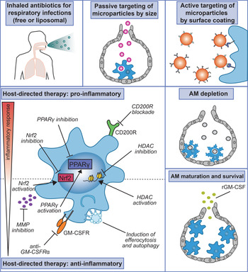
There are many strategies to deliver drugs directly to the lungs (Fig. 1). While we are unable to discuss all of the approaches here, there are several excellent and comprehensive reviews covering current strategies for pulmonary drug delivery (Refs Reference Ruge, Kirch and Lehr62, Reference Lee65). Here, we will highlight just a few of the approaches used to target AMs for therapy. Direct aerosolisation is designed to expose all cells in the airway to a drug or treatment, while other strategies such as nano- and micro-particle encapsulation and liposome formulations leverage size, shape, hydrophobicity, charge, surface ligands and the propensity of AMs to phagocytose particles to direct drugs to distal alveoli and into AMs (Ref. Reference Patel, Gupta and Ahsan66). For example, several studies have shown that particles 1–5 μm in diameter are optimal for AM uptake in vivo (Refs Reference Geiser44, Reference Pinkerton45). Particles smaller or larger are either quickly removed from the upper airway via the ‘mucociliary escalator’ or remain in the lower respiratory tract (Refs Reference Geiser44, Reference Patel, Gupta and Ahsan66–Reference Edwards68).
Therapeutic strategies to modulate alveolar macrophages. (clockwise from upper left) (1) During respiratory infections, antibiotics can be directed to AMs through inhaled antibiotics delivered either in free-form or packaged within liposomes. (2) Passive targeting modifies physiochemical characteristics such as size for optimal delivery to AMs. (3) Active targeting modifies the particle surface to express ligands that bind receptors on AMs to achieve enhanced uptake. (4) AM depletion strategies can limit the pathological effects of AM hyperinflammation. (5) Promotion of AM maturation and survival such as through rGM-CSF treatment can improve airway clearance and lung function. (6) Numerous host-directed therapies modulate specific molecular targets to either enhance or diminish the inflammatory response of AMs.

Active targeting modifies the particle surface to express ligands that bind receptors expressed by AMs to achieve enhanced uptake. Several different organic ligands have been shown to enhance uptake (Ref. Reference Kelly, Jefferies and Cryan69). Immunoliposomes are designed with antibodies on their surface to target cell-surface antigens or with Fc regions of antibodies to be recognised by Fc receptors (Ref. Reference Lawlor70). Mannosylated particles target mannose receptors and other lectins can similarly bind other scavenger receptors, such as Mincle or Dectin-1, that are highly expressed by myeloid cells.
Nanoparticles may be taken up through interaction with macrophage receptor with collagenase structure (MARCO), a scavenger receptor that is highly expressed on myeloid cells. MARCO has been shown to be a primary receptor for unopsonised aerosolised particles and bacteria both in murine and human AMs (Refs Reference Arredouani71, Reference Palecanda72).
There are additional strategies currently in development to target nanoparticles to macrophages from the field of cancer biology that could eventually be used to target AMs. For example, a lipid-based self-assembled supramolecule can be loaded with inhibitors that block the immunomodulatory and ‘don't eat me’ signals, CSF1R and SIRPα, respectively, and ultimately enhance phagocytotic activity and skew macrophage polarisation to enhance anti-tumour immunity (Refs Reference Kulkarni73, Reference Ramesh74). Myeloid-targeting approaches such as this could potentially be developed for aerosolised formulations that would specifically target and modulate AMs in the airway.
AMs are long-lived and subject to trained immunity
Recently, it has been shown that AMs are imprinted long-term by inflammatory signals in the context of several bacterial infections (Refs Reference Yao60, Reference Nemeth75). Following adenovirus exposure, AMs are reprogrammed by CD8+ T-cell-dependent IFNγ production that contributes to their enhanced ability to control Streptococcus pneumoniae infection months later (Ref. Reference Yao60). The Aderem lab (with A. Rothchild) previously showed that a contained model of Mtb infection, which generates a low-level of systemic cytokinemia including IFNγ, leads to a reprogramming of AMs resulting in an enhanced pro-inflammatory response to aerosol Mtb infection (Ref. Reference Nemeth75). Over the last few years, increasing evidence demonstrates that myeloid cells, specifically macrophages, can undergo immune training in response to inflammatory stimuli, such as β-glucan, Bacillus Calmette-Guerin (BCG) vaccination or Mtb (Refs Reference Kaufmann76–Reference Netea78). Efforts to target innate immunity for protection against respiratory infections such as Mtb are now being more seriously investigated in conjunction with traditional vaccination approaches (Ref. Reference Khader79).
Alveolar macrophage-targeted therapies
AM-directed antibiotics
Pulmonary drug delivery has become a popular and well-studied administration route. The lung possesses limited drug metabolizing enzyme activity which results in extended bioavailability and avoidance of first-pass metabolism compared to other routes. Due to the number of lower respiratory tract infections, there is great interest in adapting antibiotics for inhaled rather than oral delivery (Ref. Reference Quon, Goss and Ramsey80). Delivery of antibiotics directly to the lung, instead of systemically, should theoretically deliver a high concentration of antibiotics directly to the affected tissue and more effectively penetrate the lung parenchyma, while minimizing host toxicity (Ref. Reference Wenzler81). This strategy would be especially valuable for treating chronic respiratory infections, such as those commonly found in patients with cystic fibrosis, for which there have been several completed clinical trials (clinicaltrials.gov: NCT01519661, NCT01641822) and for infections that are resistant to first-line therapies. While several clinical trials have seen successful results using inhaled antibiotics, there is limited data on the efficacy of inhaled antibiotics in their current formulations for many lung infections.
For example, inhaled pentamidine is commonly used to treat pneumocystis pneumonia in HIV patients. A clinical trial from 1990 highlighted this as an effective therapy and suggested that success was related to direct delivery of the antibiotic to the alveolar space where P. carinii infection is concentrated (Ref. Reference Leoung82). A separate study suggested an alternative mechanism, in which inhaled pentamidine increased AM counts in BAL from pentamidine-treated HIV patients (Ref. Reference Wassermann83), which would benefit the host during infection because HIV infection can lead to partial AM deficiencies in binding and engulfment of P. carinii that impair bacterial clearance (Ref. Reference Koziel84). However, for non-HIV cases of pneumonia, inhaled antibiotic clinical trials have so far failed to show efficacy (clinicaltrials.gov: NCT01799993, NCT01969799).
Additionally, there is a desire to test the efficacy of inhaled antibiotics during TB infection, due to a shortage of new antibiotics in the R&D pipeline. Additionally, there are growing concerns that current systemic antibiotic delivery routes lead to an uneven distribution of drug in the lung, with drug concentrations within lung lesions below the Minimum Inhibitory Concentration (MIC), leading to ineffective therapy (Refs Reference Braunstein, Hickey and Ekins85, Reference Cicchese86). Despite concerns with current regimens, there have been few clinical trials conducted so far to test the efficacy of inhaled antibiotic formulations for TB, perhaps indicating that perceived limitations with feasibility, accessibility, and staff training requirements may have to be addressed before inhaled antibiotics are more widely adopted.
While previous antibiotic inhalation strategies required long administration times, recent advances in nebulisers that deliver a narrower range of particle size with dry powder drug formulations have improved treatment regimens (Ref. Reference Quon, Goss and Ramsey80). The lung possesses unique mucociliary and macrophage clearance mechanisms, which can generally impede tissue delivery and distribution (Ref. Reference Patton and Byron87); however, the latter characteristic has also been leveraged to develop specific drug treatments to target AMs.
A recent study targeted B. pseudomallei, the causative agent of melioidosis, using a polymeric ciprofloxin prodrug linked to mannose ligands. This facilitates drug uptake through binding to mannose receptors, which are highly expressed on AMs (Ref. Reference Chavas88). Using a mouse-adapted infection model of B. thailandensis and a B. pseudomallei clinical isolate, the authors showed that the therapy was significantly more effective than free drug at protecting mice from infection and showed improved pharmacokinetics in AMs.
Additionally, liposomal formulations have been shown to be effective for macrophage antibiotic delivery. Liposomal amikacin, marketed as Arikayce, is the first inhalable antibiotic via carrier formulation to be FDA approved and used clinically. Amikacin liposome inhalation suspension was able to penetrate non-tuberculous mycobacterial biofilms and showed enhanced efficacy of antibiotic uptake in infected macrophages (Ref. Reference Zhang89). Similarly, an inhaled liposomal formulation of ciprofloxin was shown to protect against a lethal infection of pneumonic plague in mice (Ref. Reference Hamblin90). In addition to the direct antibacterial effects, microparticle formulations carrying payloads of frontline antibiotics have been designed to stimulate and activate macrophages for pro-inflammatory cytokine production as a secondary host effect (Ref. Reference Sharma91).
Targets for host-directed therapy
PPARγ
PPARγ is a nuclear receptor that plays a critical role in the development of AMs as a downstream mediator of GM-CSF signalling (Ref. Reference Schneider11). PPARγ activation through intratracheal instillation of lentiviral driven gene expression has been shown to rescue lipid accumulation in AMs in GM-CSF-deficient animals (Ref. Reference Malur10). Also, PPARγ has been linked to all-trans retinoic acid (ATRA) synthesis, which may provide additional regulatory signals (Ref. Reference Gyongyosi92). During IAV infection in mice, PPARγ is downregulated in AMs through type I IFN signalling, leading to increased morbidity and inflammation, while restoration of PPARγ expression in AMs during early infection may be a novel therapy to improve host responses and limit pathology (Ref. Reference Huang93). In a bleomycin-induced lung injury model, activation of PPARγ through intratracheal instillation of apoptotic cells led to enhanced efferocytic activity of AM, TGFβ and IL-10 production by AM, and resolution of lung inflammation (Ref. Reference Yoon94). In contrast, PPARγ is induced in Mtb-infected macrophages and both knockdown and deletion lead to reduced bacterial growth of Mtb and enhanced pro-inflammatory responses, suggesting PPARγ activation during Mtb infection is detrimental for host control and may serve as a potential therapeutic target (Refs Reference Rajaram95, Reference Guirado96).
GM-CSF
GM-CSF is a growth factor that is essential for AM development (Refs Reference Dranoff8, Reference Schneider11), but it has also been explored as a therapy for limiting inflammation and damage in several pulmonary conditions (Ref. Reference Petrina, Martin and Basta97). GM-CSF can have opposing roles in both limiting and exacerbating lung inflammation depending on the stage of disease. Therefore, paradoxically, both GM-CSF activation and inhibition strategies have been explored for COVID-19 therapy (Ref. Reference Lang98). In studies of other infections, the enhancement of tissue repair following treatment with recombinant GM-CSF (rGM-CSF) was shown to be AM-dependent. Intranasal treatment with rGM-CSF led to protection against a murine H1N1 IAV infection and protection was dependent on the presence of AMs (Ref. Reference Huang99). In a mouse model of allergic airway inflammation that exacerbated RSV infection, GM-CSF treatment led to a reduction in RSV susceptibility and tissue damage dependent on AM maturation (Ref. Reference Naessens100). Inhaled Leukine® (sargramostim), an FDA-approved rGM-CSF treatment, improved oxygenation scores in the 6 patients with pneumonia-associated acute respiratory distress syndrome (ARDS) and was associated with a shift from M2 to M1 in alveolar mononuclear phagocytes (Ref. Reference Herold101). Treatment with Leukine® is currently being tested for COVID-19 patients with hypoxic respiratory failure (clinicaltrials.gov: NCT04400929, NCT04326920). In contrast, GM-CSF blockade with mavrilimumab (anti-GM-CSFRα mAb) limited lung damage associated with severe late-stage COVID-19 pneumonia and hyperinflammation (Ref. Reference De Luca102).
CD200-CD200R
CD200R1 (previously called OX-2R) is a membrane-bound receptor that is highly expressed on myeloid cells and at lower levels on T and B lymphocytes. CD200R1 is activated by CD200 binding. CD200 lacks its own intracellular domain, and CD200-CD200R1 signalling is thought to be unidirectional through the CD200R cytoplasmic domain. CD200-CD200R signalling has been shown to lead to a dampened immune response and tissue inflammation. CD200R is highly expressed on AMs, and during IAV infection CD200 expression on either epithelial cells or T cells is critical for minimizing a hyperinflammatory AM response (Ref. Reference Snelgrove27). CD200−/− mice have an overexuberant AM immune response following IAV infection with increased expression of TNF and IL-6 in response to ex vivo LPS stimulation and increased cell recruitment and pulmonary tissue damage. Restoration of CD200-CD200R signalling through treatment with a soluble fusion protein or CD200R antibody reduced inflammation and pathology without compromising viral clearance, suggesting that CD200R agonists may have therapeutic potential in reducing viral-driven inflammatory damage (Ref. Reference Snelgrove27). Interestingly, the CD200R-CD200 axis is also reported to play an important immunomodulatory role in bacterial and parasitic infections and cancer (Refs Reference Vaine and Soberman103, Reference Mahadevan104). CD200-CD200R blockade is currently being explored as an immunotherapy for cancer, with phase I results indicative that modulation of CD200 activity is well tolerated and safe in humans (Ref. Reference Mahadevan104).
Nrf2
Nrf2 is a master transcriptional regulator that is protective against inflammatory damage and oxidative stress. Pulmonary expression is primarily restricted to AMs and epithelial cells. Decreased Nrf2 expression is thought to contribute to COPD and smoking-induced emphysema through decreases in antioxidant gene expression including HO-1 (Refs Reference Boutten105, Reference Rangasamy106). Nrf2 is activated following inflammation in pulmonary conditions including lung injury (Ref. Reference Zhao107), COPD (Ref. Reference Boutten105) and chronic Mtb infection (Refs Reference Andrade108, Reference Scharn109) and aids in the resolution of pulmonary inflammation and damage (Ref. Reference Reddy110). Several Nrf2 agonists are currently being explored for the treatment of COPD and other inflammatory conditions (Ref. Reference Boutten105). We recently found that Nrf2 is up-regulated in Mtb-infected AMs early during infection (Ref. Reference Rothchild18). In the case of acute low-dose infection, Nrf2 expression in AMs was detrimental to early bacterial control, blocking rapid myeloid activation and pro-inflammatory responses. Therefore, targeting Nrf2 may be a therapeutic strategy for modulating the AM inflammatory response.
Efferocytosis
One strategy to modulate AM function has focused on improving efferocytic activity, the uptake of dead and dying cells. For inflammatory disorders, such as COPD, enhanced efferocytosis in the airways has been shown to reduce pulmonary inflammation (Ref. Reference Grabiec and Hussell111). Efferocytosis by AMs involves receptors for phosphatidylserine such as T-cell immunoglobulin and mucin domain containing 4 (Tim-4), high mobility group protein-1 (HMGB1) (Ref. Reference Liu112) and mannose-binding lectin (MBL) (Ref. Reference Ogden113). One ongoing study aims to investigate compounds that induce efferocytosis by AMs in COPD patients, with the goal of minimizing proinflammatory cytokine production and improving patient outcome (clinicaltrials.gov: NCT04775394). In another study, the antibiotic azithromycin was shown to significantly increase apoptotic cell clearance, dependent on phosphatidylserine recognition in AM from COPD patients (Ref. Reference Hodge114).
Autophagy
Autophagy is the process by which cells engulf intracellular pathogens, whole organelles, or protein aggregates in autophagosomes, which then deliver the cargo to the lysosome for clearance. Several inhaled therapies promoting autophagy in macrophages have been studied in the context of TB infection. ATRA is a metabolite of vitamin A that can be generated directly by AMs. Treatment with ATRA reduced bacterial burden in human Mtb-infected AMs in a process dependent on autophagy and TBK1 (Ref. Reference Coleman115). In another recent study, treatment of Mtb-infected mice with inhaled ATRA-loaded microparticles targeted to macrophages led to enhanced bacterial control (Ref. Reference O'Connor116).
Under standard conditions, mTORC1 inhibits autophagy by interacting with the pre-initiation complex, preventing autophagosome formation. Rapamycin, an mTOR inhibitor, has shown promise in testing for TB treatment in combination with standard antibiotic regimens (Ref. Reference Singh and Subbian117). Additionally, rapamycin-loaded PLGA microparticles can be taken up efficiently by Mtb-infected macrophages in vitro limiting bacterial growth and presenting fewer cytotoxic side effects than rapamycin solution, a therapy that is highly suitable for aerosol delivery (Ref. Reference Gupta118). Everolimus, another mTOR inhibitor, was recently tested as adjunctive host-directed therapy for TB (Ref. Reference Wallis119). Preliminary evidence suggests that it was well tolerated in patients simultaneously receiving standard-of-care antibiotic treatment and may improve lung function (clinicaltrials.gov: NCT02968927).
Matrix metalloproteases
MMPs are proteases that degrade the extracellular matrix and can induce pro-inflammatory and pro-fibrotic factors. In the lung, excess MMP secretion is associated with tissue damage and disease progression in several inflammatory conditions including COPD, emphysema, asthma and idiopathic pulmonary fibrosis (Ref. Reference Vandenbroucke, Dejonckheere and Libert120). Activated macrophages have been shown to be some of the main producers of MMPs. MMP inhibitors have been tested in several inflammatory respiratory conditions to limit inflammation. In one study investigating the increased risk for emphysema in HIV+ patients that smoke, AMs from HIV+ smokers were shown to express higher levels of several MMPs including MMP-1, MMP-9 and MMP-12 than AMs from HIV- smokers suggesting that enhanced MMP production may contribute to the increased risk for emphysema among HIV+ smokers (Ref. Reference Kaner, Santiago and Crystal121). The same group is now running a clinical trial to test whether doxycycline, a widely used antibiotic that also functions as a broad MMP inhibitor, is effective at treating COPD and/or emphysema in HIV-infected patients (clinicaltrials.gov: NCT01744093).
Histone deacetylases
Histone deacetylases (HDACs) remove acetyl groups from histones, altering the chromatin structure and ‘deactivating’ genetic loci by making them less accessible to transcription factors and enhancers. HDACs generally function in opposition to histone acetyl transferases and can be regulated during infection, for example, to dampen the immune response. The HDAC inhibitor suberanilohydroxamic acid promotes glycolysis and pro-inflammatory responses in Mtb-infected human AMs and enhances their ability to stimulate CD4+ T cells (Ref. Reference Cox122). AMs from COPD patients have significantly reduced HDAC activity, which is associated with increased release of IL-8 and TNFα. Therefore, activation of HDACs has been tested as a treatment for COPD. In one clinical trial, stimulation of HDAC activity in COPD patients led to enhanced dexamethasone-mediated suppression of IL-8 in AMs (Ref. Reference Cosio123) (clinicaltrials.gov: NCT00671151).
Resolvin D1
Resolvin D1 (RvD1), a lipid mediator that was originally described as a product of aspirin-triggered cyclooxygenase-2, has a role in resolving lung inflammation. In a mouse model of E.coli-induced pneumonia, treatment with RvD1 led to enhanced phagocytosis and clearance of neutrophils by macrophages from the BAL and was associated with increased bacterial clearance alone or in combination with antibiotics (Ref. Reference Abdulnour124). RvD1 treatment was also shown to enhance AM phagocytic ability in vivo and ex vivo and dampen pro-inflammatory cytokine production in a murine model of cigarette-smoke exposure (Ref. Reference Hsiao125). RvD1 treatment also reduced lung inflammation and tissue damage following infection with Pseudomonas aeruginosa and was associated with a decrease in pro-inflammatory gene expression from lung-sorted macrophages (Ref. Reference Codagnone126). These studies suggest that RvD1 treatment can lead to enhanced resolution of lung inflammation in part from direct actions on macrophages.
Alveolar macrophage depletion
While AM depletion is unlikely to be a viable therapeutic strategy, reduction in disease progression in animal models following AM depletion highlights the contribution of AMs to lung pathogenesis. During RSV infection, with AM RIPK3-dependent necroptosis leading to lung inflammation and immunopathology, AM depletion decreases RSV susceptibility (Ref. Reference Santos29). AM depletion also prevents progressive lung pathology in a murine smoking model of COPD (Ref. Reference Beckett58), protects against lung damage in a SARS-CoV-2 hACE-transgenic murine model (Ref. Reference Lv31), and reduces pulmonary fibrosis in a bleomycin-induced mouse model (Ref. Reference Li127).
Alveolar macrophage-targeted therapy for COVID-19
COVID-19, caused by infection with SARS-CoV-2, was first identified in late 2019 and quickly spread globally leading to a worldwide pandemic. The virus can be spread by multiple routes that lead to pulmonary infection. SARS-CoV-2 transmission can occur when an uninfected person comes in contact with droplets or aerosols that contain virus or through contact with surfaces (fomites) that have been contaminated with infected droplets (Ref. Reference Wang128). In severe cases of COVID-19, infection can lead to ARDS coupled with cytokine storm and multi-organ failure (Ref. Reference Cevik129). Ongoing studies have highlighted the critical role that AMs play during SARS-CoV-2 infection and potential therapies aimed at AMs that may impact COVID-19 disease progression.
AMs are directly infected by SARS-CoV-2
A recent study that performed both bulk and single-cell RNA-sequencing on BAL samples from 88 COVID-19 patients found that AMs could be directly infected with SARS-CoV-2 and that AMs and T cells in the airway formed a positive feedback loop resulting in exacerbated lung inflammation (Ref. Reference Grant130). The authors concluded that this cellular interaction in turn promotes alveolitis and lung pathology in severe COVID-19 patients. The finding that AMs can harbour SARS-CoV-2 virus has also been confirmed by other observational studies (Ref. Reference Bost131), and animal models have shown that AM can support SARS-CoV-2 replication (Ref. Reference Lv31). Other studies have confirmed that AMs express the receptors required for viral entry (ACE2, TMPRSS2, etc.) (Refs Reference Wang132, Reference Abassi133). These data show that when SARS-CoV-2 enters the airway and moves into the lower respiratory tract, AMs can serve as a site of viral entry and replication.
AMs exhibit dysfunctional immune responses during COVID-19
AM dysfunction potentially plays opposing roles in two different stages of COVID-19 disease, dampening the early innate response and accelerating a hyperinflammatory late stage of disease (Ref. Reference Merad and Martin134).
A dampened early AM response during SARS-CoV-2 infection is directly influenced by age, one of the biggest risk factors for COVID-19 disease severity. Ageing alters the pulmonary environment and has been shown to produce dysfunctional AM responses, including deficiencies in phagocytosis, ineffective innate sensing and a decline in number due to impaired self-renewal (Refs Reference McQuattie-Pimentel5, Reference Schneider135). The detrimental effect of ageing on AM antiviral function was demonstrated in a murine model of influenza in aged mice (Ref. Reference Wong28). Despite reports that SARS-CoV-2 generates a robust type I IFN response in the airway (Ref. Reference Zhou32), an impaired type I IFN response has been described for both monocytes and macrophages from peripheral blood and airway macrophages from BAL of COVID-19 patients (Refs Reference Bost131, Reference Hadjadj136, Reference Blanco-Melo137). The lack of an early type I IFN response is paradoxically associated with a more severe and hyperinflammatory immune response during the later stages of disease, highlighting the substantial impact that the early innate response can have on later disease (Refs Reference Carvalho, Krammer and Iwasaki138–Reference Zhang140).
Dysregulated hyperinflammatory macrophages have been observed in the airways in severe COVID-19 disease (Refs Reference Zhou32, Reference Melms34, Reference Szabo64). The presence of these highly activated and inflammatory macrophages is associated with diffuse alveolar damage, disease severity and death (Ref. Reference Melms34). A study using a high-throughput myeloid-cell receptor screening approach found that several C-type lectin receptors that are highly expressed on AMs could bind the viral spike protein and that binding led to myeloid cell activation and proinflammatory responses, rather than viral entry suggesting that AMs could become highly activated even in the absence of direct infection (Ref. Reference Lu141). The authors also tested therapeutic nanobodies that blocked these host–viral interactions and reduced disease severity (Ref. Reference Lu141). In an hACE-transgenic mouse model of SARS-CoV-2, depletion of AMs led to a reduction in viral load and lung pathology, while IFNγ activation of AM increased their susceptibility for viral replication, suggesting that in an inflammatory environment AM may become even more detrimental for SARS-CoV-2 control (Ref. Reference Lv31).
In addition to dysfunctional AM responses during early and late stages of disease, the replacement of tissue-resident AMs with a more inflammatory monocyte-derived macrophage population has been identified as one mechanism driving disease-associated lung damage. Single-cell RNA sequencing of BAL samples from SARS-CoV-2 patients revealed that during severe late-stage disease monocyte-derived macrophages replace tissue-resident AMs and the disappearance of resident AMs was associated with greater pulmonary damage (Ref. Reference Liao30). Additionally, the lack of resident macrophage proliferation and an increase in macrophage recruitment chemokine expression in the lung are highly suggestive that peripheral blood monocyte-derived cells may seed the lung with these hyperinflammatory cells during infection (Ref. Reference Szabo64). Both transcriptional and multi-omics approaches have also described dysfunctional and hyperinflammatory macrophage and monocyte populations in the blood (Refs Reference Szabo64, Reference Su142) and longitudinal immune profiling observed increases in Ki-67+ monocyte population suggestive of early release of monocytes from the bone marrow during disease (Ref. Reference Mann143). In a macaque model of SARS-CoV, CD163+ CD68+ airway macrophages accumulate in the lungs and are simultaneously polarised towards both pro-inflammatory (IL-6+) and wound-healing (TGFβ +) phenotypes, by a mechanism dependent on anti-spike IgG and FcγRs (Ref. Reference Liu144). This accumulation led to an increased risk for acute lung injury following infection. These studies suggest that a major driver of late-stage lung inflammation and damage may be the replacement of resident AMs with hyperinflammatory monocyte-derived cells that have recently emerged from the bone marrow. Strategies to retain or mature resident AM populations, in combination with approaches to manage over-activated neutrophils and dendritic cells that also contribute to pulmonary inflammatory damage (Ref. Reference Zhou32), may be able to stop the feedforward cycle of pulmonary inflammation and tissue damage.
Therapeutic strategies to modulate AMs during COVID-19
While most host-directed therapy studies for COVID-19 have focused on blocking systemic inflammatory cytokines such as IL-6 and TNF to minimise lung pathology, several studies are evaluating whether lung damage can be avoided in COVID-19 patients by either promoting or blocking GM-CSF signalling (Ref. Reference Lang98). GM-CSF has a direct role in AM development and maintenance of airway homeostasis (Ref. Reference Hamilton145) and providing rGM-CSF is thought to help mature resident AMs and improve airway clearance. In a previous clinical trial, inhaled Leukine®, an FDA-approved rGM-CSF treatment, led to significantly improved lung function for patients with pneumonia-associated ARDS and a shift to an M1 phenotype in alveolar mononuclear phagocytes (Ref. Reference Herold101). Treatment with Leukine® is currently being tested against hypoxic respiratory failure in COVID-19 patients (clinicaltrials.gov: NCT04400929, NCT04326920). In patients suffering from hyperinflammation, GM-CSF blockade through mavrilimumab (anti-GM-CSFRα mAb) therapy has also shown efficacy in inhibiting lung pathology associated with severe late-stage COVID-19 pneumonia (Ref. Reference De Luca102).
The strategy to induce trained immunity in innate cells (monocytes, macrophages and NK cells) to augment the initial response to SARS-CoV-2 has also been evaluated (Refs Reference Netea78, Reference Netea146). While BCG vaccination has been effective at boosting trained immunity in animal models to limit respiratory infections such as Mtb (Ref. Reference Kaufmann76), results from clinical trials examining BCG vaccine-induced protection from SARS-CoV-2 have yet to report significant effects.
Ongoing questions and conclusions
Concerns and challenges for therapeutic potential
There are several challenges facing AM-targeted therapies. One of these is the unintended and off-target effects of aerosol delivery systems (Ref. Reference Forbes147). Inhalation of drugs or therapeutics can lead to irritation in nasal passages and upper airways (trachea, bronchi). Pulmonary delivery, when dosed incorrectly, can lead to AM overloading that can produce a ‘foamy’ appearance associated with impaired phagocytosis and airway clearance (Ref. Reference Forbes147). Additionally, targeting the respiratory tract through drug inhalation can present difficulties in people with severely compromised and inflamed airways, often the patients that are most likely to benefit from therapy. Fortunately, respiratory side effects can now be carefully monitored during clinical trials using functional read-outs such as spirometry, forced vital capacity, forced expiratory volume or cellular read-outs from induced sputum, BAL or bronchoscopy (Ref. Reference Forbes147).
An additional obstacle for aerosol therapy is the lack of widespread patient access to intranasal or inhalation drug delivery procedures, which can require special nebulisers or equipment and specifically trained health-care workers. These distinctive requirements for therapy are an additional hurdle for access, especially for patients in areas with poor healthcare infrastructure who might reap the biggest benefits from them.
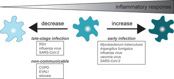
The third and most difficult impediment to AM-targeted therapies is the expected and unanticipated consequences to modulating AM function. Because AMs serve critical homeostatic roles in the lung, any efforts to either limit their responses or promote their activation could potentially lead to dysfunctional airway and pathogen clearance, hyperinflammation or tissue remodelling (Ref. Reference Hussell and Bell14). One crucial step to overcoming this hurdle is to gain a better understanding of the most effective treatment window for each of the respiratory diseases discussed above. If the effort to modify AMs is analogous to dialling up or down a thermostat of pulmonary inflammation, an effective treatment must find the right balance and timing between too little and too much inflammation. Based on many of the studies discussed above, it is possible that tuning AM inflammatory responses ‘up’ during early and acute infection, and then ‘down’ during late and chronic stages may provide the most beneficial outcome (Fig. 2).
‘Tuning’ the inflammatory response of alveolar macrophages. Increasing or decreasing AM inflammatory responses could have therapeutic benefits depending on the disease and stage of disease. Several examples of diseases that might benefit from direct targeting of AMs are shown here. RSV, respiratory syncytial virus; SARS-CoV-2, severe acute respiratory syndrome coronavirus 2; COPD, chronic obstructive pulmonary disorder; EVALI, e-cigarette or vaping product use-associated lung injury.

Remaining questions
There are many questions remaining and work to be done to fully understand the role of AMs during complex disease states and identify effective strategies to target them. While the hyperinflammatory state during chronic late-stage pulmonary diseases is well documented, additional studies that focus on the very early events in infection and initial environmental triggers of inflammatory conditions are critically needed to understand the role of tissue-resident AMs in the induction of disease. While human studies and in vivo animal models are ideal for identifying relevant cellular states, these studies are often impractical for answering mechanistic questions. As a result, there is also a substantial need to develop new in vitro models to study AM biology that more accurately recapitulate the lung microenvironment.
AMs are critical innate immune sentinels with untapped therapeutic potential due to their positioning at the airway-mucosal barrier and their role in the uptake and early response to many microbial and environmental airborne insults. However, there is still much to learn about how these cells are regulated and how their function can be modulated for clinical purposes.
Acknowledgements
We thank our colleagues in the Department of Veterinary and Animal Sciences at University of Massachusetts Amherst, including Barbara Osborne and Leonid Pobezinsky, for helpful discussions and feedback.
Financial support
This work was supported by start-up funds from the University of Massachusetts Amherst, College of Natural Sciences, Department of Veterinary and Animal Sciences.
Conflict of interest
None.