Introduction
Laryngeal framework surgery is an operation that alters the position and tension of the vocal folds and improves symptoms by manipulating the cartilaginous tissues constituting the larynx. One of the procedures used for this surgery is that devised by Isshiki et al.;Reference Isshiki, Tanabe and Sawada1 specifically, arytenoid adduction for the purpose of moving the vocal fold to the adduction position and fixing it in place. This procedure is performed primarily in patients with vocal fold atrophy or arytenoid cartilage shake because of deterioration in intralaryngeal muscle function, leading to severe glottic insufficiency.
In this operation, a nylon thread is attached to the arytenoid cartilage and then drawn, in order to move the paralysed vocal fold to the physiological adduction position. However, under normal conditions, it can be difficult to guide the arytenoid cartilage (which is controlled by multiple muscles and ligaments) to the physiological adduction site using a single nylon thread. Indeed, we have encountered cases where we were unable to move the target vocal fold to the desired position. A degree of improvement in speech quality may be obtained when the vocal fold is fixed in place, even if movement to the adduction site is insufficient; however, this partial movement is an unacceptable outcome in patients seeking restoration of the speech capacity they had before the onset of paralysis. These patients may demand further improvement.
This article describes our experience of performing revision surgery at our hospital in two patients who were not satisfied with the quality of their speech after surgery performed to treat vocal fold palsy carried out by other surgeons. We also discuss the techniques used during these surgical procedures.
Case reports
Patient one
The patient was a 65-year-old male monk who developed left-sided vocal fold palsy following surgical treatment of thyroid cancer. Ten months later, when there was no improvement in vocal fold palsy, and his ability to work was greatly impeded by hoarseness, the patient's previous surgeon performed arytenoid adduction (using a posterior thyroplasty window approachReference Maragos2) and a thyroplasty type 1 procedure. Although there was some improvement in speech quality following surgery, the hoarseness remained. Therefore, the patient still found it difficult to work at his desired level and came to our hospital requesting additional treatment to further improve the quality of his speech.
On examination, the left vocal fold was located in the middle position. We determined that the adduction effect achieved by the prior surgery was inadequate and so planned to perform revision surgery. A combination of arytenoid adduction and type 1 thyroplasty procedures had been performed at the time of the patient's initial surgery. When performing the revision surgery, it was necessary to mitigate the influence of each previous surgical procedure in order to identify the factors responsible for the unsatisfactory improvement in speech quality. For this reason, we explained to the patient that only arytenoid adduction would be performed as revision surgery. Informed consent was obtained from the patient.
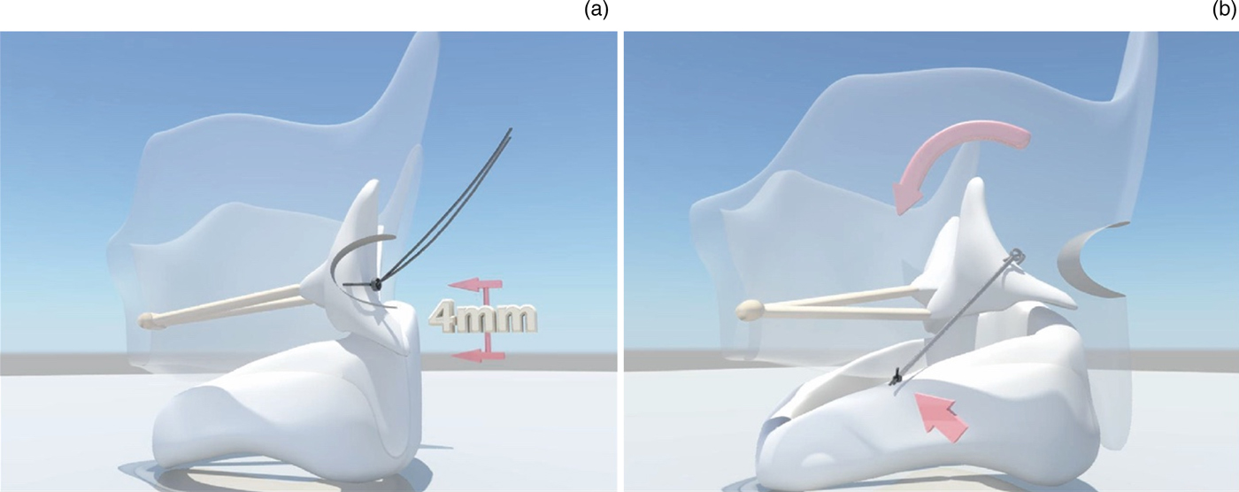
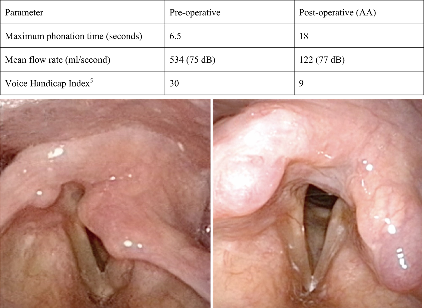
We performed the revision surgery nine months after the patient's initial surgery. The surgery was performed under general anaesthesia using a laryngeal mask. The arytenoid cartilage was identified via a posterior thyroplasty window approach. The mucosa of the piriform fossa was adherent to the arytenoid cartilage as a consequence of the initial surgery, but could be separated by careful manipulation without causing perforation. The nylon thread used in the initial surgery was checked and removed intra-operatively up to the point at which the arytenoid cartilage was exposed; thereafter, the position at which the thread had been fixed to the arytenoid cartilage and the direction of pulling could not be confirmed. After confirming the mobility of the arytenoid cartilage, a size 4-0 nylon thread was attached to the base of the muscular process (approximately 4 mm from the tip of the processReference Geo, Cui, Sun, Zhang, Zhou and Zhao3) and used as an adduction thread (Figure 1a, arrows). The adduction thread was run through the point at which the cricoid and thyroid cartilages intersect, and then fixed to the thyroid cartilage (Figure 1b, arrows). We confirmed that the vocal fold was moved to the adduction point using a flexible laryngoscope, after which the operation was completed (Figure 2).
Position of adduction thread attachment, pulling direction and fixation position. (a) The adduction thread was attached to the base of the muscular process (approximately 4 mm from the tip of the process). (b) The adduction thread was run through the point at which the cricoid cartilage and the thyroid cartilage intersect and then fixed to the thyroid cartilage.

Pre- and post-operative examination results and laryngeal findings in case one. All parameters assessed were found to have improved after surgery when compared to the pre-operative period. Post-operatively, the left vocal fold was moved closer to the adduction site, and the arytenoid mucosa covering the glottis (lower left image) was returned to its normal position (lower right image). AA = arytenoid adduction

The patient's voice was not checked intra-operatively. His vocal quality improved after surgery and he was able to work again at his desired level (Figure 2).
Patient two
The patient was a 52-year-old male lecturer who developed left-sided vocal fold paralysis following surgical treatment of a mediastinal schwannoma. No improvement in hoarseness was observed during one year of follow up, and the patient subsequently underwent arytenoid adduction (with a fenestration approachReference Tokashiki, Hiramatsu, Tsukahara, Kanebayashi, Nakamura and Motohashi4) and type 1 thyroplasty. Although his vocal quality improved post-operatively, the hoarseness remained such that the patient still found it difficult to work at his desired level, and so he came to our hospital requesting additional treatment to further improve his speech quality.
On examination, the left vocal fold was located in the middle position. We determined that the adduction effect achieved by the prior surgery was inadequate and so planned to perform revision surgery. However, as in case one, we explained that initially only arytenoid adduction would be performed. The patient's consent was obtained.
Ten months after the patient's initial surgery, we performed revision surgery under general anaesthesia using a laryngeal mask. The arytenoid cartilage was identified via the posterior thyroplasty window approach. As in case one, the mucosa of the piriform fossa was adherent to the arytenoid cartilage because of the effects of the initial surgery, but could be separated by careful manipulation without causing perforation. Moreover, although the nylon thread used in the initial surgery was checked and removed during the operation up to the point at which the arytenoid cartilage was exposed, it was not possible to confirm the position where the thread had been fixed to the arytenoid cartilage or the direction of pulling. As in case one, after confirming the mobility of the arytenoid cartilage, the adduction thread was attached to the base of the muscular process; it was then pulled in the same direction and the vocal fold was fixed in place. We confirmed that the left vocal fold was moved to the adduction point using a flexible laryngoscope, after which the operation was completed. The patient's voice was not checked during surgery.
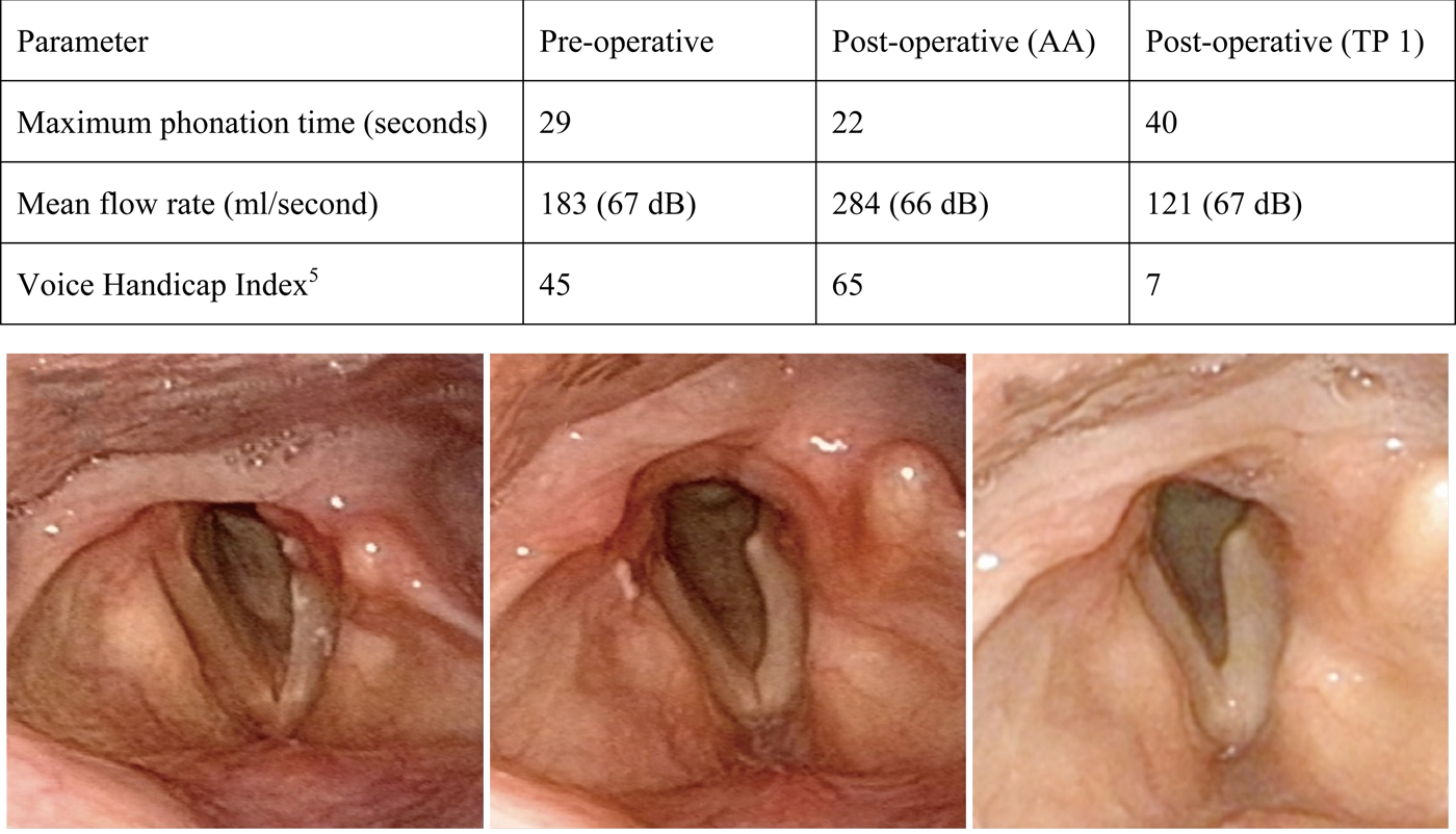
The left vocal fold was confirmed to be moved to the midline after surgery, and clear arcuate changes attributable to nerve paralysis became apparent as the post-operative swelling subsided (Figure 3). Furthermore, aerodynamic examination and subjective assessmentReference Shiromoto, Oridate, Ikui, Taguchi, Mizoguchi and Watanabe5 indicated worsening when compared with the pre-operative state. We determined that medialised movement of the arcuate vocal fold would be necessary to obtain improvement in vocal quality. Nine months after the revision surgery, a type 1 thyroplasty was performed under local anaesthesia using a specially designed titanium plate.Reference Matsushima6 After surgery, the patient's vocal capacity improved and he was able to work at his desired level (Figure 3).
Pre-operative (lower left image) and post-operative (lower middle and right images) examination results and laryngeal findings in case two. Following arytenoid adduction (lower middle image), marked arcuate changes as a result of vocal fold atrophy were observed, and all parameters assessed were worsened when compared with the pre-operative period. Thyroplasty was performed to correct these arcuate changes (lower right image) and all parameters examined showed improvement. AA = arytenoid adduction; TP 1 = type 1 thyroplasty

Discussion
Arytenoid adduction is generally indicated in patients with an oversized glottal gap during vocalisation. Awareness of cases of arytenoid cartilage shake during vocalisation is increasing, and arytenoid adduction is also indicated for this condition.Reference Hiramatsu, Tokashiki and Suzuki7 Therefore, vocal fold palsy is becoming an increasingly common indication for arytenoid adduction surgery.
The key points in this surgical technique are: the method of approaching the arytenoid cartilage, the position at which the adduction thread is attached to the arytenoid cartilage, and the direction in which this thread is pulled to move the target vocal fold.
Various approaches to the arytenoid cartilage have been reportedReference Maragos2,Reference Tokashiki, Hiramatsu, Tsukahara, Kanebayashi, Nakamura and Motohashi4,Reference Su, Lui, Lin, Chiu and Cheng8,Reference Murata, Yasuoka, Shimada, Shino, Iida and Takahashi9 since the aforementioned report by Isshiki et al.Reference Isshiki, Tanabe and Sawada1 At our hospital, we utilised the posterior thyroplasty window approach, and maintained the external laryngeal and pharyngeal constrictor muscle groups, which play an important role in swallowing and work to supplement and compensate during vocalisation. These muscles also work to preserve the outer branches of the superior laryngeal nerve and maintain the function of the cricothyroid muscle.
There have been a few reports published on the fixation position and direction of traction for the adduction thread. Su et al.Reference Su, Tsai, Chuang and Chiu10 reported that the speech improvement achieved as a result of sharply tracking the adduction thread towards the caudal side was superior to that achieved when using the method described by Isshiki et al.Reference Isshiki, Tanabe and Sawada1 The arytenoid cartilage forms a cylindrical joint with the cricoid cartilage and allows for rotational motion.Reference von Leden11 Therefore, in order to move the vocal process of the arytenoid cartilage of the paralysed vocal fold inward and downward, we can infer that the application of force to the arytenoid cartilage towards the caudal side will be effective. With this notion in mind, in our revision surgery cases, we attached an adduction thread to the base of the muscular process, passed the thread through the point where the cricoid cartilage and thyroid cartilage intersect, and fixed the thread to the thyroid cartilage. The effectiveness of this pulling method was verified by demonstrating that the vocal fold could be moved to the adduction position by pulling at an angle equal to or more acute than the pulling angle described in the report by Su et al.Reference Su, Lui, Lin, Chiu and Cheng8
There have been sporadic reports comparing the efficacy of surgical procedures performed in cases of vocal fold palsy. However, no significant differences in the degree of improvement in speech quality have been observed between groups that underwent type 1 thyroplasty alone and groups that underwent a combination of type 1 thyroplasty and arytenoid adduction.Reference Siu, Tam and Fung12–Reference Benninger, Manzoor and Ruda14 These reports did not consider whether each type of surgery used the optimal procedures because they only examined quality of speech post-operatively.
Both of the cases reported here involved patients who had undergone a combination of both arytenoid adduction and type 1 thyroplasty. In case one, because the effect of type 1 thyroplasty was found to be sufficient, we were able to obtain further improvement in vocal quality by performing revision surgery using only an adduction procedure. However, in case two, the patient's speech worsened as a result of adduction. Our experience with these two cases suggests that both surgical procedures should be sufficiently able to obtain favourable improvement in vocal quality when performed to treat vocal fold palsy.
While we also considered a method for incorporating adductive movement prior to the type 1 thyroplasty procedure, we determined that the physiological rotation of the arytenoid cartilage could not be reproduced by applying lateral pressure alone. As such, in order to move the vocal fold to the adduction position and reduce the size of the glottal gap during vocalisation, we decided to perform an adduction procedure first.
• This study investigated the position of adduction thread attachment, pulling direction and fixation position in revision arytenoid adduction surgery
• The adduction thread was attached to the base of the muscular process
• The adduction thread was run through the point at which the cricoid and thyroid cartilages intersect, and fixed to the thyroid cartilage
• Arytenoid adduction and type 1 thyroplasty can improve vocal quality when performed to treat vocal fold palsy
In conclusion, we performed revision surgery in two patients in whom vocal quality had not improved to the desired level despite the patients having undergone a combination of the standard treatments for vocal fold palsy, specifically arytenoid adduction and type 1 thyroplasty. We were able to achieve the desired level of vocal improvement in both patients by performing arytenoid adduction procedures with or without type 1 thyroplasty. Although no consensus has been reached regarding arytenoid adduction, the surgical procedures described in this paper are effective and could become index procedures.
Acknowledgement
The authors would like to express their sincere gratitude to the surgeons who performed the initial procedures in these patients and who agreed to the publication of this report.
Competing interests
None declared