Introduction
Folate is a water-soluble B vitamin essential for one-carbon metabolism; it participates in DNA methylation by serving as the primary source of methyl group donors in homocysteine-to-methionine conversion and the subsequent S-adenosylmethionine (SAM) synthesis. Reference Bailey, Stover and McNulty1,Reference Stover2,Reference Ebara3 Because SAM is the universal methyl donor for DNA methylation reactions, folate availability during critical developmental windows directly shapes the capacity of methyltransferases to establish and maintain epigenetic marks. Reference Stover2,Reference Ebara3 This biochemical dependency becomes especially consequential during embryogenesis and fetal growth, Reference Reik, Dean and Walter4,Reference Tamura and Picciano5 when the genome undergoes extensive reprogramming and when the epigenetic landscape governing gene expression throughout postnatal life is being established. Reference Crider, Yang and Berry6,Reference Van Vliet, Schoenmakers and Gribnau7
Among the genomic regions most sensitive to disruptions in this process are imprinted loci, where allele-specific methylation patterns enable parent-of-origin-dependent gene expression. Reference Tucci, Isles and Kelsey8 The IGF2-H19 locus on chromosome 11p15.5 is one of the most extensively studied imprinted domains in the human genome. Reference Soejima and Higashimoto9 It primarily governs insulator function through allele-specific binding of the CTCF zinc-finger protein, as a result of methylation of the imprinting control region (ICR). Reference Bell and Felsenfeld10,Reference Hark, Schoenherr and Katz11 On the maternal allele, the unmethylated state of the ICR allows for CTCF binding thereby preventing downstream enhancers from interacting with IGF2 and promoting expression of H19. Conversely, the methylated state of the ICR on the paternal allele prevents CTCF binding thereby allowing enhancer access to IGF2 while silencing H19. Reference Bell and Felsenfeld10,Reference Hark, Schoenherr and Katz11 This coordinated mechanism regulates fetal growth, placental development, and postnatal metabolic programming. Reference Constancia, Hemberger and Hughes12,Reference Sandovici, Georgopoulou and Pérez-García13 Disruption of methylation at this locus has been associated with growth disorders as Beckwith-Wiedemann and Silver-Russell syndromes, and with altered risk for childhood cancers and metabolic disease in later life. Reference Soejima and Higashimoto9
Given this architecture, pregnancy represents a plausible window during which maternal folate intake could influence methylation at the IGF2-H19 locus in offspring tissues. Reference Reik, Dean and Walter4 Experimental animal data demonstrate that dietary methyl-group donors alter imprinted gene methylation at epigenetically labile loci in a dose-dependent manner; moreover, early embryogenesis, particularly the periconceptional period and first trimester, is when de novo methylation of DMRs is actively established. Reference da Mota, Ribeiro and Carvalho14–Reference Mahajan, Sapehia and Rahat16 Public health recommendations for periconceptional folic acid supplementation are among the most robustly established in reproductive medicine, Reference Soejima and Higashimoto9,17,Reference Czeizel and Dudas18 yet the downstream epigenetic consequences of folate exposure at specific imprinted loci in human offspring remain far less clear.
Over the past two decades, a growing body of epidemiological and interventional research has examined whether variation in maternal folate intake, from dietary sources, supplementation, or circulating biomarkers, is associated with DNA methylation changes at the IGF2-H19 locus in placenta, cord blood, or other birth-related tissues. These studies have employed diverse designs across multiple populations, and their findings have been heterogeneous, reflecting substantial variation in the regions analyzed, tissues sampled, timing of exposure assessment, and statistical approaches. Reference Steegers-Theunissen, Obermann-Borst and Kremer19–Reference Hoyo, Murtha and Schildkraut22 No prior systematic review has synthesized this evidence with the methodological rigor appropriate to the complexity of this question. The present study addresses this gap through a narrative systematic review of the available literature on maternal folate intake during pregnancy and DNA methylation at the IGF2-H19 locus in the offspring.
Methods
Study design
This systematic review was conducted and reported in accordance with the Preferred Reporting Items for Systematic Reviews and Meta-Analyses 2020 guidelines. Reference Page, McKenzie and Bossuyt23 The objective of this review was to investigate whether maternal folate intake before or during pregnancy is associated with DNA methylation at the IGF2-H19 locus in offspring tissues. This review was not prospectively registered.
Eligibility criteria
Studies were eligible for inclusion if they satisfied five prespecified criteria: (1) a pregnant or maternal human population; (2) assessment of DNA methylation in pregnancy products, including placenta, umbilical cord blood, or other fetal or neonatal tissues; (3) documented maternal exposure to folate or folic acid, whether through dietary intake, supplementation, or objectively measured circulating biomarkers; (4) quantitative measurement of DNA methylation; and (5) specific analysis of the IGF2 and/or H19 locus. Studies were excluded if they focused exclusively on non-pregnant populations, if they assessed global DNA methylation without locus-specific analysis of IGF2 or H19, if they used animal or in vitro models, or if they were narrative reviews, editorials, or conference abstracts without peer-reviewed full-text data.
Information sources and search strategy
Three electronic databases were searched: PubMed, Embase, and Epistemonikos. Searches were conducted on October 30, 2025, without language restrictions and covered all records available through the date of the search. In PubMed, MeSH terms and free-text keywords were organized around three conceptual domains: folate exposure (including folic acid, dietary supplements, vitamin B9, and related intake and serum level terms), epigenetic regulation at the IGF2-H19 locus (including DNA methylation, genomic imprinting, differentially methylated regions, CTCF, and gene-specific terms for IGF2 and H19), and the maternal-offspring context (including pregnancy, prenatal exposure, placenta, cord blood, and related terms). Equivalent strategies were adapted for Embase using Emtree vocabulary, and a simplified keyword-based strategy was applied in Epistemonikos. The complete search strategies for all databases are provided in Supplementary Table S1. The PubMed and Embase searches yielded 421 and 640 records respectively, and Epistemonikos returned 8 records, for a total of 1069 records identified.
Study selection
Records were imported into a reference management system and deduplicated, yielding 834 unique records for title and abstract screening. Two reviewers independently screened all records. Records were excluded at this stage if they were animal studies or narrative reviews, accounting for 293 and 314 exclusions respectively. A total of 227 full-text articles were assessed for eligibility. Full-text exclusions were made for the following reasons: population not relevant, defined as absence of pregnant women or pregnancy-related tissues (n = 72); no assessment of maternal folate exposure (n = 61); no DNA methylation outcome (n = 9); DNA methylation not assessed at the IGF2 or H19 locus (n = 69); and maternal folate intake evaluated only as a secondary exposure (n = 1). Fifteen studies met all inclusion criteria and were included in the final synthesis.
Data extraction
Two reviewers independently extracted data using a prespecified extraction form. For each study, the following variables were collected: study identifier, first author and year, country of origin, study design, cohort or trial name, eligibility criteria, analytic sample size, type of folate exposure, exposure window, exposure contrast, biological tissue analyzed, genomic region assessed within the IGF2-H19 locus, methylation measurement technique, statistical method, effect estimate with standard error and 95% confidence interval where reported, primary and secondary outcomes, and relevant supplementary findings including effect modification by maternal BMI, offspring sex, or genetic variants such as MTHFR C677T genotype.
Genomic region classification
Given the biological and methodological heterogeneity in the regions assessed across studies, a functional classification framework was developed prior to data synthesis. Assessed regions were grouped into five categories based on their regulatory function within the IGF2-H19 domain: Group A (IGF2 promoter region DMR0), Group B (intragenic IGF2 DMR2, somatic differentially methylated region), Group C (H19 ICR), Group D (IGF2 intermediate region), and Group E (H19 promoter). This classification was grounded in published evidence demonstrating high within-DMR correlation of methylation levels across CpG sites, supporting the biological comparability of studies targeting different portions of the same functional region. Reference Soejima and Higashimoto9,Reference Murrell, Heeson and Reik24,Reference Sélénou, Brioude and Giabicani25 For two studies the exact region could not be confirmed from published coordinates, for one of them a conservative assumption was made based on the cited methodology and contemporaneous literature. To assess the robustness of the synthesis, the study was also considered separately from the main narrative interpretation.
Risk of bias assessment
A design-specific tool was used to evaluate the risk of bias for each type of study. For observational studies, the Risk of Bias in Non-Randomised Studies of Exposures (ROBINS-E) was used. This is a seven-domain assessment tool that includes: bias due to confounding, bias in participant selection into the study, bias in how interventions are classified, bias resulting from unintended changes to interventions, bias resulting from missing data, bias resulting from differences in measuring outcomes, and bias in reporting results. Reference Higgins, Morgan and Rooney26 These domains were then evaluated as either at “low risk,” “some concerns” or “high risk.” Based on these individual ratings, an overall risk assessment was made. The two randomized controlled trials that were reviewed were also appraised using the Cochrane Collaboration’s RoB 2. This assessment tool evaluates studies based on five categories including; the randomization process, deviations from planned interventions, missing data related to the primary outcome, quality of measurements related to the primary outcome, and selection of which results were reported. Reference Sterne, Savović and Page27 Both evaluations were performed independently by two reviewers who would meet to resolve any disagreements regarding their assessments.
Certainty of evidence assessment
A formal assessment of the overall certainty of evidence was not performed. Given the substantial heterogeneity across included studies in design, folate exposure assessment, biological tissues analyzed, genomic regions evaluated, and effect measures reported, the evidence was synthesized narratively.
Data synthesis
A narrative synthesis was conducted for all included studies. The primary organizational structure crossed exposure type (Dimension 1) with genomic region (Dimension 3), yielding a matrix of evidence blocks within which findings were described and compared. Three exposure categories were distinguished: folic acid supplementation, blood folate biomarkers (serum, plasma, or erythrocyte folate concentrations), and dietary folate intake. Within each exposure category, findings were organized by functional genomic region using the five-group classification framework described above, covering the IGF2 promoter region (Group A), the intragenic IGF2 DMR2 somatic differentially methylated region (Group B), the H19 imprinting control region (Group C), the IGF2 intermediate region (Group D), and the H19 promoter (Group E).
Within each exposure-by-region block, the exposure window (Dimension 2) served as the primary narrative thread, allowing assessment of whether associations were consistent across periconceptional, first trimester, second trimester, third trimester, and whole-pregnancy windows, or whether evidence suggested a window-specific pattern of effect. Tissue source (Dimension 4) and the type of effect estimator reported (Dimension 5) were used as internal consistency checks within each narrative block. Where heterogeneity in findings across studies could plausibly be attributed to differences in tissue type rather than to true biological variation, this was explicitly noted. Where studies reported non-comparable estimators, including correlation coefficients, odds ratios for dichotomized methylation outcomes, or group mean differences without extractable variance, these were described qualitatively and distinguished from studies contributing beta coefficients with standard errors that permitted more direct cross-study comparison.
Effect estimates were tabulated for each exposure-region combination and summarized according to direction, magnitude, statistical significance, and degree of confounder adjustment. Risk of bias ratings from the ROBINS-E and RoB 2 assessments were incorporated into the interpretation of findings within each narrative block.
Results
Study selection
The systematic search identified 1,069 records across PubMed, Embase, and Epistemonikos. After removal of duplicates, 834 unique records underwent title and abstract screening. Of these, 607 were excluded at this stage, primarily comprising animal studies and narrative reviews. The remaining 227 records were assessed by full-text review. Full-text exclusions were made for the following reasons: population not relevant, no assessment of maternal folate exposure, DNA methylation not assessed at the IGF2 or H19 locus, no DNA methylation outcome, and maternal folate intake evaluated only as a secondary exposure. Fifteen reports met all inclusion criteria and were included in the final synthesis (Figure 1).
PRISMA 2020 flow diagram.

Characteristics of included studies
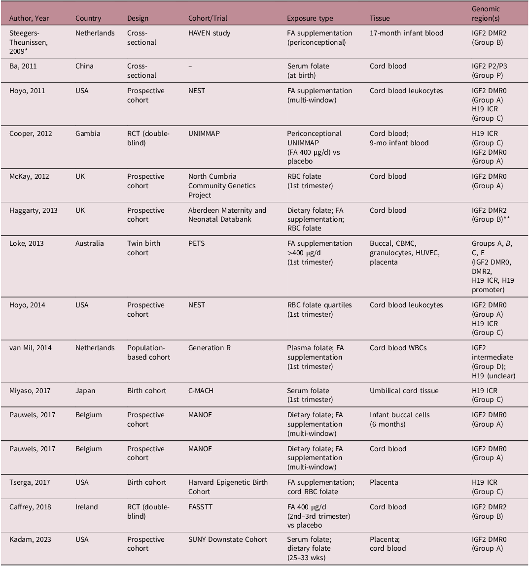
The 15 included reports were published between 2009 and 2023 (Table 1). Two were based on the same Belgian cohort (MANOE) but reported results in different tissue types, and two from the United States cohort (NEST) were derived from the same underlying cohort. Accordingly, the included reports represented 13 distinct study populations across nine countries: the United States (n = 3), the United Kingdom (n = 2), the Netherlands (n = 2), Belgium (n = 1), Australia (n = 1), China (n = 1), The Gambia (n = 1), Ireland (n = 1), and Japan (n = 1). Study designs included prospective birth cohorts (n = 8), twin birth cohorts (n = 1), cross-sectional studies (n = 2), and double-blind randomized controlled trials (n = 2). Analytic sample sizes ranged from 47 to 913 mother-offspring pairs.
Characteristics of included studies

Table 1. Long description
A table comparing studies on folate and genomic regions across different countries. The table has six columns: Author, Year, Country, Design, Cohort/Trial, Exposure type, Tissue, and Genomic region(s). It includes 15 rows of data. Row 1: Author, Steegers-Theunissen; Year, 2009; Country, Netherlands; Design, Cross-sectional; Cohort/Trial, HAVEN study; Exposure type, FA supplementation (periconceptional); Tissue, 17-month infant blood; Genomic region(s), IGF2 DMR2 (Group B). Row 2: Author, Ba; Year, 2011; Country, China; Design, Cross-sectional; Cohort/Trial, —; Exposure type, Serum folate (at birth); Tissue, Cord blood; Genomic region(s), IGF2 P2/P3 (Group P). Row 3: Author, Hoyo; Year, 2011; Country, USA; Design, Prospective cohort; Cohort/Trial, NEST; Exposure type, FA supplementation (multi-window); Tissue, Cord blood leukocytes; Genomic region(s), IGF2 DMR0 (Group A), H19 ICR (Group C). Row 4: Author, Cooper; Year, 2012; Country, Gambia; Design, RCT (double-blind); Cohort/Trial, UNIMMAP; Exposure type, Periconceptional UNIMMAP (FA 400 µg/d) vs placebo; Tissue, Cord blood; 9-mo infant blood; Genomic region(s), H19 ICR (Group C), IGF2 DMR0 (Group A). Row 5: Author, McKay; Year, 2012; Country, UK; Design, Prospective cohort; Cohort/Trial, North Cumbria Community Genetics Project; Exposure type, RBC folate (1st trimester); Tissue, Cord blood; Genomic region(s), IGF2 DMR0 (Group A). Row 6: Author, Haggarty; Year, 2013; Country, UK; Design, Prospective cohort; Cohort/Trial, Aberdeen Maternity and Neonatal Databank; Exposure type, Dietary folate; FA supplementation; RBC folate; Tissue, Cord blood; Genomic region(s), IGF2 DMR2 (Group B). Row 7: Author, Loke; Year, 2013; Country, Australia; Design, Twin birth cohort; Cohort/Trial, PETS; Exposure type, FA supplementation >400 µg/d (1st trimester); Tissue, Buccal, CBMC, granulocytes, HUVEC, placenta; Genomic region(s), Groups A, B, C, E (IGF2 DMR0, DMR2, H19 ICR, H19 promoter). Row 8: Author, Hoyo; Year, 2014; Country, USA; Design, Prospective cohort; Cohort/Trial, NEST; Exposure type, RBC folate quartiles (1st trimester); Tissue, Cord blood leukocytes; Genomic region(s), IGF2 DMR0 (Group A), H19 ICR (Group C). Row 9: Author, van Mil; Year, 2014; Country, Netherlands; Design, Population-based cohort; Cohort/Trial, Generation R; Exposure type, Plasma folate; FA supplementation (1st trimester); Tissue, Cord blood WBCs; Genomic region(s), IGF2 intermediate (Group D); H19 (unclear). Row 10: Author, Miyaso; Year, 2017; Country, Japan; Design, Birth cohort; Cohort/Trial, C-MACH; Exposure type, Serum folate (1st trimester); Tissue, Umbilical cord tissue; Genomic region(s), H19 ICR (Group C). Row 11: Author, Pauwels; Year, 2017; Country, Belgium; Design, Prospective cohort; Cohort/Trial, MANOE; Exposure type, Dietary folate; FA supplementation (multi-window); Tissue, Infant buccal cells (6 months); Genomic region(s), IGF2 DMR0 (Group A). Row 12: Author, Pauwels; Year, 2017; Country, Belgium; Design, Prospective cohort; Cohort/Trial, MANOE; Exposure type, Dietary folate; FA supplementation (multi-window); Tissue, Cord blood; Genomic region(s), IGF2 DMR0 (Group A). Row 13: Author, Tserga; Year, 2017; Country, USA; Design, Birth cohort; Cohort/Trial, Harvard Epigenetic Birth Cohort; Exposure type, FA supplementation; cord RBC folate; Tissue, Placenta; Genomic region(s), H19 ICR (Group C). Row 14: Author, Caffrey; Year, 2018; Country, Ireland; Design, RCT (double-blind); Cohort/Trial, FASSTT; Exposure type, FA 400 µg/d (2nd–3rd trimester) vs placebo; Tissue, Cord blood; Genomic region(s), IGF2 DMR2 (Group B). Row 15: Author, Kadam; Year, 2023; Country, USA; Design, Prospective cohort; Cohort/Trial, SUNY Downstate Cohort; Exposure type, Serum folate; dietary folate (25–33 wks); Tissue, Placenta; cord blood; Genomic region(s), IGF2 DMR0 (Group A).
Note: FA = folic acid; RCT = randomized controlled trial; WBC = white blood cell; MNC = mononuclear cell; CBMC = cord blood mononuclear cells; HUVEC = human umbilical vein endothelial cells; DMR = differentially methylated region; ICR = imprinting control region. Group A = IGF2 promoter/DMR0; Group B = IGF2 intragenic DMR2; Group C = H19 ICR; Group D = IGF2 intermediate region; Group E = H19 promoter; Group P = IGF2 promoters P2/P3 (non-canonical).
*Cross-sectional design with methylation measured in infant blood at the standardized 17-month postnatal visit; periconceptional exposure ascertained retrospectively.
**Genomic region assumed as Group B based on cited methodology (see Methods).
Cord blood, including cord blood leukocytes or whole cord blood, was the most frequently analyzed tissue (n = 10 studies), followed by placenta (n = 3), umbilical cord tissue (n = 1), infant buccal epithelial cells at six months of age (n = 1), and 17-month infant blood (n = 1). DNA methylation was quantified using bisulfite pyrosequencing in the majority of studies (n = 9), with mass spectrometry-based approaches, including Sequenom MassARRAY EpiTyper, used in five studies, and methylation-sensitive high-resolution melting analysis in one study. Assessed genomic regions spanned all five functional groups within the IGF2-H19 domain, with the IGF2 promoter region (Group A) and the H19 ICR (Group C) most frequently examined.
Folate exposure was characterized as folic acid supplementation in eight studies, as objectively measured blood folate biomarkers, including serum, plasma, or erythrocyte concentrations, in six studies, and as dietary folate intake in three studies. Several studies assessed more than one exposure type, contributing multiple independent comparisons. Across the 15 included reports, a total of 14 exposure-region comparisons were identified and organized into the evidence matrix described in the Methods, defined as unique combinations of exposure category and genomic subregion: five under folic acid supplementation (Groups A, B, C, D, and E/unclassified H19), six under blood folate biomarkers (Groups A, B, C, D, P, and unclassified H19), and three under dietary folate intake (Groups A, B, and unclassified H19).
Risk of bias
Of the 13 observational reports assessed using ROBINS-E, (Figure 2) five were rated as overall high risk of bias (Tserga 2017, Reference Tserga, Binder and Michels28 McKay 2012, Reference McKay, Groom and Potter29 Haggarty 2013, Reference Haggarty, Hoad and Campbell30 Pauwels 2017, Reference Pauwels, Ghosh and Duca31,Reference Pauwels, Ghosh and Duca32 [both publications]) and eight were rated as some concerns (Steegers 2009, Reference Steegers-Theunissen, Obermann-Borst and Kremer19 Ba 2011, Reference Ba, Yu and Liu33 Hoyo 2011, Reference Hoyo, Murtha and Schildkraut22 Kadam 2023, Reference Kadam, Bhatt and Bhatt34 Hoyo 2014, Reference Hoyo, Daltveit and Iversen35 Loke 2013, Reference Loke, Galati and Morley21 van Mil 2014, Reference Van Mil, Bouwland-Both and Stolk36 Miyaso 2017 Reference Miyaso, Sakurai and Takase37 ). No observational study was rated as low overall risk. The most common domains driving concern were confounding (D1), missing data (D5), and selection of the reported result (D7). The two randomized controlled trials were assessed using RoB 2, (Figure 3) Caffrey 2018 Reference Caffrey, Irwin and McNulty20 (FASSTT trial) and Cooper 2012 Reference Cooper, Khulan and Owens38 (UNIMMAP trial) were rated as some concerns, primarily due to selective reporting across multiple loci, windows, and subgroup analyses.
Risk of Bias — observational studies (ROBINS-E)%. Legend: ROBINS-E = Risk of bias in non-randomized studies of exposures. D1 = Confounding; D2 = Selection of participants into the study; D3 = Classification of interventions; D4 = Deviations from intended interventions; D5 = Missing data; D6 = Measurement of outcomes; D7 = Selection of the reported result. Overall risk of bias ratings are also reported per comparison in Table 2.

Risk of Bias — Randomized controlled trials (Cochrane RoB 2). Legend: RoB 2 = Cochrane Risk of Bias tool version 2. D1 = Randomization process; D2 = Deviations from intended interventions; D3 = Missing outcome data; D4 = Measurement of the outcome; D5 = Selection of the reported result. Overall risk of bias ratings are also reported per comparison in Table 2.

Narrative synthesis
Folic acid supplementation
IGF2 promoter region (Group A)
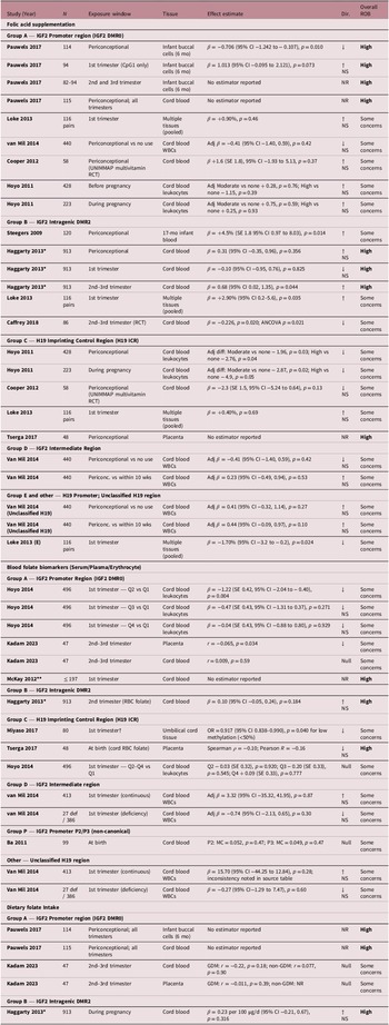
Four cohorts and one randomized controlled trial examined folic acid supplementation in relation to IGF2 promoter methylation, one study provides separate analyses in cord blood and infant buccal epithelial cells (Table 2). Loke 2013, used a pooled multi-tissue model in newborn twins and reported a near-null and non-significant association for first trimester supplementation. Van Mil 2014 reported no significant association between periconceptional supplementation and methylation in cord blood white blood cells. Hoyo 2011 found no association between before and during pregnancy supplementation and methylation in cord blood leukocytes. And Cooper 2012 similarly found no difference in cord blood after periconceptional supplementation with a folic-acid-containing multimicronutrient formulation (UNIMMAP). Pauwels 2017 also reported no association in cord blood for periconceptional supplementation, but in infant buccal epithelial cells at 6 months the same study observed lower IGF2 methylation across all measured CpGs with higher supplemental folic acid intake before pregnancy (β = −0.706, 95% CI −1.242 to −0.107, p = 0.0101), however no significant associations were observed for first, second, or third trimester supplementation at this region. Overall, evidence at Group A for folic acid supplementation was heterogeneous and predominantly null, with the single significant finding from Pauwels 2017 limited to infant buccal cells and rated from a study with high overall risk of bias.
Summary of findings by exposure type and genomic region

Table 2. Long description
The table presents findings from studies on folic acid supplementation and IGF2 promoter methylation. It includes columns for study, exposure, effect, and overall findings. The studies are grouped by genomic region and include various cohorts and a randomized controlled trial. Key studies mentioned are Loke 2013, Van Mil 2014, Hoyo 2011, Cooper 2012, and Pauwels 2017. Each row details the specific exposure type, the effect measured, and the overall findings, with some studies showing significant associations while others do not. The table highlights the heterogeneity in findings and the predominantly null results, with a notable significant finding from Pauwels 2017 in infant buccal cells.
Note: Dir. = direction of association. ↑ = positive (higher folate associated with higher methylation); ↓ = inverse (higher folate associated with lower methylation); Null = no association; NS = not statistically significant. Overall ROB: Low = low risk of bias; Some concerns = some concerns; High = high risk of bias; NR = not reported; entries with “No estimator reported” should not be interpreted as fully reported null estimates. ROB assessed using ROBINS-E for observational studies and Cochrane RoB 2 for randomized controlled trials (see Figures 2 and 3 for domain-level detail). β = regression coefficient (absolute change in % DNA methylation per unit exposure); r = partial correlation coefficient; OR = odds ratio; pp = percentage points; MC = mean change.
*Genomic region assumed as Group B (see Methods).
†Subgroup restricted to women with caloric intake <2,000 kcal/day.
**Exact analytic N not reported (maternal RBC folate measured in 197 mothers overall).
IGF2 intragenic DMR2 (Group B)
Four studies examined this region, including one randomized trial, one observational study and two cohorts, one in which the DMR2 classification could not be confirmed. This group produced the most heterogeneous findings across the review. Steegers 2009, in the earliest included study, reported a significant positive association between periconceptional supplementation and IGF2 DMR methylation in 17-month infant blood (+4.5%, SE 1.8, p = 0.014). Loke 2013 similarly reported a significant positive association for first trimester supplementation in the pooled multi-tissue model in newborn twins (β = +2.90%, 95% CI 0.2 to 5.6, p = 0.035). In contrast, Caffrey 2018, the only randomized controlled trial contributing to this region, reported a significant reduction in cord blood methylation associated with second and third trimesters supplementation (β = −0.226, p = 0.020; ANCOVA p = 0.021), with the effect reaching significance only in female offspring in sex-stratified analyses. Additionally, Haggarty 2013 was rated as having a high overall risk of bias, and was considered separately from the main narrative interpretation because its classification as Group B was based on assumption. The authors found no significant association for periconceptional and first trimester supplementation but reported a borderline significant positive association for supplementation after 12 weeks of gestation (β = 0.68, p = 0.044). The divergence in direction of effect associations likely reflects differences in exposure window, tissue type, and the biological distinction between periconceptional and post-first-trimester supplementation.
H19 imprinting control region (Group C)
Four studies contributed evidence for this region under folic acid supplementation. Hoyo 2011 provided the most robust evidence in this group, reporting significant inverse associations between moderate and high supplement users relative to non-users and H19 ICR methylation in cord blood leukocytes for both periconceptional (adjusted difference: −1.96 to −2.76 percentage points, p < 0.05) and pregnancy windows (adjusted difference: −2.87 to −4.9 percentage points, p ≤ 0.05). This study also reported that moderate folic acid use before pregnancy was associated with reduced odds of H19 hypermethylation (OR 0.47, 95% CI 0.22 to 0.91), a finding directionally consistent with the continuous estimates. Cooper 2012, in a periconceptional Randomized controlles trial (RCT) using multimicronutrient supplementation including 400 µg folic acid versus placebo, reported a non-significant inverse difference in cord blood H19 methylation, that is directionally consistent with Hoyo 2011 but insufficiently powered to reach significance. In contrast, Tserga 2017, examining placental methylation, reported no significant association with periconceptional supplementation and was rated high overall risk of bias. And Loke 2013 found no association in pooled twin tissues in first trimester association.
Other regions (groups D, E and unclassified H19 region)
Van Mil 2014 contributed comparisons at the IGF2 intermediate region (Group D) and an unverified H19 region, reported no significant association in cord blood white blood cells, for any window examined. Loke 2013 reported a significant inverse association between first trimester supplementation and H19 promoter methylation in pooled twin tissues (Group E, β = −1.70%, 95% CI − 3.2 to −0.2, p = 0.024).
Blood folate biomarkers
IGF2 promoter region (Group A)
Three studies contributed evidence for circulating folate biomarkers in relation to this region. Hoyo 2014, found that erythrocyte folate in the second quartile was significantly associated with lower methylation compared to the first quartile (β = −1.22, p = 0.004), but associations for the third and fourth quartiles were non-significant, suggesting a non-linear pattern that was attenuated at higher folate levels. Stratified analyses revealed that this pattern was present only among non-obese women (BMI<30), with no significant associations observed among obese women, indicating potential effect modification by maternal pre-pregnancy BMI. Kadam 2023, in a smaller cohort with and without gestational diabetes pregnancies, reported a significant inverse association between serum folate and methylation in placenta group (r = −0.065, p = 0.034), but no significant associations in cord blood. McKay 2012 reported no significant association between maternal erythrocyte folate and IGF2 promoter methylation in cord blood, but the exact estimate was not reported and the study was rated high overall risk of bias. Overall, Group A biomarker evidence suggested at most weak and tissue-specific inverse associations, but not a consistent dose–response pattern across studies. And comparability was limited by the use of quartile contrasts, continuous correlations, and incompletely reported screening-based analyses.
H19 imprinting control region (group C)
Three comparisons from three studies examined this region. Miyaso 2017 reported a significant association between higher first trimester serum folate and lower odds of H19 ICR hypomethylation (defined as methylation below 50%) in umbilical cord tissue among women with caloric intake below 2,000 kcal/day (OR = 0.917, 95% CI 0.838 to 0.990, p = 0.040), suggesting that this association of folate on H19 ICR methylation may be conditioned by overall nutritional adequacy. Hoyo 2014, found no significant associations in first trimester between erythrocyte folate quartiles and methylation in cord blood leukocytes across any quartile comparison. And Tserga 2017, using cord blood erythrocyte folate at birth as a proxy for maternal exposure, reported weak inverse non-significant correlation with placental methylation. For this group the evidence was methodologically heterogeneous.
Other regions
Haggarty 2013 reported no significant association between second trimester maternal erythrocyte folate and IGF2 DMR2 methylation in cord blood, additionally it was judged at high risk of bias with an explicitly uncertain region assignment. Van Mil 2014 found no significant associations between first trimester plasma folate, whether treated as a continuous variable or categorized as deficient versus adequate, and IGF2 intermediate region or H19 methylation in cord blood white blood cells. Ba 2011 examined maternal serum folate at delivery in relation to IGF2 promoter P2 and P3 methylation in cord blood, reporting non-significant associations at both sites, given that this study assessed IGF2 promoter regions biologically distinct from the canonical imprinted DMRs, its findings are not directly comparable to other Group A studies and were analyzed separately.
Dietary folate intake
Three studies examined dietary folate intake. Evidence in this category was entirely null. Pauwels 2017, assessed across periconceptional and all trimester-specific windows in both cord blood and infant buccal cells, reported no significant associations between dietary folate and IGF2 promoter methylation at any window; these results were explicitly noted by the authors as not shown due to lack of significance, which raises selective reporting concerns consistent with the high overall risk of bias. As well in Kadam 2023, dietary folate intake was also not clearly associated with IGF2 promoter methylation. Comparability was limited because reporting was often incomplete and the dietary assessment methods differed substantially. Haggarty 2013 reported no significant association between habitual dietary folate intake across pregnancy and IGF2 DMR2 methylation in cord blood, but the exact region could not be confirmed.
Discussion
This systematic review synthesized human evidence on the association between maternal folate intake during pregnancy and DNA methylation at the IGF2-H19 locus in offspring, evaluating 15 reports published between 2009 and 2023. The included studies were a heterogeneous group, varying in exposure type, genomic region assessed, tissue source, and gestational window, making direct comparison across them difficult without an explicit organizing framework. To our knowledge, this is the first review to apply a functional subregion classification of the IGF2-H19 domain as a structure for evidence synthesis, giving the fragmented literature a shape that a study-by-study reading would not have revealed. The evidence that follows is insufficient to support definitive causal inference. But it is structured enough to permit biologically grounded hypotheses about when, where, and in whom maternal folate is most likely to leave an epigenetic mark at this locus Reference Bell and Felsenfeld10,Reference Hark, Schoenherr and Katz11 .
The evidence points to a regionally and temporally specific pattern rather than a uniform epigenetic effect across the locus. The H19 ICR produced the most suggestive evidence in this review. Hoyo 2011, the largest observational study included, reported inverse associations between folic acid supplementation and H19 ICR methylation in cord blood leukocytes across both periconceptional and pregnancy windows, with adjusted differences ranging from roughly two to five percentage points. Cooper 2012, a randomized trial, pointed the same way despite being underpowered for this endpoint, whereas Miyaso 2017 supported the nutritional sensitivity of this region using serum folate and a dichotomized low-methylation outcome in umbilical cord tissue. Together, these findings provide a meaningful basis for cautious inference despite the absence of low-risk observational studies.
These findings matter beyond their statistical consistency. IGF2 is a primary regulator of fetal growth, and the H19 ICR is the region that makes this regulation work in an allele-specific way. A shift in its methylation, even a modest one, could plausibly influence growth outcomes, and there is at least a preliminary suggestion of this in the data: Miyaso 2017 reported that lower H19 ICR methylation in cord tissue was associated with smaller head circumference in the newborn, though the authors acknowledged limited statistical power and this was a secondary exploratory finding. This direction is biologically plausible: reduced H19 ICR methylation would be expected to attenuate IGF2 expression through the known CTCF-mediated insulator mechanism, consistent with a growth-restricting effect. Reference Bell and Felsenfeld10,Reference Hark, Schoenherr and Katz11 What the available evidence cannot yet tell us is whether the observed reduction in methylation reflects a meaningful perturbation of imprinting or normal variation within an acceptable biological range, since none of the included studies assessed methylation with allele-specific resolution.
The IGF2 intragenic DMR2 showed a different pattern from the H19 ICR, one that depended heavily on when during pregnancy folate exposure occurred. Steegers 2009 and Loke 2013 both reported positive associations between periconceptional and first trimester supplementation and DMR2 methylation, suggesting that early folate exposure may promote methylation at this somatic DMR. But Caffrey 2018, the only randomized trial specifically targeting this region and the study with the lowest overall risk of bias in the review, found the opposite: supplementation in the second and third trimesters was associated with significantly lower methylation, with the effect reaching significance only in female offspring. The IGF2 DMR2 is a somatic rather than germline-established DMR, and its methylation continues to be dynamically regulated during fetal development well beyond the periconceptional window, which may make it responsive to folate availability in different ways depending on whether methylation is still being established or already being maintained. This raises a question that current guidance does not address, if folate supplementation continues beyond the first trimester, as many women do in practice, its epigenetic consequences at this locus may differ from those observed in the periconceptional window.
The IGF2 promoter region was predominantly null across all three exposure categories and study designs, and dietary folate intake showed no significant associations anywhere in the locus. These null findings are not uninformative. The promoter region is not a germline-imprinted DMR, and its methylation operates through mechanisms distinct from the allele-specific insulation that characterizes the H19 ICR. That it behaved differently from the ICR and DMR2 is actually consistent with the biological logic underlying the functional classification applied in this review. The absence of dietary folate associations is also interpretable: most included populations were well-nourished, which means there was little exposure variation to detect an effect against, and food frequency questionnaires are known to measure dietary folate with considerable imprecision.
These findings are broadly consistent with the experimental and epidemiological literature on methyl-donor availability and imprinted locus methylation. Animal models have established that periconceptional methyl-donor supplementation can shift methylation at imprinted loci in a dose-dependent manner with lasting phenotypic consequences Reference da Mota, Ribeiro and Carvalho14,Reference Downing, Johnson and Larson15,Reference Mahajan, Sapehia and Rahat16 . The Dutch Hunger Winter studies, the most compelling natural experiment available in this area, showed that periconceptional famine exposure was associated with hypomethylation of the IGF2 DMR in adult offspring Reference Heijmans, Tobi and Stein39,Reference Tobi, Lumey and Talens40 , providing a directional anchor that is consistent with the inverse associations observed at the H19 ICR in this review. That human observational and interventional data point in a similar direction, even if imprecisely, supports the biological plausibility of this pathway.
This review has several methodological strengths: a comprehensive three-database search, prespecified eligibility criteria applied independently by two reviewers, risk of bias assessment using design-appropriate tools, and a functional classification framework that gave biological structure to a fragmented literature. The limitations are also real. This systematic review was not prospectively registered; the analytical approach was nevertheless prespecified and applied consistently throughout. No observational study achieved low overall risk of bias, and five were rated high risk, mostly due to confounding and selective reporting. Neither trial was powered for the methylation endpoint, which limits the precision of their estimates. Technical variability across measurement platforms and bisulfite protocols introduces noise that the classification framework can reduce but not eliminate. The evidence base is also heavily dominated by cord blood leukocytes, while placental tissue, arguably the most relevant matrix for studying imprinting, is underrepresented. And almost all included populations were from high-income settings where folate deficiency is uncommon, limiting generalizability to deficiency-prevalent populations. The heterogeneity of the included evidence also precluded a formal certainty-of-evidence rating.
The field needs better studies, and it is fairly clear what they should look like. Randomized trials with IGF2-H19 methylation as a primary outcome, placental tissue collection alongside cord blood, and prespecified sex-stratified analyses would go a long way. Of these, resolving the window-dependency question through repeated biomarker measurement across pregnancy is the most urgent priority, as it is the dimension most directly implicated in the divergent findings this review identified and the one most feasible to implement in existing prospective cohorts. Studies in populations with genuine folate deficiency would also substantially strengthen causal inference in ways that well-nourished cohorts simply cannot. Finally, standardized reporting of genomic coordinates and assay specifications across all future studies is a basic prerequisite for the kind of quantitative synthesis this literature is not yet ready to support.
Conclusion
The available evidence, though heterogeneous and largely limited by risk of bias, suggests that maternal folic acid supplementation is associated with small but directionally consistent reductions in H19 ICR methylation in offspring tissues across observational and experimental designs. The IGF2 intragenic DMR2 shows a window-dependent pattern in which early pregnancy supplementation may increase methylation while later exposure appears to have the opposite effect, a distinction that requires confirmation but carries potential biological significance. Taken together, these findings demonstrate that folate does not exert a uniform epigenetic effect across the IGF2-H19 domain, and that the nature, timing, and magnitude of any association are specific to the genomic subregion and exposure window assessed. Whether these methylation differences translate into functional consequences for IGF2 expression, fetal growth, or long-term offspring health remains an open and critical question for future investigation.
Supplementary material
The supplementary material for this article can be found at https://doi.org/10.1017/S2040174426100932.
Acknowledgements
The authors thank Universidad El Bosque for its support during this research project.
The authors used ChatGPT (OpenAI; GPT-5 series models, April 2026) for language editing and manuscript organization. All scientific content, study selection, interpretation of findings, and final manuscript decisions were performed and verified by the authors.
Financial support
This research received no specific grant from any funding agency, commercial, or not-for-profit sectors.
Competing interests
None.