Introduction
Haemonchus contortus, a common trichostrongylid parasite of sheep, goats and cattle, is one of the most important blood-sucking parasites. Extensive production losses have been reported in animals in tropical regions due to this parasite (Miller & Horohov, Reference Miller and Horohov2006). In the past, morphological features were used for identification of different species of this parasite but currently the molecular signature of parasite genes is utilized (Kia et al., 2007). In order to study the genetic variation within a species, a multicopy gene family of ribosomal DNA (rDNA) is the ideal choice due to its ubiquitous nature in all living organisms and its relative easy amplification via polymerase chain reaction (PCR). rDNA is characterized by multiple tandemly repeated copies of highly conserved genes coding for ribosomal RNA. It is interrupted by variable external and internal transcribed ‘spacer’ regions (ETS, ITS-1 and ITS-2). Of these spacer regions, the internal transcribed spacer 1 (ITS-1) located between 18S and 5.8S genes is the most conserved and therefore recognized as a valuable genetic marker at species level (Pontecorvo et al., Reference Pontecorvo, Fantaccione and Woodrow2008).
Previously it was accepted that the nucleotide sequence homogeneity reported among rDNA multigene families is maintained within individuals and even within species by ‘concerted evolution’ (Dover, Reference Dover1982; Gandolfi et al., Reference Gandolfi, Bonilauri, Rossi and Menozzi2001). In contrast to this theory, a certain degree of intra-species variation has been found in the nucleotide sequence of ITS-1 and/or ITS-2 gene regions of different isolates of a species (Harris & Crandall, Reference Harris and Crandall2000).
In view of the above, the main objective of this study was to ascertain the existence of different isolates of H. contortus prevailing in West Bengal, India, by studying the variation in nucleotide sequences of ITS-1. Primers for amplification of ITS-1 were designed from the published ITS-1 sequence of H. contortus (accession #AF044927). The amplified product was further analysed through single-strand conformation polymorphism (SSCP). Different SSCP conformations were sequenced and their variation among isolates of H. contortus was determined.
Materials and methods
Sampling of parasites
During the period of June to August 2009, goat intestines were collected from slaughter houses in and around Kolkata, WB, India. Haemonchus contortus was isolated from abomasums of these intestines. Male and female worms were identified microscopically, washed with phosphate-buffered saline (PBS) (pH 7.2) and preserved separately in 70% alcohol at − 20°C for DNA extraction.
DNA extraction and polymerase chain reaction
Genomic DNA was extracted from randomly selected male and female worms. One-third of the body part of males and females was broken up and added to 30 μl nuclease-free water (NFW) (Promega, India). Samples were then centrifuged at 10,000 rpm for 15 min and supernatants were eliminated. The resultant pellets were resuspended in NFW and centrifuged at 10,000 rpm for another 10 min, and supernatants were eliminated once again. Thereafter pellets were resuspended in 30 μl NFW, boiled at 100°C in a water bath for 30 min, cooled on ice and centrifuged at 10,000 rpm for 10 s before being stored at − 20°C (Queipo-ortuno et al., Reference Queipo-ortuno, Colmenero, Macias, Bravo and Morata2008). The integrity of the extracted DNA was determined by running it on a 1% agarose gel containing ethidium bromide and then visualizing it under a transluminator (Bio-imaging system, Jerusalem, Israel).
PCR was carried out in 25 μl volumes using 5 μl DNA as template (1:5 dilution), 2 μl dNTPs (100 μm), 2.5 μl 10 × PCR buffer (with MgCl2), 0.5 μl (20 pmol) ITS-1 forward (5′-TATGACATGAGCCGTTCGAG-3′), 0.5 μl reverse primer (5′-TGATCATTAAGGTTCCCCGA-3′), 0.5 μl (1.5 units) Taq DNA polymerase (Fermentas, Glen Burnie, Maryland, USA) and 14 μl NFW. The temperature profile included 5 min at 95°C, 45 s at 95°C, 45 s at 55°C (annealing temperature) and 2 min at 72°C for a total of 35 cycles. Further extension of the temperature profile was carried out for 10 min at 72°C on the PCR machine (Applied Biosystems, Carlsbad, California, USA). Finally the PCR products were separated by electrophoresis in 1.5% agarose gel containing ethidium bromide and visualized under a transluminator (Bio-imaging system).
Single-stranded conformational polymorphism (SSCP)
SSCP analysis was performed according to techniques described by Xie et al. (Reference Xie, Ho and Ma1997). Some modifications to the technique were, however, applied. In brief, the process was carried out in 24 μl volumes, using 15 μl 2 × SSCP buffer (98% formamide, 0.5 m EDTA and 0.5 m NaOH) and 9 μl of PCR amplified product. In order to denature the double-stranded DNA, mixtures were heated for 10 min at 95°C in a thermocycler (Eppendorf, Mastercycler, Hamburg, Germany). After snap cooling, denatured samples were run on a 30% acrylamide gel at 60 V for 12 h and observed under a transluminator (Bio-imaging system).
Cloning and sequencing
ITS-1 PCR amplified products with different SSCP conformations (Lau & Sheu, Reference Lau and Sheu1992) were ligated into the PTZ57R/T vector (Fermentas) and transformed into Escherichia coli DH5α according to the manufacturer's instruction (InsTA Clone™ PCR Cloning Kit, Fermentas). Transformants (white colonies) were selected from Luria–Bertani plates containing isopropyl thiogalactoside (IPTG) (0.2 mm), bromo-chloro-indolyl-galactopyranoside (X-Gal) (20 mg/ml) and ampicillin (100 μg/ml), and were confirmed to contain ITS-1 inserts by PCR (Rahman et al., Reference Rahman, Hamid and Othman2007). Selected clones were sent to an outside laboratory (Department of Biochemistry, University of Delhi South Campus, India) for sequencing.
Sequences from different clones were aligned along with the original sequence of H. contortus ITS-1 (accession #AF044927) using the Mega 4.1 program (http://www.megasoftware.net/mega41.html). The phylogenetic tree was obtained by unweighted pair group method with arithmetic mean (UPGMA) analysis. The phylogenetic distance was estimated by Kimura's two-parameter method (Kimura, Reference Kimura1980) using 1000 bootstrap replicates (Gandolfi et al., Reference Gandolfi, Bonilauri, Rossi and Menozzi2001).
Predicted RNA secondary structure of each clone along with the original sequence of H. contortus ITS-1 was depicted using RNA fold server (Gruber et al., Reference Gruber, Lorenz, Bernhart, Neuböck and Hofacker2008).
Results
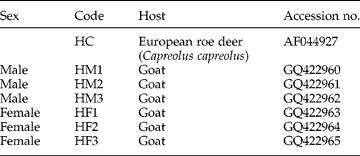
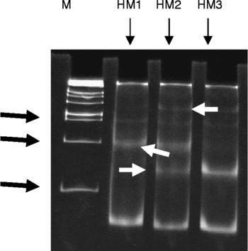
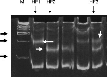
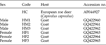
The PCR amplification of ITS-1 from different isolates of H. contortus (table 1) yielded a fragment of 198 bp in size. Amplified products obtained from 34 male and 30 female H. contortus were subjected to SSCP analysis. The analysis revealed the presence of three distinct conformations in males (out of 34 different PCR amplified products) (fig. 1) as well as in females (out of 30 different PCR amplified products) (fig. 2).
Genotypes of male and female H. contortus parasites collected from goats from West Bengal, India and the specific genotype (HC) of the parasite from which primers were designed.
SSCP analysis of PCR amplified products obtained from male H. contortus. Lanes: HM1, HM2, HM3, three male genotypes; M, 1 kb DNA ladder. Black arrows indicate 100, 200 and 300 bp; white arrows indicate three distinct conformations.

SSCP analysis of PCR amplified products obtained from female H. contortus. Lanes: HF1, HF2, HF3, three female genotypes; M, 1 kb DNA ladder. Black arrows indicate 100, 200 and 300 bp; white arrows indicate three distinct conformations.

Analysis of the SSCP gel exhibited six distinct conformations but after sequencing of these conformations, only two and one SNP were found in male and female parasites, respectively. Some point mutations were observed arbitrarily in almost all the sequences, but these were ignored as PCR artefacts.
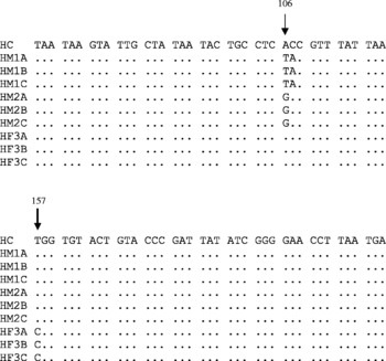
Alignment of sequences of six different conformations (three each from male and female H. contortus) with the original H. contortus sequence (accession #AF044927) (HC) from which primers of ITS-1 were designed, revealed three single nucleotide polymorphisms (SNPs) which were distinctly different from the original sequence. In the case of male H. contortus genotypes (original genotype), two SNPs were found, at codons 106 (A to T, transversion (HM1A, HM1B, HM1C); A to G, transition (HM2A, HM2B, HM2C)) and 107 (C to A, transversion (HM1A, HM1B, HM1C)). But in female H. contortus genotype, only one SNP was found at codon 157 (T to C, transition (HM3A, HM3B, HM3C)) (fig. 3).
Sequence alignment of SSCP conformers of male and female genotypes of H. contortus; arrows indicate nucleotide sequences.

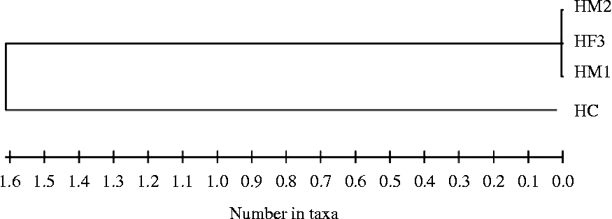
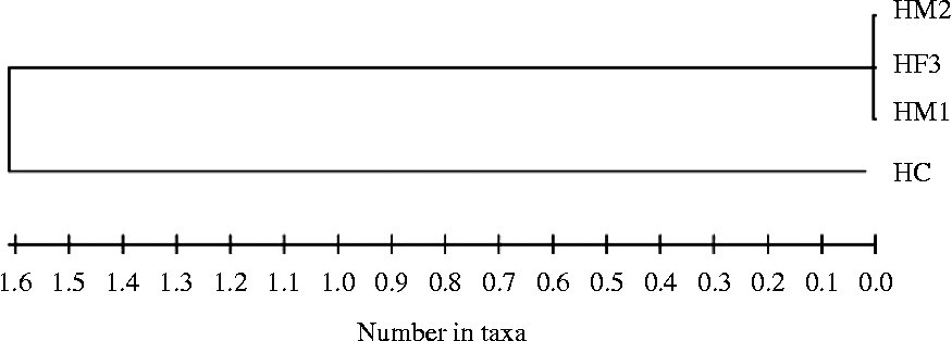
The ITS-1 neighbour-joining tree is clearly differentiated into two clades on the basis of presence/absence of SNPs in the nucleotide sequences. Although three SNPs occurred in male and female genotypes at different nucleotide positions (106, 107 and 157), they clustered into one clade and the ITS-1 sequence of H. contortus (HC) formed a different clade (fig. 4).
Relationship between sequences of H. contortus male (HM1, HM2) and female (HF3) genotypes and the original H. contortus ITS-1 sequence (HC) from which the primer was designed.
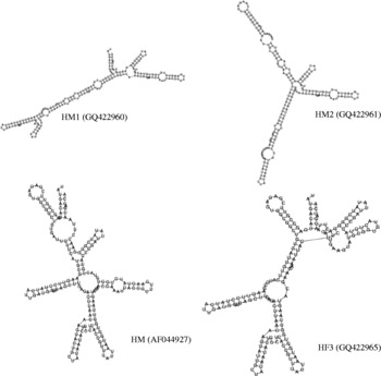
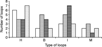
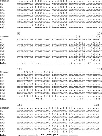
RNA secondary structures of each isolate (HM1, HM2 and HM3) along with the original ITS-1 sequence of H. contortus (HC) were predicted using RNA fold software (Vienna RNA webserver, Gruber et al., Reference Gruber, Lorenz, Bernhart, Neuböck and Hofacker2008). Several rearrangements of the predicted RNA secondary structures were observed (fig. 5) due to SNPs, which signified differences in stability of the RNA molecule (Smit et al., Reference Smit, Widmann and Knight2007). Several local differences and their distribution, specifically hairpin loops, bulge loops, interior loops and multiple branched loops of RNA structures were identified (fig. 6). Comparative analysis showed clear evidence of conservation of several secondary structural elements shared by four ITS-1 folds (fig. 7).
Secondary structural models for the 5′ end of the ITS-1 sequence of different genotypes of H. contortus.

Distribution of different types of loops of ITS-1 sequences of H. contortus. H, hairpin loops; B, bulge loops; I, interior loops; M, multiple branched loops. HC, open bars; HM1, dotted bars; HM2, horizontally shaded bars; HF3, bars shaded with dashed lines.

Comparative analysis of conservation of secondary structural element of ITS-1. *, >90%;+, >80%; − , >50%; and ·, ≤ 50%, indicate the reliability of common structural motifs.

Discussion
SSCP analysis of the male ITS-1 amplified product showed three distinct conformations (HM1, HM2 and HM3) and sequence analysis of these conformations revealed the presence of two SNPs (nucleotide positions 106 and 107 of HM1 and HM2 conformations). In the case of the female, out of six haplotypes, only three conformations (HF1, HF2 and HF3) were detected by SSCP analysis. Of these three conformations, only one SNP (HF3) was detected (nucleotide position 157).
In this study, only a fraction of ITS-1 variants were identified. This may be either due to a small number of clones per individual examined (Van Herwerden et al., Reference Van Herwerden, Blair and Agatsuma1999) or due to the role of spacers in the rRNA secondary structure which is of key importance in post-transcriptional processing (Lalev & Nazar, Reference Lalev and Nazar1999). Low variability, mostly found within individuals, has been reported for ribosomal DNA spacers, particularly in nematodes (Blok et al., Reference Blok, Malloch, Harrower and Phillips1998), corn root beetle (Szalanski et al., Reference Szalanski, Roehrdanz, Taylor and Chandler1999), mites (Navajas et al., Reference Navajas, Lagnel, Fauvel and De Moraes1999) and Anopheles nuneztovari (Fritz et al., Reference Fritz, Conn, Cockburn and Seawright1994). Our result supported the observation of Harris & Crandall (Reference Harris and Crandall2000), who also observed that intragenomic variation in ITS-1 and ITS-2 is common but this variation is significant enough to obscure phylogenetic relationships at the population level.
Although HM1 and HF1 were collected from the intestine of the same goat, sequence-level variations were observed between two haplotypes. Similar results were also obtained from HM2 and HF2 haplotypes. Therefore, it may be assumed that this goat was exposed to different genotypes of parasite larval population during their characteristic migratory grazing.
The biological function of an RNA molecule is determined by its secondary structure (Fang et al., Reference Fang, Luo, Yuan and Wang2007). This RNA function is preserved by sequence variations, and these variations also contribute to differences between species (Smit et al., Reference Smit, Widmann and Knight2007). Key functional elements of ITS secondary structures are different unpaired regions, which are sites of nucleation for overall three-dimensional folding and protein recognition (Bhargavi et al., Reference Bhargavi, Vishwakarma and Murty2005; Campbell et al., Reference Campbell, Wright, Cox, Vining, Major and Arsenault2005; Smit et al., Reference Smit, Widmann and Knight2007). The probability profiling approach revealed that variation of the ITS-1 sequences among different isolates does not affect the stability of the interior loop, which is an essential prerequisite for correct RNA processing (Bhargavi et al., Reference Bhargavi, Vishwakarma and Murty2005). Thus, SNPs seen in different isolates of H. contortus may not influence critical changes in the rRNA folding pattern which influences evolutionary changes among isolates.