Chest tube placement during paediatric cardiac surgery is common. The management and monitoring of the patient after chest tube removal is not standardised across paediatric cardiac surgery programmes. Obtaining a chest radiograph after chest tube removal to determine the presence of a pneumothorax after removal varies among institutions. Reference Taylor, Bates and Kipps1 There are limited studies describing the incidence of pneumothorax in this population, or studies describing patient and clinical characteristics of patients that develop a clinically significant pneumothorax requiring intervention, such as chest tube reinsertion. Of the small number of studies that exist, the data suggest paediatric post-operative cardiac patients are unlikely to develop a pneumothorax following chest tube removal. Reference Pacharn, Heller and Kammen2–Reference Woodward, Dowling, Taylor and Salvin4 Of the patients who did develop pneumothoraces, the post-removal chest radiograph did not provide any diagnostic or therapeutic advantage over the clinical exam. Close monitoring for respiratory distress such as changes in baseline vital signs or arterial blood gases, requirement of or a need for an increase in supplemental oxygen were deemed sufficient in determining the need for chest tube replacement. Reference Pacharn, Heller and Kammen2–Reference Woodward, Dowling, Taylor and Salvin4
Paediatric patients with complex congenital heart disease are exposed to cumulative effects of low-dose ionising radiation due to necessary diagnostic investigations over the course of their disease trajectory. Cumulative low-dose ionising radiation has been independently associated with cancer and predisposes the most complex (congenital heart disease) patients to an increased risk of developing cancer as adults. Reference Cohen, Liu and Gurvitz5–Reference Johnson, Hornik and Li7 While the radiation exposure from a chest radiograph may be low, this exposure may contribute to the overall cumulative risk of malignancy.
The purposes of this study were to describe the incidence of pneumothorax after chest tube removal in paediatric post-operative cardiac surgical patients and describe the patient and clinical characteristics of patients who developed a pneumothorax after chest tube removal.
Materials and methods
This retrospective, descriptive study was approved by the Institutional Review Board from the investigator’s academic institution and hospital. The study was conducted at a large quaternary, free-standing urban children’s hospital that serves as a global referral centre for patients with congenital and acquired heart disease and performs over 1000 cardiac surgeries each year. The standard of care at this hospital is to obtain a chest radiograph following chest tube removal in all post-operative cardiac surgical patients. Utilising combinations of diagnoses and the International Classification of Disease-9 codes, the investigator queried the hospital’s Department of Cardiovascular Surgery’s surgical database to identify all paediatric post-operative cardiac surgical patients from newborn to 18 years old diagnosed with a pneumothorax, admitted from 1 January, 2010 through 31 December, 2018. A pneumothorax was diagnosed based on interpretation by a board-certified radiologist.
Inclusion criteria were patients who developed a pneumothorax following chest tube removal and did not have a documented pneumothorax prior to chest tube removal. For the purposes of this examination, we excluded patients with a known pneumothorax prior to chest tube removal.
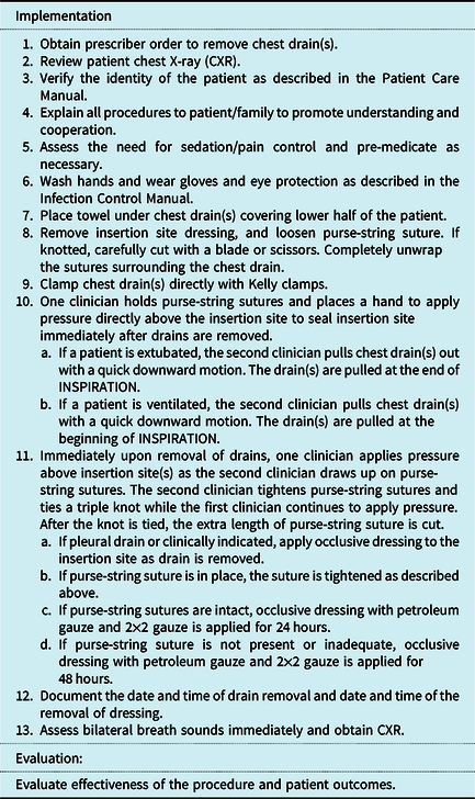
Knowing that the development of a pneumothorax is a potential vulnerability for all patients, there were no deviations from the usual practice made for differences in patient demographics, surgical repair, or chest tube type. All chest tubes were removed according to the hospital’s standard chest tube removal protocol by either advanced practice registered nurses on the inpatient cardiac floor or by senior-level staff nurses within the cardiac ICU (Table 1). The level of patient monitoring by the nurse after chest tube removal was the same, regardless of the location in which the chest tube was removed. Once the chest tube was removed, the patient’s breath sounds were immediately assessed. Vital signs were continuously monitored until the post-removal chest radiograph was obtained. The chest radiograph was obtained immediately in the ICU and within 4 hours after removal for patients who were on the inpatient floor. If a purse string was present, the occlusive dressing was removed 24 hours later. If there was no purse string, the dressing remained in place for 48 hours before removal.
Standardised protocol for the removal of chest drains

Note: Tachypnea, dyspnea, increased cyanosis, decreased oxygen saturation, agitation, and diminished breath sounds may be signs of respiratory compromise related to a pneumothorax. Notify the prescriber for signs of respiratory distress.
Removal of Chest Drains. The Cardiovascular and Critical Care Clinicians’ Manual © Boston Children’s Hospital, 2020. All rights reserved. Reprinted with permission.
The following demographic data was collected and summarised on cases that met study inclusion criteria: age at the time of surgery, history of prematurity, gender, body surface area, primary cardiac diagnosis, presence of non-cardiac anomaly(s), surgical procedure, prior cardiac surgery, Risk-Adjusted Congenital Heart Surgery-1 Category, and required intervention(s) to treat a pneumothorax post-chest tube removal. The investigator reviewed each patient’s electronic medical record to determine if the patient exhibited or reported any changes in baseline clinical signs or symptoms after the chest tube was removed up until the post-removal chest radiograph was obtained.
Results
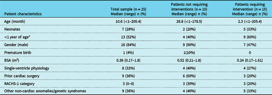
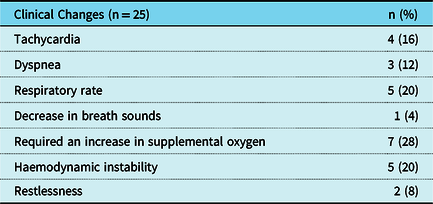
Of the 11,651 cardiac surgical cases that were eligible for consideration, 25 (0.2%) patients met inclusion criteria. Select demographic and clinical characteristics for the inclusion patients are as follows: median patient age at the time of surgery was 10.6 months (with a range from <1 to 205 months); 13 (52%) were less than 1 year of age, of which 7 (28%) were neonates; 16 (64%) were male; 1 (4%) was premature; median body surface area was 0.38 m2 (with a range from 0.17 to 1.8 m2); 8 (32%) had single-ventricle palliation physiology; 9 (36%) had a prior cardiac surgery; median Risk-Adjusted Congenital Heart Surgery-1 Category was 3 (with a range from 0 to 6); and 9 (36%) had other non-cardiac anomalies/genetic syndromes (Table 2). The median chest tube duration was 4 days. Of the study population, 9 (36%) had a change in respiratory status, 7 (28%) required supplemental oxygen, and 5 (20%) had haemodynamic instability (Table 3). None had experienced death related to the pneumothorax.
Characteristics of patients diagnosed with pneumothorax after chest tube removal

* Neonates included (BSA = body surface area; RACHS-1 = Risk-Adjusted Congenital Heart Surgery-1 Category).
Clinical presentation of patients who developed a pneumothorax

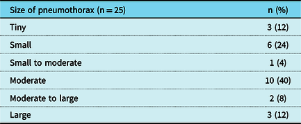
Fifteen (60%) were diagnosed with a moderate, moderate to large, or large pneumothorax by chest radiograph (Table 4). Of these 15 patients, specifically, 1 underwent a needle decompression, 14 underwent chest tube placement. Of the patients requiring intervention, 9 (60%) were less than 1 year of age; 4 (27%) had single-ventricle physiology; the median Risk-Adjusted Congenital Heart Surgery-1 Category was 3, and 5 (33%) had other non-cardiac anomalies/genetic syndromes. Of the patients requiring intervention, eight patients (53% of this group) did not exhibit a change in clinical status or required increased supplemental oxygen; 5 (33%) had a changed respiratory exam; and 7 (47%) had increased oxygen requirement.
Pneumothorax size

Discussion
Findings in this study are consistent with previously published studies on the incidence of pneumothorax after chest tube removal in the paediatric cardiac population. Woodward and colleagues Reference Woodward, Dowling, Taylor and Salvin4 studied paediatric cardiac surgical patients, newborn to 7 years old. and found the incidence of pneumothorax was less than 2%, which is similar to the incidence of <1% found in our study. Yet, none of the patients in Woodward’s study developed a pneumothorax requiring chest tube reinsertion and the authors were unable to conclude if changes in vital signs or clinical examination were adequate in triggering the need for a chest radiograph. Pacharn et al, found that all paediatric cardiac surgical patients who had moderate and large pneumothoraces had changes in respiratory status, that alerted clinicians of the need to reinsert a chest tube. Reference Pacharn, Heller and Kammen2 Our findings, however, showed those patients who had a clinically significant pneumothorax, diagnosed as moderate or larger on chest radiograph, did not reliably demonstrate a change in respiratory status. Of this group, all but one patient had an intervention to evacuate the pneumothorax by either needle decompression or chest tube reinsertion. This finding is especially concerning since almost two-thirds of these patients were less than a year old and almost a third had single-ventricle physiology who may have a poor cardiac or respiratory reserve.
Our experience and review of these cases identified a need for the development of criteria for a chest radiograph following chest tube removal for patients at increased risk of developing a pneumothorax. Based on our data, high-risk patients included patients less than 1 year of age, those who have single-ventricle physiology and/or have other congenital abnormalities. Balancing the desire to limit low-dose ionising radiation exposure to this medically complex population with diagnostic confirmation of pneumothorax following chest tube removal will continue to be a challenge to bedside clinicians. Given the lack of confirmatory evidence, clinical consideration of patient risk factors is required to determine the need for post-chest tube removal chest radiograph in this population.
Limitations
This study is a single-centre experience with a retrospective study design. Due to the low incidence of pneumothorax, a prospective study with adequate power was not achievable. Therefore, independent risk factors associated with pneumothorax were unable to be identified. The investigator utilised the hospital’s cardiac surgical departmental database, which incorporates a number of quality control processes to ensure accurate and complete patient data and outcomes. This database is used to review a number of patient outcomes at the departmental level as well as the cardiac ICU. For each case identified in the cardiac surgical database, the investigator confirmed this information using the electronic medical record. While it may be possible to have missed patients who developed a pneumothorax after chest tube removal, we can confirm each patient identified was corroborated in both databases.
Conclusion
The incidence of post-chest tube removal pneumothoraces is extremely low in paediatric posto-perative cardiac surgical patients. Clinically significant post-chest tube removal pneumothoraces can be asymptomatic and unidentifiable based solely on clinical symptoms. Given the lack of conformational evidence, our results do not specifically support a pathway change at this time. We will continue to examine a select population of patients that may not require chest radiographs after chest tube removal. This patient population may include patients with lower RACHS-1 category, patients who are older and who are able to verbalise changes in their respiratory status, and patients without chromosomal abnormalities. Based on patient clinical characteristics, the development of criteria for identifying patients at increased risk of developing a pneumothorax and who should receive a routine chest radiograph following chest tube removal is recommended.
Acknowledgements
The authors would like to acknowledge Meena Nathan MD, FRCS, MPH and Kim Gauvreau ScD for their support in designing the study as well as statistical support.
Financial Support
This research received no specific grant from any funding agency, commercial, or not-for-profit sectors.
Conflicts of Interest
None.
Ethical Standards
The authors assert that all procedures contributing to this work comply with the ethical standards of the relevant national guidelines on human experimentation (United States of America) and with the Helsinki Declaration of 1975, as revised in 2008, and had been approved by the Institutional Committees (Boston Children’s Hospital).