Ventricular septal defect is the most common CHD.Reference Hoffman and Kaplan1 It may be located in the muscular, perimembranous, inlet, or supracristal portion of the ventricular septum.Reference Wells, Lindesmith and Arciniegas2 It may occur as an isolated anomaly or in association with other cardiac malformations. Spontaneous closure of muscular and perimembranous ventricular septal defect is common.Reference Zhang, Ko, Guileyardo and Roberts3 We report a case of unusual mechanism of spontaneous closure of a large muscular ventricular septal defect by growth of right ventricular muscle bundles that created a blind pouch around the defect.
Case presentation
An 18-year-old Bengali male was referred to paediatric cardiology clinic for evaluation because of history of cardiac septal defect. Parents reported that patient was diagnosed with a large cardiac septal defect in early infancy. However, he did not receive any treatment or intervention. There was no history of cyanosis, dyspnea, activity intolerance, or syncope. Medical history is significant for development delay with severe cognitive impairment, pectus excavatum, and scoliosis.
On examination, he appeared comfortable with no respiratory distress. His vital signs were normal. Oxygen saturation was 99% on room air. Pectus excavatum was noted. Palpation revealed a non-displaced apical impulse and no right ventricular heave. Cardiac auscultation revealed normal cardiac sounds and no heart murmur. There was no brachiofemoral delay. His peripheral pulses and capillary refill time were normal.
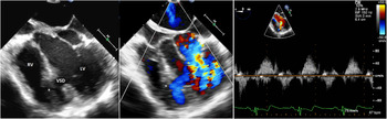
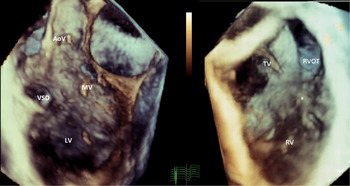
A transthoracic echocardiogram was performed. The echocardiogram images were limited due to lack of patient’s cooperation, poor acoustic windows, and presence of pectus excavatum. It showed a large muscular ventricular septal defect. Color flow Doppler showed bi-directional flow across the defect. Because of limited assessment by transthoracic echocardiography, subsequently a transoesophageal echocardiogram was performed to further assess his cardiac anatomy. The transoesophageal echocardiogram confirmed the presence of a large anterior muscular ventricular septal defect. Color Doppler interrogation of the defect showed bidirectional flow. Muscle bundles on the right ventricular side of the defect formed a pouch-like structure that totally covered the defect. There was no evidence of interventricular shunt (Fig 1, Supplementary video S1). Additional findings of mild mitral valve prolapse, mild mitral valve regurgitation, absence of the right superior vena cava, and persistent left superior vena cava draining into coronary sinus were noted. Three-dimensional transoesophageal echocardiogram was obtained to better visualise the ventricular septal defect and mechanism of closure (Fig 2, Supplementary video S2).
Two-dimensional and color Doppler transoesophageal echocardiogram, mid esophageal four-chamber view, shows a large muscular ventricular septal defect. A muscle bundle (*) in the right ventricle is covering the defect. Pulse-wave Doppler at the level of the defect indicates bidirectional flow. LV = left ventricle; RV = right ventricle; VSD = ventricular septal defect.

Three-dimensional transoesophageal echocardiogram shows a large anterior muscular ventricular septal defect from the left ventricular side (image to the left) and from the right ventricular side (image to the right). Note the muscle bundles (*) covering the defect on the right ventricular side. AoV = aortic valve; LV = left ventricle; MV = mitral valve; VSD = ventricular septal defect; RVOT = right ventricular outflow tract; TV = tricuspid valve; RV = right ventricle.

Discussion
Ventricular septal defect is the most common congenital heart defect. Patients born with this anomaly are followed regularly and may require intervention depending on the size of the defect and associated cardiac anomalies.Reference Hoffman and Kaplan1, Reference Wells, Lindesmith and Arciniegas2 Sometimes, the defect might close spontaneously. There are various mechanisms of spontaneous closure of the defect depending on its location within the ventricular septum.Reference Zhang, Ko, Guileyardo and Roberts3 In case of perimembranous ventricular septal defect, tricuspid valve leaflet adhesions to the edges of the defect or extrusion of folds of tissue from the ventricular aspect of the valve leaflets in the environs of the defect is the most common mechanism of spontaneous closure. Less commonly, spontaneous closure of perimembranous ventricular septal defect results from growth of aneurysmal membranous septum.Reference Anderson, Lenox and Zuberbuhler4 In case of muscular ventricular septal defect, the proposed mechanism of spontaneous closure is growth of muscular and fibrous tissue causing complete obliteration of the defect.Reference Zhang, Ko, Guileyardo and Roberts3, Reference Glancy and Roberts5
Our case report shows an unusual mechanism of spontaneous closure of a large anterior muscular ventricular septal defect by growth of muscle bundles on the right ventricular side. These bundles formed a pouch-like structure that completely covered the defect from the right ventricular side. A similar case report has been published by Dasgupta and Aly. The authors presented transthoracic echocardiogram and cardiac Magnetic resonance imaging images of a case of midmuscular ventricular septal defect that closed spontaneously by growth of right ventricular trabeculations without obliteration of the defect.Reference Dasgupta and Aly6 Our case confirms that this rare mechanism of spontaneous closure of muscular ventricular septal defect does exist. We provide additional imaging by three-dimensional echocardiography.
Conclusion
In rare cases, spontaneous closure of muscular ventricular septal defects may occur by growth of right ventricular muscle bundles forming a pouch-like structure on the right ventricular side of the defect. Echocardiography is a valuable tool to visualise and confirm this rare mechanism of spontaneous closure.
Supplementary Material
To view supplementary material for this article, please visit https://doi.org/10.1017/S1047951119002920
Financial Support
This research received no specific grant from any funding agency, commercial, or not-for-profit sectors.
Conflict of Interest
None.
Ethical Standards
The authors assert that all procedures contributing to this work comply with the ethical standards of the relevant national guidelines on human experimentation and with the Helsinki Declaration of 1975, as revised in 2008, and has been approved by the ethical committee of the Children’s Hospital of Michigan and Wayne State University School of Medicine.