The RAF family of serine/threonine protein kinases (cellular homologues of the product of the murine viral oncogene Raf1) are components of a kinase signalling cascade that links extracellular signals to downstream cellular effectors. The family includes three members – A-RAF, B-RAF (BRAF) and C-RAF – which are translated from distinct genes on different chromosomes and are expressed in various tissues (Ref. Reference Naumann, Eisenmann-Tappe and Rapp1). The regulatory mechanisms of the various RAF isoforms differ in that A-RAF and C-RAF require additional phosphorylation events for activity and possess a lower basal kinase activity than BRAF (Refs Reference Beeram, Patnaik and Rowinsky2, Reference Emuss3). C-RAF and A-RAF also require two mutational events for oncogenic transformation, whereas BRAF requires one (Ref. Reference Pritchard4). Given that dual mutation of a single gene is an unlikely event, it is not wholly surprising that oncogenic CRAF mutations are rare in human cancers and activating ARAF mutations in human cancers have not been described to date (Ref. Reference Emuss3).
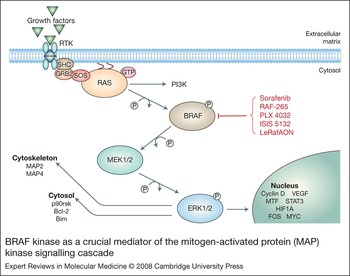
The BRAF gene encodes a serine/threonine kinase involved in signalling from RAS to mitogen-activated protein kinase (MAPK; also known as ERK) through the MAPK pathway (Fig. 1). BRAF signalling regulates a variety of cellular processes, including growth, differentiation and apoptosis (Refs Reference Peyssonnaux and Eychène5, Reference Cruz6). Since RAF activation is essential and sufficient to activate the MAPK cascade, RAF is a critical link between RAS and the MAPK pathway (Ref. Reference Marshall7). RAS is a mediator of several downstream kinase cascades within the cell, and so its activity is very tightly regulated. It is inactive when bound to GDP; however, binding to GTP promotes RAS activation, which leads to the phosphorylation and activation of BRAF kinase and activation of the MAPK pathway. BRAF subsequently activates MEK (gene symbol MAP2K) through a phosphorylation event. The cascade culminates in the activation of ERK (Ref. Reference Kohno and Pouyssegur8). ERKs transmit survival signals through of a variety of cytoplasmic and cytoskeletal targets. Nuclear transcription factors are also activated by ERKs, resulting in the expression of a variety of cancer-associated genes, including cyclin D1, cyclin D2 and cyclin D3 (cell-cycle genes associated with growth promotion), vascular endothelial growth factor (VEGF; angiogenesis), MYC (insensitivity to antigrowth signals), β3 integrin (tissue invasion and metastasis) and mouse double minute 2 (MDM2; apoptosis evasion, limitless replicative potential and angiogenesis) (Refs Reference Kohno and Pouyssegur8, Reference Domingo and Schwartz9).
BRAF kinase as a crucial mediator of the mitogen-activated protein (MAP) kinase signalling cascade. Cell-surface signalling is mediated through receptor tyrosine kinase (RTK) signalling following binding to extracellular growth factor ligands including epidermal growth factor, vascular endothelial growth factor, platelet-derived growth factor and fibroblast growth factor. Signals are transmitted to RAS via the SHC, GRB2 and SOS complex, which results in initiation of the RAF–MEK–ERK kinase cascade. Signalling through this mitogen-activated protein kinase cascade results in growth-promoting effects at the cytoskeletal, cytosolic and nuclear levels through upregulation of proteins involved in transcription, translation, cell trafficking, motility and cell proliferation. Activating BRAF mutations allow for the bypass of extracellular signalling events to promote cell growth. Current therapeutic interventions under investigation to target BRAF kinase in melanoma are indicated in red (see also Table 2). Abbreviations: Bcl-2, B-cell leukaemia 2 protein; BIM, Bcl-2 interacting mediator; BRAF, BRAF oncogene product; FOS, protein product of c-fos; GRB2, growth factor receptor bound protein 2; ERK, extracellular-signal-regulated kinase; HIF-1α, hypoxia-inducible factor 1α; MAP2, microtubule-associated protein 2; MAP-4, microtubule-associated protein 4; MEK, MAPK extracellular-signal-regulated kinase; MTF, membrane-bound transferrin-like protein; p90rsk, 90 kDa ribosomal s6 kinase; PI3K, phosphoinositide 3-kinase; SHC, SRC-homology-2-containing protein; STAT 3, signal transducer and activator of transcription 3; SOS, son of sevenless; VEGF, vascular endothelial growth factor.

Somatic oncogenic mutations of BRAF have been identified most commonly in primary human melanomas, where mutational frequencies have been reported to be as high as 70% (Refs Reference Davies10, Reference Edlundh-Rose11). BRAF mutations have also been found to occur at a lower frequency in several other types of primary human malignancies, including ovarian cancers, colorectal cancers, non-small-cell lung cancers and papillary thyroid carcinomas (Refs Reference Cohen12, Reference Rajagopalan13, Reference Singer14, Reference Brose15). Interestingly, all known BRAF mutations in primary human cancers result in changes within the kinase domain, with the V600 single substitution accounting for 80% of such mutations (Ref. Reference Davies10). The mutated BRAF found in melanoma is almost exclusively a substitution of valine by glutamic acid at codon 600 (V600E; originally designated V599E) (Ref. Reference Wan16). Hydrophobic interactions between residues in the activation loop and those in the ATP-binding site normally maintain BRAF in an inactive conformation; however, the V600E mutation results in an acidic substitution in the kinase domain that disrupts these hydrophobic interactions, promoting novel folding of the kinase into a catalytically competent, constitutively active form (Ref. Reference Wan16). This mutated kinase has been found to have 10- to 12-fold increased kinase activity compared with that of wild-type BRAF (Ref. Reference Davies10). Although somatic mutations of BRAF have been frequently reported in melanoma and other malignancies, this gene does not seem to play a role in familial melanoma susceptibility per se and germline mutations of BRAF have not been identified to date (Refs Reference Jackson17, Reference Meyer18, Reference Laud19, Reference Lang, Boxer and MacKie20, Reference Casula21).
BRAF and melanoma
Previous studies have demonstrated that BRAF kinase induces many of the biological hallmarks of cancer. Expression of exogenous BRAF V600E in cultured immortalised melanocytes stimulated constitutive activation of MEK and ERKs, and promoted tumourigenicity in nude mice (Refs Reference Wellbrock22, Reference Bennett, Cooper and Hart23). Conversely, BRAF V600E suppression by small interfering RNA (siRNA) inhibited the downstream activation of MAPK, leading to reversal of the transformed phenotype, growth arrest and apoptosis (Ref. Reference Hingorani24). Additionally, in vivo studies of human skin reconstructs in nude mice demonstrated a significant proliferative effect of NRAS or phosphoinositide 3-kinase (PI3K), as well as cyclin-dependent kinase 4 (CDK4) activation, dominant-negative p53 expression and telomerase activation, in primary human melanocytes. Melanocytic tumours with only mild junctional melanocytic growth were induced by BRAF V600E, suggesting important functional contributions of the PI3K pathway to melanoma development and invasion, which are not supported by BRAF activation alone (Ref. Reference Chudnovsky25).
Interestingly, a high frequency of BRAF V600E mutations (73–82%) has also been reported in common benign naevi, suggesting that BRAF activation might be the critical inducer of these benign ‘precursor’ lesions (Refs Reference Uribe, Wistuba and Gonzalez26, Reference Pollock27, Reference Poynter28). BRAF activation appears to be an early event in the development of melanomas that arise from benign naevi as it is functionally preserved throughout the course of the disease, and continued expression of BRAF V600E is necessary to maintain tumour growth and progression (Ref. Reference Hoeflich29). Since mutational activation of BRAF kinase is not required for ERK activation in NRAS-transformed melanocytes, NRAS and BRAF mutations provide overlapping functions, which have been shown to be mutually exclusive events in melanoma development (Refs Reference Edlundh-Rose11, Reference Wellbrock22). Therefore, activation of the RAS–RAF–MAPK pathway is a critical step in the initiation of benign and malignant melanocytic neoplasms, which most commonly occurs through mutation of either BRAF or NRAS (Refs Reference Uribe, Wistuba and Gonzalez26, Reference Pollock27). It is interesting to note that pigmented lesions that are rapidly growing or undergoing structural change are more likely to harbour a BRAF mutation (Ref. Reference Loewe30). Furthermore, although most melanomas are not histologically contiguous with a naevus and appear to develop de novo (Ref. Reference Yazdi31), tumours with BRAF mutations are more likely to occur in association with a contiguous naevus, supporting an evolutionary event in some cases (Refs Reference Edlundh-Rose11, Reference Poynter28, Reference Bevona32). BRAF mutations in naevi have been suggested to play an indirect role in melanoma development and are thought to serve as markers of melanoma susceptibility in an individual. Notably, melanocytic naevi remain inactive for decades despite the presence of oncogenic BRAF mutations, and rarely progress to melanoma (Refs Reference Kuwata, Kitagawa and Kasuga33, Reference Chin, Merlino and DePinho34). Therefore, it is of interest to define the mechanisms responsible for allowing these benign tumours to remain in check, in addition to the molecular events necessary to transform a naevus containing a BRAF mutation into a melanoma. Sustained BRAF V600E expression in cultured primary human melanocytes has recently been shown to induce cell-cycle arrest accompanied by the induction of both p16INK4a and senescence-associated acidic β-galactosidase activity, a commonly used senescence marker (Ref. Reference Michaloglou35). The expression of p16INK4a was not strictly required for the induction of this ‘senescence’ in primary human fibroblasts, indicating the existence of a p16INK4a-independent growth arrest in these cells (Ref. Reference Michaloglou35). Thus, growth arrest of melanocytic naevi may result from oncogene-induced ‘senescence’ acting as an effective cellular brake against BRAF-mediated oncogenic signalling (Refs Reference Michaloglou35, Reference Bennett36). Although such data suggest a putative role for BRAF kinase in inducing oncogene-associated senescence in benign melanocytic naevi (Ref. Reference Hoeflich29), the precise molecular mechanisms allowing for the escape from senescence and progression to malignant melanoma remain to be elucidated.
Defining the role of BRAF in melanoma onset and progression
Although evidence suggests that activation of the MAPK pathway promotes the proliferation, invasion and survival of melanoma cells in vitro, the exact role of BRAF mutation and activation in melanoma tumour progression, maintenance and outcome remains controversial. Notably, BRAF mutations were increased in vertical-growth-phase and metastatic melanomas compared with radial-growth-phase melanomas, suggesting a progression-associated mutational activation of BRAF kinase in melanoma (Ref. Reference Dong37). Since this mutational rate in radial-growth-phase melanomas would also be significantly lower than that seen in benign naevi, an evolutionary progression from naevi through radial growth with fewer BRAF mutations would be difficult to reconcile. Since large radial-growth-phase melanomas often occur on chronic sun-damaged skin and are more frequently associated with NRAS versus BRAF mutations (Ref. Reference Curtin38), a simple explanation for this discrepancy would be increased NRAS mutations in these lesions; however, no NRAS mutations were seen in the 20 primary radial-growth-phase melanomas analysed. Further studies have noted a higher incidence of BRAF mutations in metastatic lesions compared with primary melanomas (Ref. Reference Shinozaki39), and mutated BRAF has been identified in circulating DNA from stage III (lymph node involvement) and IV (metastatic disease) patients, with the level correlating with the stage of disease (Ref. Reference Daniotti40).
The above data therefore support a model of melanoma development that might occur through a precursor lesion, or naevus, with sustained activation of BRAF kinase. Additionally, melanomas may occur de novo without the pre-existence of a naevus through activation of BRAF kinase and additional tumourigenic events. Since the majority of benign naevi possess activating BRAF kinase mutations, these lesions must have a low risk for malignant progression because of a BRAF-associated tumour suppressor mechanism that remains to be elucidated. The clear association of increased numbers of naevi and melanoma risk suggests that naevi may serve as ‘markers’ of malignant risk and susceptibility to activating BRAF kinase mutations either within the context of a benign naevus or within a malignant melanocytic lesion itself. Thus, oncogenic BRAF may play a role in the progression of the malignant phenotype in melanoma in addition to its role in tumour initiation. Of note, recent investigations have sought to determine the prognostic significance of BRAF mutations in melanoma; however, studies to date have failed to identify a link between BRAF mutational status and disease outcome (Ref. Reference Shinozaki39). Although the notion of BRAF kinase as a ‘driver’ for the malignant phenotype in melanoma remains to be clarified, it appears that mutated BRAF kinase alone, although necessary for the development of the majority of melanomas, is insufficient to induce the onset of this malignancy. Clearly, additional molecular events are required in order for melanocytic transformation to occur in the context of BRAF kinase activation. The nature of these additional molecular changes is an area of active investigation at the present time, and results are eagerly anticipated by the melanoma research community.
Clinicopathological features associated with BRAF V600E melanomas
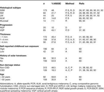
Recent studies have helped to define the genetic and environmental risk factors associated with BRAF mutational status in melanoma and support the existence of susceptibility loci that increase the likelihood for individuals to acquire BRAF mutations depending on the type of ultraviolet radiation (UVR) exposure to the skin. Previous studies have looked at the frequencies of BRAF V600E in histological subtypes, finding that superficial spreading melanomas (SSMs) and nodular melanomas (NMs) had higher frequencies of mutated BRAF than lentigo maligna melanomas (LMMs) or acral lentiginous melanomas (ALMs) (Table 1). However, the use of such a classification scheme is controversial because a substantial number of melanomas do not fit the classic histological subtypes, and these subtypes are not independent prognostic factors for melanoma. Recently, melanomas were segregated into four groups according to the site of the primary lesion and sun exposure (Ref. Reference Curtin38). Two groups were based on high UVR exposure – either chronic sun damage throughout life (CSD) occurring on the face and neck, or as a consequence of intermittent exposure (non-CSD), which occurs on the trunk, back and extremities. The other two groups were from sites of little to no UVR exposure, such as the palms and soles or mucosal and uveal sites, respectively. Using array-based comparative genomic hybridisation (CGH), researchers were able to identify significant differences in the frequencies of regional changes in the number of copies of DNA and mutation frequencies in BRAF among the four groups of melanomas. BRAF or NRAS mutations were seen in 81% of melanomas from non-CSD sites as mutually exclusive events, but were found to be rare in the other three groups (Refs Reference Curtin38, Reference Curtin41). It was concluded that BRAF mutations are associated with melanomas originating in areas of repetitive acute sun exposure, but not in areas with chronic UVR exposure or those that are protected from UVR altogether. It is notable that neither the NRAS nor BRAF mutations observed in melanomas demonstrate characteristic UVR-induced signature changes of cytosine/thymine (C/T) or CC/TT, and that the target of UVR injury leading to such mutations remains unclear (Refs Reference Davies10, Reference Albino43, Reference Hocker and Tsao44).
Summary of BRAF V600E mutation frequencies in melanoma based on clinical and pathological characteristics

Abbreviations: A, allele-specific PCR; ALM, acral lentiginous melanoma; C, array comparative genomic hybridisation; CSD, chronic sun-damaged skin; D, PCR and dHPLC; LM, lentigo maligna melanoma; NM, nodular melanoma; P, PCR sequence analysis; R, PCR-RFLP; RGP, radial growth phase; S, PCR:SSCP; SSM, superficial spreading melanoma; VGP vertical growth phase.
The observation that congenital naevi rarely harbour BRAF V600E, whereas the mutation is very common in acquired naevi, provides further support for the notion that UVR exposure is associated with acquired BRAF mutations (Ref. Reference Bauer45). One possible explanation for the UVR-associated alterations of BRAF kinase without usual UVR-signature changes is that a combination of reactive oxygen species and cytokines, such as those released from the inflammatory reaction induced by sunburn, can lead to replicative errors in DNA and, ultimately, to base substitutions without classical UV-signature mutations (Refs Reference Balch46, Reference Jhappan, Noonan and Merlino47). Although the V600E mutation of T to A is not at a dipyrimidine site and is not generally viewed as resulting from error-prone replication of UVR-damaged DNA, it has been proposed that common BRAF mutations could be linked to the presence of nearby pyrimidine dimers and an error-prone polymerase (Ref. Reference Thomas, Berwick and Cordeiro-Stone48). Thus, although a link between UVR exposure and BRAF mutational status in melanomas exists, the precise nature of UVR effects that lead to BRAF mutation are not so straightforward. Interestingly, BRAF V600E is not seen in basal or squamous cell skin cancers, which are associated with chronic sun-damaged skin (Ref. Reference Libra42).
The genetic variations identified in melanomas at various sites with different levels of sun exposure indicate that there are distinct genetic pathways associated with melanoma development. Interestingly, in melanomas without BRAF or NRAS mutations, copies of the genes for CDK4 or cyclin D1 (CCND1) were increased. Additionally, of the melanomas with CDK4 amplification, none included NRAS or BRAF mutations, or CCND1 amplification. Since CDK4 and CCND1 are both downstream effectors of the MAPK pathway (Ref. Reference Curtin38), the results suggest that activation of these cell-cycle-regulatory genes serves an independent oncogenic function in melanomas lacking BRAF or NRAS mutations. In addition, these findings further support the notion that divergent molecular pathways exist during melanoma development, which would explain the heterogeneous nature of this malignancy that has been observed clinically. Further support for divergent pathway activation in melanoma comes from uveal melanomas in which BRAF mutations are not responsible for activation of the MAPK pathway, indicating that these ocular melanomas arise though a fundamentally distinct set of genetic alterations compared with primary cutaneous melanomas (Refs Reference Cruz6, Reference Zuidervaart49, Reference Edmunds50, Reference Cohen51, Reference Rimoldi52, Reference Weber53).
Additional genetic events that predispose to BRAF mutation
Since melanomas on anatomical sites exposed to UVR predominantly affect Caucasians, and non-CSD melanomas occur at relatively low UVR doses in Caucasian populations, it has been hypothesised that the high frequency of BRAF mutations in non-CSD melanomas is due to susceptibility factors that occur at higher frequencies in Caucasian populations (Ref. Reference Landi54). The melanocortin 1 receptor (MC1R) accounts for normal pigment variation in the skin. When activated in response to UVR, the MC1R protein is responsible for the conversion of pheomelanin (yellow-red pigment) to eumelanin (black-brown pigment) (the two major melanin classes) (Ref. Reference Landi54). The MC1R gene is highly polymorphic and many alleles have been observed that account for varying degrees of loss of function, leading to red hair, freckling and sun sensitivity. Most people with red hair are homozygous for alleles of the MC1R gene that show varying degrees of diminished function. Recently, investigators found that MC1R variants are strongly associated with BRAF mutations in non-CSD melanomas. BRAF mutations are a characteristic feature of more than 80% of the non-CSD melanomas in individuals with two variant MC1R alleles but only in 30% of individuals with wild-type MC1R (Ref. Reference Landi54). Thus, mutated BRAF was found to be more common in melanocytes with impaired MC1R signalling. Although MC1R variants are associated with melanoma risk and progression, that risk seems to be independent of skin type or hair colour (Refs Reference Rees55, Reference Fargnoli56, Reference Kennedy57, Reference Palmer58). Interestingly, in uveal melanocytes, where BRAF mutations are not seen, there is also no MC1R protein expressed (Ref. Reference Li59). Therefore, when MC1R is absent, mutated BRAF does not drive tumourigenicity, and melanocytic neoplasia in such cases is related to activation of alternative growth-stimulatory pathways. Although these observations imply that germline events (such as heritable MC1R locus variants) can largely influence acquired genetic events leading to tumourigenesis in response to environmental exposures (such as UVR), the precise link between MC1R function, UVR exposure and BRAF mutation remains to be elucidated.
Clinical implications and applications
In an attempt to gain more knowledge of the functional role of BRAF in melanoma development and progression, previous studies have evaluated the clinical and pathological features associated with BRAF V600E melanomas. It was found that characteristics of primary melanomas with BRAF V600E included those normally associated with a favourable prognosis (Ref. Reference Liu60). These include tumours <1 mm in Breslow's thickness (a measurement of the actual depth of penetration of the tumour into the skin) and those with a lower mitotic rate, detected by the Ki67 active cell-cycle marker, and a lower rate of cell proliferation (Refs Reference Liu60, 61). These observations suggest that BRAF V600E may be associated with a more-differentiated form of melanoma, compared with melanomas possessing wild-type BRAF. It has also been suggested that melanomas with mutated BRAF respond to treatment more readily and that such patients have a longer disease-free survival following therapy (Ref. Reference Kumar62). The precise molecular basis for these observations remains to be determined.
Targeting BRAF kinase in melanoma
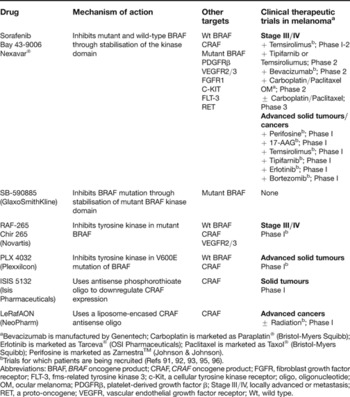
Knowledge of the genetic differences that subclassify melanomas is valuable in the design of targeted therapeutic strategies for this disease. Owing to the high frequency of BRAF mutations in melanoma, many studies have sought to assess the significance of BRAF as a therapeutic target in melanoma. Sorafenib is a small-molecule inhibitor of both mutant and wild-type BRAF, which binds to the kinase domain, rendering it inactive (Refs Reference Adnane63, Reference Strumberg64). Further characterisation of this molecule has shown that it inhibits other protein kinases including the VEGF receptors and platelet-derived growth factor (PDGF) receptor family members (PDGFR-β and KIT), which play a role in angiogenesis and tumour progression (Ref. Reference Adnane63). Given this additional activity of sorafenib, and the fact that the MAPK pathway is one of two major endothelial-cell signalling pathways responsible for angiogenesis, sorafenib has been proposed to possess dual mechanisms of activity in the treatment of melanoma: by inhibiting tumour growth directly (through BRAF and KIT) and by inhibiting angiogenesis (via VEGF receptors and PDGFR-β) (Ref. Reference Adnane63). Preclinically, sorafenib has been documented to cause decreased microvessel density, decreased branching of vessels and massive regional tumour cell death in murine melanomas (Ref. Reference Murphy65), and death of human melanoma tumour xenografts (Ref. Reference Karasarides66). Four Phase I studies using oral sorafenib as a single agent have been completed, and the agent showed a positive safety profile, with mild to moderate diarrhoea and hand/foot skin rash being the most common adverse events attributable to treatment (Refs Reference Strumberg64, Reference Strumberg67). Sorafenib showed moderate activity in a Phase II study involving 20 patients with refractory stage IV melanoma, with one partial response and three stable patients after treatment for 12 weeks with 400 mg twice daily (BID).
Another Phase II, randomised discontinuation study found that sorafenib had no significant activity in human metastatic melanoma as a single agent, with the best response being stable disease in 19% of patients (Ref. Reference Eisen68). However, in a Phase I/II study of sorafenib in combination with carboplatin and paclitaxel, 31% of patients had an objective response and 54% demonstrated progression-free survival of at least 3 months (Ref. Reference Flaherty69). Of note, all responses have been with previously treated patients and are not dependent on BRAF mutational status (Refs Reference Eisen68, Reference Flaherty69). Two Phase III studies are under way comparing sorafinib, carboplatin and paxitaxel with carboplatin and paclitaxel alone in both chemotherapy-naive patients and previously treated patients (Table 2). Additional clinical trials are under development to evaluate the use of sorafenib in combination with other chemotherapies or targeted agents. A randomised Phase II trail of temozolimide with sorafenib is in progress, and the preliminary results are promising (Ref. Reference Amaravadi70). In addition, a newer targeted therapy that selectively inhibits oncogenic RAF kinases with a higher potency for BRAF is currently in preclinical studies and has shown promising specific activity against BRAF-mutant melanomas (Ref. Reference King71). Although sorafenib gained accelerated approval by the US Food and Drug Administration for the treatment of advanced renal cell carcinoma in 2005 based on improved survival benefit in randomised clinical trials (Refs Reference Eisen72, Reference Escudier73), it must be noted that renal cell carcinomas lack activating BRAF kinase mutations, suggesting that the antitumour activity of sorafenib in this malignancy is through a non-BRAF-dependent mechanism. Other kinase inhibitors with enhanced selectivity for BRAF V600E are under investigation for the specific treatment of BRAF mutant melanomas.
Current clinical trials targeting BRAF kinase in melanoma
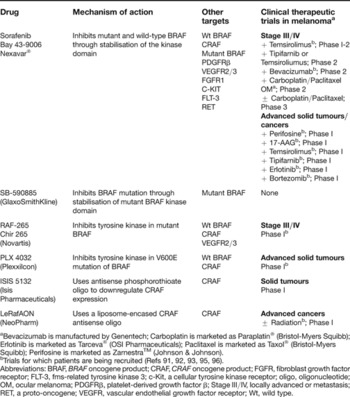
aBevacizumab is manufactured by Genentech; Carboplatin is marketed as Paraplatin® (Bristol-Myers Squibb); Erlotinib is marketed as Tarceva® (OSI Pharmaceuticals); Paclitaxel is marketed as Taxol® (Bristol-Myers Squibb); Perifosine is marketed as ZarnestraTM (Johnson & Johnson).
bTrials for which patients are being recruited (Refs Reference Omholt91, Reference Maldonado92, Reference Davison93, Reference Cunningham95, Reference King96).
Abbreviations: BRAF, BRAF oncogene product; CRAF, CRAF oncogene product; FGFR, fibroblast growth factor receptor; FLT-3, fms-related tyrosine kinase 3; c-Kit, a cellular tyrosine kinase receptor; oligo, oligonucleotide; OM, ocular melanoma; PDGFRβ, platelet-derived growth factor β; Stage III/IV, locally advanced or metastasis; RET, a proto-oncogene; VEGFR, vascular endothelial growth factor receptor; Wt, wild type.
Additional therapies with indirect activity against BRAF kinase include 17-AAG (17-allylamino-17-demethoxygeldanamycin), an anticancer drug that disrupts the binding of heat shock protein 90 (Hsp90) to its targets, causing ubiquitylation and proteasomal degradation of target proteins (Refs Reference Neckers and Neckers74, Reference Maloney and Workman75). BRAF is an Hsp90 target protein that requires binding for proper folding and stability. BRAF V600E has been shown to be more sensitive to degradation by 17-AAG treatment than wild-type BRAF (Ref. Reference da Rocha76) and Phase II studies are currently recruiting.
Towards defining a BRAF signature in melanoma
Melanomas are notorious malignancies owing to their well-known resistance to standard cancer therapies. This therapeutic resistance has largely been attributed to the complex heterogeneous nature of this disease. Although it is likely that multiple molecular alterations contribute to the complex nature of this malignancy, a major advance in the understanding of melanoma has ensued from the discovery of frequent BRAF kinase mutations in this disease. Microarray studies and genetic profiling have provided insight into the spectrum of melanomas in which BRAF plays a role, revealing new data that have implications in the diagnosis and treatment of this disease. Multiple studies support the existence of distinct genetically defined melanoma groups, mirroring different molecular and clinical behaviour (Refs Reference Daniotti77, Reference Haqq78, Reference Bittner79, Reference Seftor80, Reference de Wit81). By comparing genetic profiles, researchers were able to differentiate between benign naevi, primary melanomas and metastatic melanomas. Furthermore, they were also able to differentiate between less-aggressive, radial-growth-phase and more-aggressive, vertical-growth-phase melanomas (Ref. Reference Haqq78). It has been proposed that BRAF mutations may influence tumour progression and give rise to distinct gene expression signatures in melanomas. To define these ‘BRAF-specific’ gene signatures, investigators have evaluated melanoma gene expression profiles and sorted them on the basis of BRAF kinase mutational status. Using such a data-mining strategy, two studies were able to identify 65 and 83 genes, respectively, that distinguished BRAF-mutant melanomas from the wild type (Refs Reference Bloethner82, Reference Pavey83). Although such studies will help to define a BRAF signature in melanoma, the lack of concordance of BRAF-signature genes in these two studies suggests the need for additional selection parameters to subclassify BRAF-mutant melanomas. Furthermore, in a follow-up study analysing three separate DNA microarray cohorts, investigators identified only a single gene whose expression changes correlated with BRAF mutational status (Ref. Reference Hoek84). Of note, three recognisably discrete gene signatures for melanoma were identified in this study that distinguished melanomas of variable proliferative and metastastic potential; however, mutant BRAF was not specific to any of the melanoma signature groups (Ref. Reference Hoek84).
Although gene expression profiling studies failed to define a clear BRAF signature in melanomas, evaluation of BRAF kinase mutational status in combination with array CGH to evaluate DNA copy number changes in mutant BRAF versus wild-type BRAF melanomas has proven quite informative. Using this approach, recent studies revealed patterns of genomic aberrations that could distinguish CSD, non-CSD, mucosal and acral melanomas. Array CGH data and defined genetic profiles from the melanomas evaluated allowed investigators to classify melanomas with respect to BRAF mutational status with 70% confidence (Ref. Reference Curtin38). These studies have further distinguished particular genomic events that allowed for the subclassification of melanoma phenotypes and mutations in BRAF, NRAS, PTEN or TP53 (Ref. Reference Jonsson85). Thus, BRAF-mutant melanomas display specific genomic aberrations that correlate with BRAF mutational status and primary tumour site and/or sun-exposure patterns.
The above data suggest that BRAF kinase mutational status affords a particular genetic profile in subsets of melanomas; however, such genomic data have not fully translated to a particular molecular signature for BRAF-mutant melanomas because expression profiling studies have shown inconsistencies. Clearly, more work needs to be done in this realm and it is expected that, as additional genetic, epigenetic and expression profiling studies are performed, data will be cumulatively assessed in this regard. Ultimately it is expected that a more thorough investigation of molecular alterations in melanomas in the context of BRAF kinase mutational status will allow for improved diagnosis, classification and treatment of this disease.
Conclusion
Although much progress has been made in understanding the molecular basis of melanoma development and progression since the discovery of frequent activating BRAF kinase mutations in melanomas (Ref. Reference Davies10), it is clear that BRAF kinase has primarily served as an important starting point for subsequent molecular studies of this complex malignancy. Although current clinical trials targeting BRAF kinase have shown some therapeutic efficacy, it is unlikely that this therapeutic strategy alone will provide a significant benefit to large numbers of patients with advanced-stage disease. More complete analyses are therefore required to define the specific contribution of BRAF kinase to melanoma progression and tumour maintenance, which will allow for a clearer understanding of its importance as a therapeutic target in this disease. Knowledge of the BRAF kinase mutational status has underscored the fact that a classification scheme for melanoma based on histological subtype is limiting, because it is of little prognostic value (Refs Reference Balch46, Reference Barnhill86). As recent studies have allowed the identification of genetically distinct melanoma subtypes that are influenced by tumour site and degree of sun exposure, such a tumour classification scheme is likely to be of significant clinical value, as it is expected to result in improved targeted therapies for such melanoma subgroups. The identification of gene expression signatures for BRAF-mutant melanomas will allow for the further refinement of such targeted therapies. Thus, although the identification of frequent activating mutations of BRAF kinase in melanomas has provided much insight into the molecular defects associated with this disease, many unanswered questions still remain. Clearly, sun-exposure patterns and heritable genetic influences help to determine the precise genetic and molecular defects that occur in melanomas, including BRAF kinase mutations; however, additional molecular events must occur in order for malignant transformation to take place. We eagerly anticipate further clarification of the role of BRAF kinase in melanoma and its influence on additional molecular and genetic events leading to melanoma development and progression with the expectation that such data will allow for improved targeted therapies for this aggressive malignancy for which few therapeutic options currently exist.
Acknowledgements and funding
We thank the reviewers of this manuscript for their critical evaluation and insightful comments. Research in the Alani Lab is support by the National Institutes of Health, the American Skin Association, the Flight Attendant Medical Research Institute, The Joanna M. Nicolay Melanoma Foundation, The Murren Family Foundation, and The Henry and Elaine Kaufman Foundation.