Hypertrophic cardiomyopathy may be caused by many mutations in genes involved in the cardiac sarcomere function. Such mutations may affect the myosin heavy chain, actin, tropomyocin, and titin components of the contractile unit.Reference Maron1 Penetrance is incomplete and unpredictable with variable expression.1 The resultant pathology includes cardiac hypertrophy, myocardial disarray, ventricular tachycardia, sudden death, and left ventricular outflow obstruction on the basis of systolic anterior motion of the anterior mitral leaflet. Non-physiological left ventricular hypertrophy is driven by a combination of genetic and environmental factors. Physiological hypertrophy may co-exist with these factors in response to outflow obstruction, contributing to the overall phenotype. The interplay between physiological and non-physiological hypertrophy is poorly understood, and is likely to be complex. We report the case of a young woman with a troponin mutation of C to T nucleotide substitution in exon 17 of troponin 2 (TNNT2; c.868C>T; p.Arg288Cys) leading to hypertrophic obstructive cardiomyopathy. This mutation has previously been reported in association with familial hypertrophic cardiomyopathy and left ventricular outflow obstruction.Reference Watkins, McKenna and Thierfelder2 Following surgical resection of the outflow obstruction, she had near-complete resolution of her asymmetric left ventricular hypertrophy, such that cardiomyopathy could no longer be diagnosed on echocardiographic grounds. We believe that this unusual case shows important aspects relating to the interplay between genetic and environmental mechanisms and the overlap in the phenotypic spectrum between primary subaortic stenosis and obstructive hypertrophic cardiomyopathy.
Case report
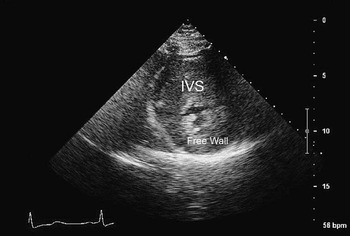
A 33-year-old morbidly obese woman with a body mass index of 38.6 was referred for assessment during pregnancy following the discovery of a heart murmur. She was found to have severe asymmetric septal hypertrophy measuring 3.5 centimetres in width, with a posterior wall measurement of 17 millimetres. She had associated left ventricular outflow obstruction partly due to systolic anterior motion of the mitral valve, but also due to the abnormal attachment of the subchordal apparatus of the anterior mitral valve leaflet within the left ventricular outflow. The peak gradient in the left ventricular outflow was 130 millimetres of mercury. She did not have any associated dysrhythmia or syncope. Her effort tolerance was severely impaired due to dyspnoea. There was no family history of cardiac disorder or sudden cardiac death and no other family member had a diagnosis of cardiomyopathy, despite subsequent screening. Genetic analysis revealed a well-recognised troponin mutation (C to T nucleotide substitution in exon 17 of TNNT2 (c.868C>T; p.Arg288Cys). She was commenced on β-blocker therapy during the remainder of her pregnancy. The course of the pregnancy was uncomplicated apart from worsening effort intolerance leading to a caesarean section being carried out at 38 weeks gestation following a failed induction of labour. Following pregnancy she had a trial of disopyramide therapy, which resulted in only modest relief of the outflow obstruction with a gradient of 100 millimetres of mercury. There was no change in her left ventricular thickness at this stage. At 3 months after delivery the patient underwent reparative surgery. At surgery the left ventricle was hypertrophied and the interventricular septum severely enlarged resulting in significant distortion of the anterior right ventricular free wall. The aortic valve was normal. but inferior to it there was a fibrous ring consisting of secondary chords from the anterior mitral valve leaflet and a fibro-proliferative membrane. In addition, there was constrictive bulging from the thickened interventricular septum causing a complex and severe outflow obstruction. A Morrow procedure was performed with the resection of left ventricular septal muscle, the secondary chords in the outflow tract, and resection of the right side of the ventricular septum through the right ventricular outflow tract. Subsequent histology showed a striking variation in the size and shape of the myocyte nuclei, many being enlarged and hyperchromatic. There was also evidence of myocyte fibre disarray.
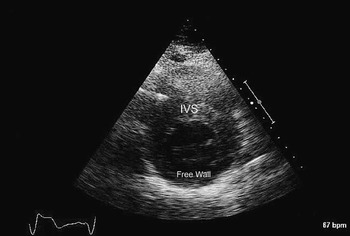
She tolerated surgery well, but the procedure was complicated by complete heart block for which she had an endocardial dual-chamber system inserted. At 18-month follow-up she had notable symptomatic improvement. Her repeat echocardiogram showed near-complete resolution of the asymmetric left ventricular hypertrophy, and the peak gradient across the left ventricular outflow was now 27 millimetres of mercury, with a mean gradient of 15 millimetres of mercury. The 12-lead electrocardiogram has shown a sensed atrial rhythm and paced ventricular rhythm with a left bundle branch block morphology. QRS duration was 200 milliseconds.
Discussion
The case described in this report represents an unexpected clinical course of near total post-operative left ventricular remodelling in a well-documented hypertrophic obstructive cardiomyopathy geno- and phenotype, comprising asymmetric septal hypertrophy, systolic anterior motion of the anterior mitral leaflet, and a known troponin mutation.
Multiple-point and frameshift mutations in genes’ coding for proteins of the sarcomeric unit have been implicated in the pathogenesis of hypertrophic obstructive cardiomyopathy. There is poor genotype–phenotype correlation of these genes due to incomplete penetrance and variable expressivity.Reference Forissier, Carrier, Farza, Bonne, Bercovici and Richard3 This variability is compounded by the fluctuant biophysical properties of mutant and wild-type sarcomeric peptides, and the co-existence of environmental modifiers that may drive unfavourable cardiac remodelling in a dose-dependent manner.Reference Arad4
Our case suggests a possible “environmental” triggering mechanism provided by the anatomic pre-existing subaortic stenosis, and also shows that once this trigger had been removed, there was a striking reverse remodelling of not only the left ventricle but also asymmetric septal hypertrophy. These observations suggest that the hypertrophic obstructive cardiomyopathy phenotype is neither persistent nor inevitably progressive once present in such circumstances. Left ventricular remodelling following septal myectomy is not a new observation, but to this degree, is novel. MazurReference Mazur5 observed a 37% reduction in myocardial mass at 2 years following surgical septal myectomy. Other reports have documented comparable left ventricular remodelling, suggesting that a substantial portion of the left ventricular mass is related to obstruction.Reference Deb, Schaff, Dearani, Nishimura and Ommen6–Reference Nielsen, Killip and Spencer10 However, in none of the reported cases was there complete resolution of the hypertrophic obstructive cardiomyopathy phenotype as there was in our case.
An additional potential contributory factor to left ventricular resolution is the post-operative introduction of atrioventricular sequential pacing; in this case, right atrium and right ventricular pacing. Pacing reorientates and synchronises electrical activation to originate in the right ventricular apex spreading to the rest of the myocardium. Theoretically, the septum is recruited to right ventricular contraction and thereby the left ventricular outflow gradient may be reduced. In reality, however, there is little evidence that pacing reduces the outflow gradient in a consistent manner.Reference Maron, Nishimura, Mckenna and Rakowski11 Indeed, the effect on left ventricular remodelling also is only limited. The effect of pacing appears to be concentrated on the reduction of segmental left ventricular hypertrophy rather than on reduction in global left ventricular hypertrophy.Reference Fananapazir, Epstein, Curiel and Panza12
This case suggests an overlap in the spectrum between primary subaortic stenosis and hypertrophic obstructive cardiomyopathy at both the genetic and phenotypic levels. It is tempting to speculate that the obstructive forms of hypertrophic obstructive cardiomyopathy and primary subaortic stenosis, particularly in the presence of a fibroproliferative membrane and/or abnormal mitral valve attachments in the outflow, form part of the same disease spectrum. Further studies are necessary to evaluate this notion (Figs 1 and 2).
Pre-operative short-axis view showing severe asymmetric left ventricular hypertrophy; IVS = interventricular septum; free wall = left ventricular free wall.

Post-operative short-axis view showing complete resolution of left ventricular hypertrophy; IVS = interventricular septum; free wall = left ventricular free wall.