

1. Introduction
The Dyrosauridae are a successful family of meso- to longirostrine, aquatic crocodiles that could reach more than 8 m in length (Hastings et al. Reference Hastings, Bloch, Cadena and Jaramillo2010). Although remains of dyrosaurids have been known for over 100 years now and have been frequently found in marginal marine sediments of the Tethys, they have not been a focal point of crocodyliform evolutionary research despite their survival across the Cretaceous–Palaeogene (K–Pg) boundary. In terms of phylogenetic relationships, Dyrosauridae has been considered as either an independent distinct lineage of marine piscivorous mesoeucrocodylians (Benton & Clark, Reference Benton, Clark and Benton1988; Norell & Clark, Reference Norell and Clark1990) or as belonging to a longirostrine mesoeucrocodylian clade with Thalattosuchia and Pholidosauridae (Clark, Reference Clark, Frazer and Sues1994; Wu, Sues & Dong, Reference Wu, Sues and Dong1997; Buckley & Brochu, Reference Buckley and Brochu1999; Wu, Russell & Cumbaa, Reference Wu, Russell and Cumbaa2001; Brochu et al. Reference Brochu, Bouaré, Sissoko, Roberts and O'Leary2002; Pol, Reference Pol2003). Owing to recent discoveries and descriptions of a large number of new dyrosaurid taxa, general interest in this enigmatic clade of Crocodyliformes has been raised and knowledge about the anatomy, palaeobiology and biogeography of dyrosaurids has been improved considerably (Jouve, Bouya & Amaghzaz, Reference Jouve2005, Reference Jouve2008; Jouve et al. Reference Jouve, Bouya and Amaghzaz2005, Reference Jouve, Iarochène, Bouya and Amaghzaz2006; Jouve, Reference Jouve2007; Barbosa, Kellner & Sales Viana, Reference Barbosa, Kellner and Sales Viana2008; Hill et al. Reference Hill, McCartney, Roberts, Bouaré, Sissoko and O'Leary2008; Hastings et al. Reference Hastings, Bloch, Cadena and Jaramillo2010; Hastings, Bloch & Jaramillo, Reference Hastings, Bloch and Jaramillo2011). But despite these new discoveries, recent phylogenetic analyses could still not clarify the systematic position of Dyrosauridae beyond the level of Neosuchia and an often supported close relationship to Pholidosauridae (Sereno et al. Reference Sereno, Larsson, Sidoe and Gado2001, Reference Sereno, Sidor, Larsson and Gado2003; Wu, Russell & Cumbaa, Reference Wu, Russell and Cumbaa2001; Jouve et al. Reference Jouve, Iarochène, Bouya and Amaghzaz2006).
Remains of dyrosaurids have been found from the Upper Cretaceous to the Lower Eocene around the margins of the Tethyan sea. Most localities are known from Africa (e.g. de Stefano, Reference de Stefano1903; Swinton, Reference Swinton1930, Reference Swinton1950; Buffetaut, Reference Bronzati, Montefeltro and Langer1979a ,Reference Buffetaut b ; Moody & Buffetaut, Reference Moody and Buffetaut1981; Buffetaut, Bussert & Brinkmann, Reference Buffetaut, Bussert and Brinkmann1990; Langston, Reference Langston1995; Brochu et al. Reference Brochu, Bouaré, Sissoko, Roberts and O'Leary2002; Jouve & Schwarz, Reference Jouve and Schwarz2004; Jouve, Reference Jouve2005; Jouve, Bouya & Amaghzaz, Reference Jouve2005, Reference Jouve, Bouya and Amaghzaz2008; Jouve et al. Reference Jouve, Bouya and Amaghzaz2005, Reference Jouve, Iarochène, Bouya and Amaghzaz2006; Jouve, Reference Jouve2007; Hill et al. Reference Hill, McCartney, Roberts, Bouaré, Sissoko and O'Leary2008), but they occur also in North America (e.g. Troxell, Reference Troxell1925; Parris, Reference Parris1986; Denton, Dobie & Parris, Reference Denton, Dobie, Parris, Callaway and Nicholls1997), South America (e.g. Argollo et al. Reference Argollo, Buffetaut, Cappetta, Fornari, Herail, Laubacher, Sige and Vizcarra1987; Gasparini & Spalletti, Reference Gasparini and Spalletti1990; Barbosa, Kellner & Sales Viana, Reference Barbosa, Kellner and Sales Viana2008; Hastings et al. Reference Hastings, Bloch, Cadena and Jaramillo2010; Hastings, Bloch & Jaramillo, Reference Hastings, Bloch and Jaramillo2011), Europe (Buffetaut & Lauverjat, Reference Buffetaut and Lauverjat1978) and Asia (Buffetaut, Reference Buffetaut1977, Reference Buffetaut1978a Reference Buffetaut,b ; Gingerich et al. Reference Gingerich, Russell, Sigogneau-Russell, Hartenberger, Shah, Hassan, Rose and Ardrey1979; Storrs, Reference Storrs1986; Gingerich, Reference Gingerich2003). Dyrosaurid-bearing localities comprise often shallow-marine, near-costal sediments, such as the phosphate deposits of Tunisia (Moody & Buffetaut, Reference Moody and Buffetaut1981) and Morocco (Bardet et al. Reference Bardet, Pereda Superbiola, Jouve, Bourdon, Vincent, Houssaye, Rage, Jalil, Bouya and Amaghzaz2010), but also transitional sediments with brackish influence and lagoonal (Hastings et al. Reference Hastings, Bloch, Cadena and Jaramillo2010) or estuarine (Langston, Reference Langston1995) conditions, and, especially in the Eocene, also fluvial (Buffetaut, Reference Buffetaut1978 a,b) environments.
One of the most interesting aspects of dyrosaurids is that they are, together with gavialoid crocodylians and marine turtles, the only marine reptiles that survived the K–Pg boundary. Their evolutionary success has been explained by their presumed role as major predators after the extinction of mosasaurs (S. Jouve, unpub. Ph.D. thesis, Muséum National d'Histoire Naturelle de Paris, 2004; Jouve & Schwarz, Reference Jouve and Schwarz2004; Barbosa, Kellner & Sales Viana, Reference Barbosa, Kellner and Sales Viana2008; Jouve, Bouya & Amaghzaz, Reference Jouve, Bouya and Amaghzaz2008). Dyrosaurids possess a peculiar anatomy of the postcranial skeleton that distinguishes them fundamentally from all other crocodyliforms (Salisbury & Frey, Reference Salisbury, Frey, Grigg, Seebacher and Franklin2001; Schwarz, Frey & Martin, Reference Schwarz, Frey and Martin2006; Schwarz-Wings, Frey & Martin, Reference Schwarz-Wings, Frey and Martin2009). Analyses of their anatomy and their bracing system showed that dyrosaurids lacked the fully aquatic adaptations that are present in metriorhynchid crocodyliforms (Salisbury & Frey, Reference Salisbury, Frey, Grigg, Seebacher and Franklin2001; Georgi, Reference Georgi2006; Schwarz, Frey & Martin, Reference Schwarz, Frey and Martin2006; Schwarz-Wings, Frey & Martin, Reference Schwarz-Wings, Frey and Martin2009), but were still capable of terrestrial locomotion, as in extant crocodylians, which is additionally underlined by their transgression into fluvial ecosystems in the Eocene. Dyrosaurids furthermore differ from all other crocodyliforms in possessing a unique, extremely elongated retroarticular process of the mandible (Jouve et al. Reference Jouve, Iarochène, Bouya and Amaghzaz2006), which must have had implications for the jaw musculature and consequently for the mechanisms of food capturing. The feeding apparatus of dyrosaurids is reconstructed in this study with its anatomy and related functional aspects to get a better idea of the possible spectrum of prey and prey acquisition options available to them in order to gain a better understanding of their evolutionary success.
Anatomical abbreviations. Caps art – capsula articularis; cart trans – cartilago transiliens; Cr A, B – quadrate crest A, B; Cr lsph – laterosphenoid crest; glen – mandibular glenoid fossa of the articular; Lig arccorp – ligamentum arcuocorporale; Lig elast – ligamentum elasticum interlaminare; Lig intc – ligamentum intercostale; Lig intart – ligamentum interarticulare; Lig n – ligamentum nuchae; Lig patarc – ligamentum proatlantoarculae; Lig ssp – ligamentum supraspinale; mATC – m. atloïdo-capitis; mAME – m. adductor mandibulae externus; mAMEM – m. adductor mandibulae externus medialis; mAMEP – m. adductor mandibulae externus posterior; mAMES – m. adductor mandibulae externus superficialis; mAMP – m. adductor mandibulae posterior; mAMI – m. adductor mandibulae internus; mCAPST – m. capitisternalis; mCSCs – m. colloscapularis superficialis; mDM – m. depressor mandibulae; mEPC – m. epistropheo-capitis; mIM – m. intermandibularis; mIRA – m. intramandibularis; mmIART – mm. interarticulares; mIlCOSTcap – m. iliocostalis capitis; mILCOSTcer – m. iliocostalis cervicis; mmISP – mm. interspinales; mLCAPp – m. longus capitis posterior; mLCAP – m. longus capitis; mLCAPs – m. longus capitis superficialis; mLCER – m. longus cervicis; mLONcoll – m. longus colli; mmf – medial mandibular fossa; mPST – m. pseudotemporalis; mPSTp – m. pseudotemporalis profundus; mPSTs – m. pseudotemporalis superficialis; mPTa – m. pterygoideus anterior; mPTp – m. pterygoideus posterior; mRCAM – m. rectus capitis anticus major; mRHO – m. rhomboideus; mSPCp – m. spinocapitis posticus; mSERRp – m. serratus profundus; mSERRs – m. serratus superficialis; mTRcap – m. transversospinalis capitis; mTRcer – m. transversospinalis cervicis; sa p – surangular process; T add cran – cranial adductor tendon; T add mand – mandibular adductor tendon; T mPTp – tendon of m. pterygoideus posterior.
Institutional abbreviations. AMNH – American Museum of Natural History, New York; BMNH/NHM – The Natural History Museum, London; IPFUB – Fachrichtung Paläontologie des Instituts für Geologische Wissenschaften der Freien Universität Berlin; MNHN – Museum National d'Histoire, Paris; MRAC – Musée Royal d'Afrique Centrale, Tervuren; NMB – Naturhistorisches Museum Basel; OCP DEK-GE – Office Chérifien des Phosphates, Direction de l'Exploitation de Khouribga, Geologie-Exploitation, Khouribga; SMNK-PAL – Staatliches Museum für Naturkunde Karlsruhe, Paläontologische Sammlung; YPM – Yale Peabody Museum, New Haven.
2. Material and methods
2.a. Nomenclature and material included
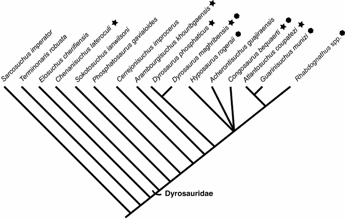
The term Crocodylia is used here exclusively for the crown group of extant and fossil Crocodyloidea, Alligatoroidea and Gavialoidea (Benton & Clark, Reference Benton, Clark and Benton1988; Sereno et al. Reference Sereno, Larsson, Sidoe and Gado2001; Brochu, Reference Brochu2003; Brochu et al. Reference Brochu, Wagner, Jouve, Sumrall and Densmore2009; Bronzati, Montefeltro & Langer, Reference Bronzati, Montefeltro and Langer2012). The family Dyrosauridae is, in accordance with recent phylogenetic analyses (Jouve et al. Reference Jouve, Bouya and Amaghzaz2005; Jouve, Bouya & Amaghzaz, Reference Jouve, Bouya and Amaghzaz2008; Hastings et al. Reference Hastings, Bloch, Cadena and Jaramillo2010; Hastings, Bloch & Jaramillo, Reference Hastings, Bloch and Jaramillo2011), used here as a term for a node constituted by the genera Chenanisuchus, Sokotosuchus, Phosphatosuchus, Cerrejonisuchus, Arambourgisuchus, Dyrosaurus, Hyposaurus, Acherontisuchus, Congosaurus, Atlantosuchus, Guarinisuchus and Rhabdognathus, which share several synapomorphies (Fig. 1). For the soft-tissue reconstructions and functional morphological analysis presented here, information on the osteology of the skull and mandible of dyrosaurids is needed, supplemented by information on the cervical vertebral column and scapula. In all included genera of dyrosaurids, the skull is preserved in a good condition that allows detailed reconstructions of the jaw musculature. More problematic is the preservation of the mandible, which in dyrosaurids possesses a very long and caudodorsally curved retroarticular process (Jouve et al. Reference Jouve, Bouya and Amaghzaz2005, Reference Jouve, Iarochène, Bouya and Amaghzaz2006; Jouve, Bouya & Amaghzaz, Reference Jouve, Bouya and Amaghzaz2008; Hastings et al. Reference Hastings, Bloch, Cadena and Jaramillo2010). The retroarticular process is preserved only in four genera of dyrosaurids, which nevertheless are members of successively more inclusive groups within Dyrosauridae, such that its similar morphology is assumed here for the whole group (Fig. 1). Detailed information on the cervical vertebral column and scapula is available for six genera, all of a more inclusive clade within Dyrosauridae, but isolated skeletal remains in other specimens show that there was most probably a general postcranial ‘bauplan’ that can be adopted for all dyrosaurids (Fig. 1).
Strict consensus cladogram from phylogenetic analyses of Dyrosauridae, modified from Hastings, Bloch & Jaramillo (Reference Hastings, Bloch and Jaramillo2011). Taxa with preserved retroarticular processes are marked with an asterisk; taxa with information on the morphology of the cervical vertebral column and scapula are marked with a polygon.

The material examined personally includes the partial skeleton of Congosaurus bequaerti (one individual with skeletal elements having the numbers MRAC 1741–1743, 1745, 1796, 1797, 1802, 1803, 1806, 1809–1811, 1813–1819, 1823, 1828, 1839, 1852, 1854, 1895, 1870, 1871, 1887, 1894), the partial skeleton of Dyrosaurus sp. (SMNK-PAL 3826) and isolated elements of Dyrosaurus spp. (MNHN), Hyposaurus natator (YPM), Hyposaurus spp. (AMNH, BMNH, MNHN, YPM), Rhabdognathus sp. (BMNH, MNHN), cf. Phosphatosaurus (MNHN) and cf. Sokotosaurus (MNHN). Information on the Moroccan taxa Chenanisuchus lateroculi (OCP DEK-GE 262, Jouve, Bouya & Amaghzaz, Reference Jouve, Iarochene, Bouya and Amaghzaz2005), Dyrosaurus maghribensis (OCP DEK-GE 43, Jouve et al. Reference Jouve, Iarochène, Bouya and Amaghzaz2006), Arambourgisuchus khouribgaensis (OCP DEK-GE 18, Jouve et al. Reference Jouve, Bouya and Amaghzaz2005) and Atlantosuchus coupatezi (OCP DEK-GE 51, Jouve, Bouya & Amaghzaz, Reference Jouve, Bouya and Amaghzaz2008) was taken from high-quality photographs provided personally by Dr Stéphane Jouve. Information on cf. Rhabdognathus (Langston, Reference Langston1995), cf. Hyposaurus (Storrs, Reference Storrs1986), Cerrejonisuchus improcerus (Hastings et al. Reference Hastings, Bloch, Cadena and Jaramillo2010), Acherontisuchus guajiraensis (Hastings, Bloch & Jaramillo, Reference Hastings, Bloch and Jaramillo2011) and Guarinisuchus munizi (Barbosa, Kellner & Sales Viana, Reference Barbosa, Kellner and Sales Viana2008) was taken from the literature. Osteological data on the postcranial skeleton of the dyrosaurid genera Dyrosaurus, Hyposaurus, Congosaurus and Rhabdognathus are provided in Schwarz, Frey & Martin (Reference Schwarz, Frey and Martin2006) and therefore not repeated in detail in this work.
For comparisons with extant crocodylians, complete skeletons of Crocodylus porosus (IPFUB, OS 38) and Tomistoma schlegelii (NMB, no collection number), as well as skull remains of Gavialis gangeticus (NMB 1193) were used. A skeleton of Crocodylus intermedius was dissected for comparisons of the jaw and neck musculature.
2.b. Methods
Soft-tissue reconstructions were done by one-way phylogenetic comparisons between the skeletons of dyrosaurids and extant crocodylians, using osteological correlates and topographical similarities and differences. The soft-tissue reconstructions are presented only as qualitative data, as the maximum extension of the muscle bulk is speculative and no percentage of size differences in comparison with extant crocodylians can be given. The direct comparison of all structures with extant crocodylians makes it possible to estimate deviations from the normal crocodylian bauplan in terms of considerable size differences apparent from the osteology and differences in the course and extension of muscles and ligaments. Ranges of motion between single elements were tested directly on the bones, with the examples of the skeletons of Congosaurus bequaerti (MRAC) and Dyrosaurus sp. (SMNK).
In the context of the functional morphological analysis, bone is regarded as being resistant mainly to compression (Carter & Beaupré, Reference Carter and Beaupré2001). The morphology of the studied bones allows conclusions on their loading ability and the possible force transmission from inserting muscles or ligaments to be made. The implications for the operation of the feeding apparatus and the bracing of the cervical vertebral column are elucidated by these reconstructions and involve corresponding models for extant crocodylians (Frey, Reference Frey1988 a; Cleuren & De Vree, Reference Cleuren and Vree1990, Reference Cleuren and De Vree1991, Reference Cleuren, Vree and Schwenk2000; Salisbury & Frey, Reference Salisbury, Frey, Grigg, Seebacher and Franklin2001). The mechanics of the cranium and mandible are reconstructed using a lever model that provides an estimate of the orientation of the muscles, their relative forces, and bite force (Sinclair & Alexander, Reference Sinclair and Alexander1987; Pooley, Reference Pooley and Ross1989; Busbey, Reference Busbey and Thomasson1995; Cleuren, Aerts & De Vree, Reference Cleuren, Aerts and Vree1995; Greaves, Reference Greaves and Thomasson1995; Preuschoft & Witzel, Reference Preuschoft and Witzel2002). This method does not allow prediction of absolute forces or detailed stress distribution in the skull (Porro et al. Reference Porro, Holliday, Anapol, Ontiveros, Ontiveros and Ross2011). Thus, the lever model allows the interpretation of present structures, but does not aim for detailed reconstruction of the absolute amount of force of the jaw muscles. Information on behavioural aspects of crocodylian feeding was taken from the literature, personal observation and video material freely available through several web-based media.
3. Soft-tissue reconstructions
3.a. Reconstruction of jaw adductors and abductors
3.a.1. Cranial and mandibular adductor tendon, cartilago transiliens
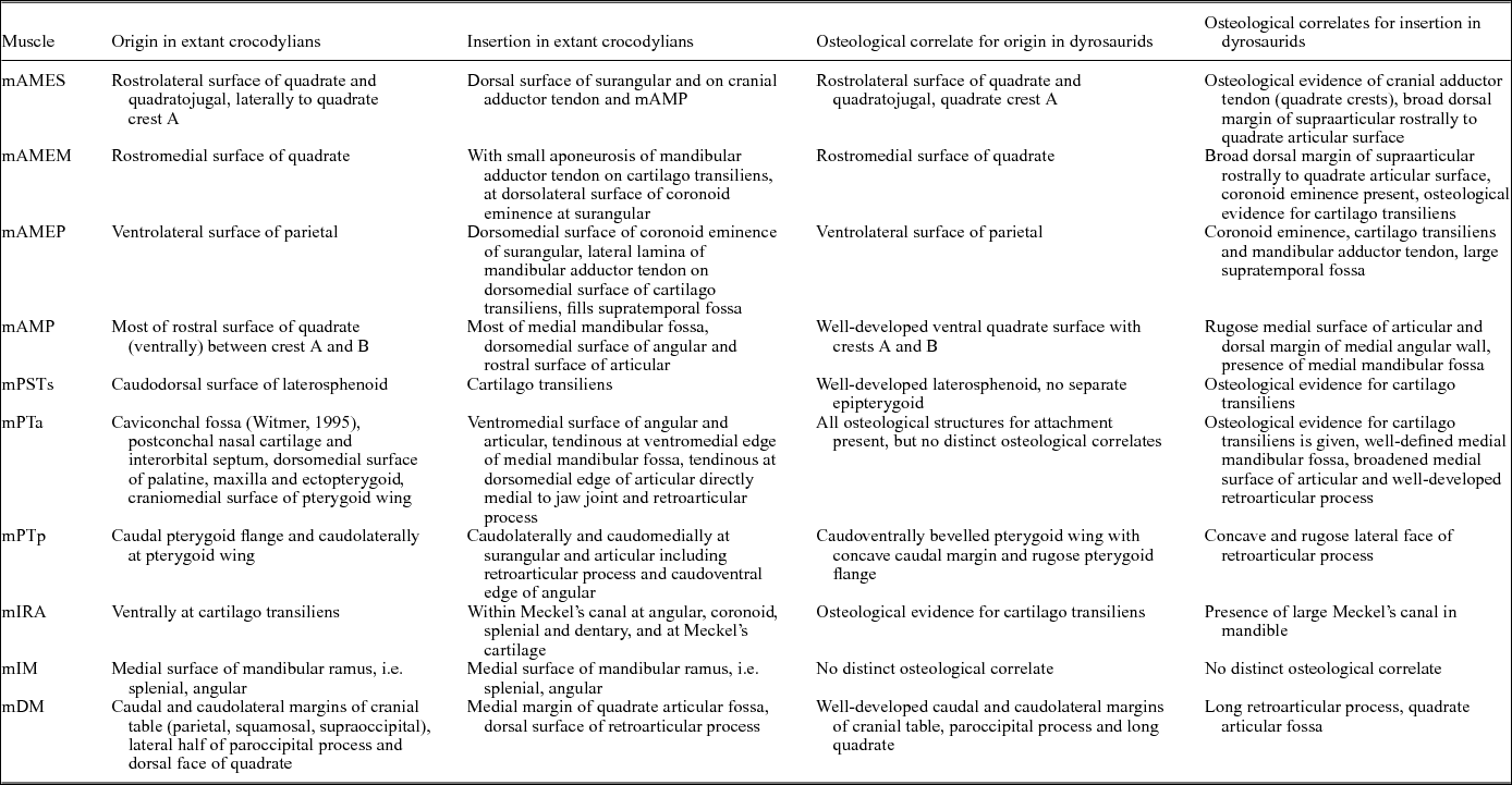
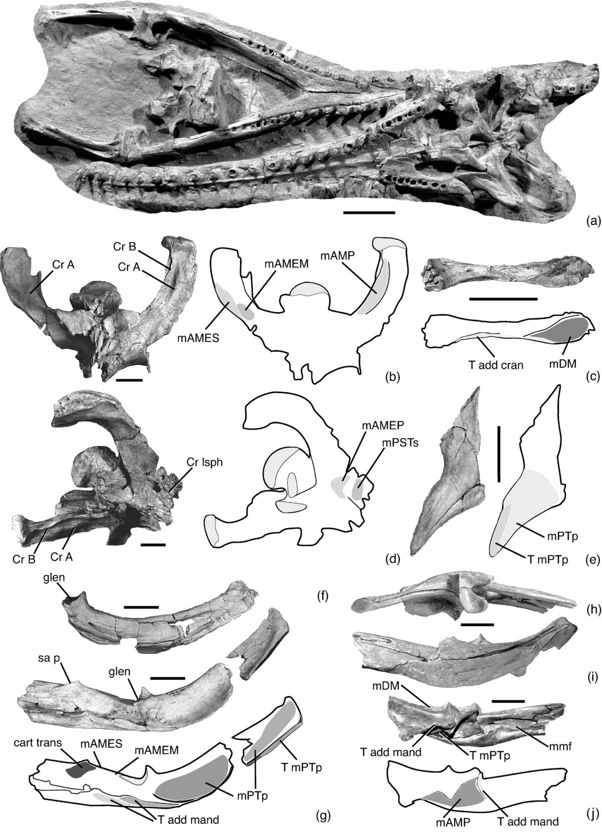
The ventral face of the quadrate of dyrosaurids possesses well-developed crests A and B (Fig. 2b, d). These crests and the rugose internal surface of the lateral margin of the skull roof represent the insertion area of the cranial adductor tendon at which the mAME and mAMP insert, as in extant crocodylians (Iordansky, Reference Iordansky1964; Schumacher, Reference Schumacher, Gans, Bellairs and Parsons1973; Busbey, Reference Busbey1989). The dorsal margin of the surangular of dyrosaurids bears a rugosity dorsal to the mandibular fenestra and bears a coronoid eminence on the medial face of the mandible (Table 1). In a specimen of Dyrosaurus sp. the surangular rugosity is enlarged into a surangular process (Fig. 2g), as in some extant crocodylians (Van Drongelen & Dullemeijer, Reference Van Drongelen and Dullemeijer1982, p. 341). The surangular rugosity and coronoid eminence are osteological correlates for the cartilago transiliens (Schumacher, Reference Schumacher, Gans, Bellairs and Parsons1973; Van Drongelen & Dullemeijer, Reference Van Drongelen and Dullemeijer1982; Busbey, Reference Busbey1989; Iordansky, Reference Iordansky1994; Cleuren & De Vree, Reference Cleuren, Vree and Schwenk2000; Holliday & Witmer, Reference Holliday and Witmer2007), from which the mandibular adductor tendon and aponeuroses for the mAME and mAMI originate. A rugosity on the dorsal margin of the medial angular wall and the crest on the medial face of the retroarticular process of dyrosaurids were most probably further insertion areas of the mandibular adductor tendon as in extant crocodylians (Schumacher, Reference Schumacher, Gans, Bellairs and Parsons1973; Busbey, Reference Busbey1989) (Fig. 2; Table 1).
Origin and insertion of jaw muscles in extant crocodylians, based mainly on Holliday & Witmer (Reference Holliday and Witmer2007) and Cleuren & De Vree (Reference Cleuren, Vree and Schwenk2000), and osteological correlates for these muscles present in dyrosaurids

(a) Skull and mandible of Dyrosaurus maghribensis (OCP DEK-GE 253) modified from Jouve et al. (Reference Jouve, Iarochène, Bouya and Amaghzaz2006, fig. 8), scale bar is 10 cm. (b–g) Osteological characters in the skull of Dyrosaurus sp. (SMNK-PAL 3826) allowing soft-tissue reconstructions, (b) braincase (SMNK-PAL 3826 27/94) in ventral aspect, (c) isolated postorbital and squamosal (SMNK-PAL 3826 23/94) in ventral aspect, (d) braincase (SMNK-PAL 3826 27/94) in left ventrolateral aspect, (e) isolated pterygoid and partial ectopterygoid (SMNK-PAL 3826 20/94) in ventral aspect, (f) left retroarticular process (SMNK-PAL 3826 32/94) in lateral aspect, (g) caudal part of right mandible with retroarticular process (SMNK-PAL 3826 34/94 & 35/94) in medial aspect. (h–j) Caudal part of left mandibular ramus of Congosaurus bequaerti (MRAC R.G.1744) with separate retroarticular process, (h) in dorsal aspect, (i) in lateral aspect, (j) without complete retroarticular process in medial aspect. Scale bar is 5 cm, for abbreviations see text.

3.a.2. M. adductor mandibulae externus
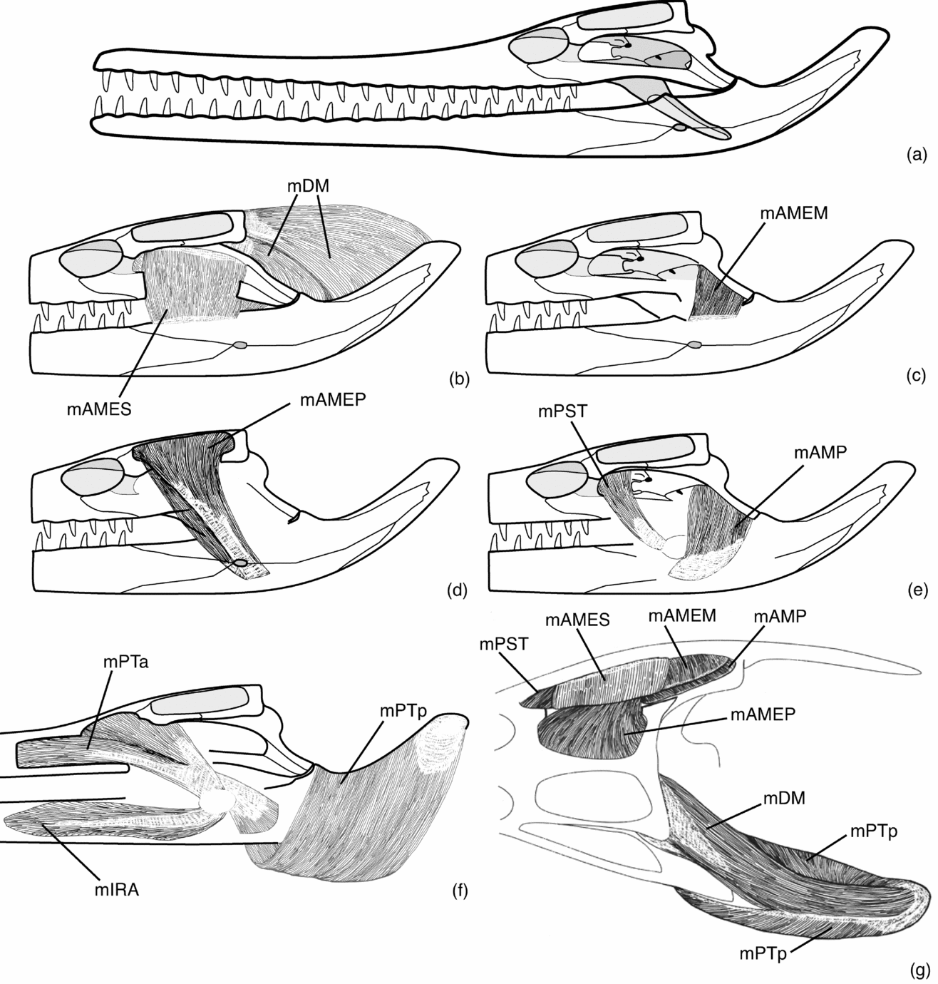
The mAMES in dyrosaurids most probably extended by parallel fibres as in extant crocodylians (Iordansky, Reference Iordansky1964; Schumacher, Reference Schumacher, Gans, Bellairs and Parsons1973; Van Drongelen & Dullemeijer, Reference Van Drongelen and Dullemeijer1982; Holliday & Witmer, Reference Holliday and Witmer2007) from ventrally to dorsally and filled the infratemporal fenestra (Table 1; Fig. 3b). The infratemporal fenestra of dyrosaurids was at least three times as large as that of extant crocodylians (Fig. 3a), which indicates that the physiological cross-section of the mAMES was larger than in extant crocodylians. In extant crocodylians, the superficial and medial parts of the mAME are not clearly separable from each other, such that these parts are characterized by unequal portions in Caiman, Crocodylus and Alligator (Busbey, Reference Busbey1989). The mAMEM of dyrosaurids inserted as in extant crocodylians (Iordansky, Reference Iordansky1964; Schumacher, Reference Schumacher, Gans, Bellairs and Parsons1973) medially to the mAMES (Fig. 3c), but the size of this muscle cannot be compared with extant crocodylians.
Reconstruction of jaw musculature of dyrosaurids based on the example of Dyrosaurus. (a) Schematic drawing of the skull and mandible of Dyrosaurus in lateral aspect. (b–f) Jaw adductors and abductor in lateral aspect; (g) reconstructed jaw musculature in dorsal aspect. Not to scale, for abbreviations see text.

The mAMEP of dyrosaurids inserted as in extant crocodylians (Iordansky, Reference Iordansky1964, Reference Iordansky2000; Van Drongelen & Dullenmeijer, Reference Van Drongelen and Dullemeijer1982; Busbey, Reference Busbey1989; Holliday & Witmer, Reference Holliday and Witmer2007, Reference Holliday and Witmer2009; Bona & Desojo, Reference Bona and Desojo2011) in the supratemporal fossa and at the mandibular adductor tendon, filling the supratemporal foramen completely (Fig. 3d, g). Schumacher (Reference Schumacher, Gans, Bellairs and Parsons1973, for Alligator, Caiman and Crocodylus) and Endo et al. (Reference Endo, Aoki, Taru, Kimura, Sasaki, Yamamoto, Arishima and Hayashi2002, for Gavialis, Tomistoma and Mecistops) determined most of the muscle belly within the supratemporal fossa as mPST instead of mAMEP. In contrast, Holliday & Witmer (Reference Holliday and Witmer2009) provided osteological evidence for the restriction of the mPSTs to a position ventral to the supratemporal foramen in dyrosaurids: in particular, the morphology of the laterosphenoid with a large epipterygoid fossa in Rhabdognathus (Brochu et al. Reference Brochu, Bouaré, Sissoko, Roberts and O'Leary2002; Holliday & Witmer, Reference Holliday and Witmer2009) and possibly also in Dyrosaurus (Jouve, Reference Jouve2005; Holliday & Witmer, Reference Holliday and Witmer2009) suggests that the mAMEP filled the supratemporal foramen completely. In longirostrine extant crocodylians (Gavialis, Tomistoma, Mecistops, Crocodylus johnstoni), the mAMEP is larger than in meso- and brevirostrine taxa and has a circular cross-section (Iordansky, Reference Iordansky and Gans1973; Langston, Reference Langston and Gans1973; Endo et al. Reference Endo, Aoki, Taru, Kimura, Sasaki, Yamamoto, Arishima and Hayashi2002; Holliday & Witmer, Reference Holliday and Witmer2007). During ontogeny, an increase in the relative width of the supratemporal fossa in Alligator is related to an increase in the cross-section of the mAMEP (Dodson, Reference Dodson1975; Erickson, Lappin & Vliet, Reference Erickson, Lappin and Vliet2003). The supratemporal foramen of dyrosaurids is at least three times as large as in extant crocodylians, including longirostrine ones (Fig. 3a, g), which suggests a significantly larger physical cross-section of the mAMEP.
3.a.3. M. adductor mandibulae posterior
The mAMP of dyrosaurids extended as in extant crocodylians from the quadrate into the infratemporal fenestra (Fig. 3e) and from there to the medial surface of the mandible (Iordansky, Reference Iordansky1964; Schumacher, Reference Schumacher, Gans, Bellairs and Parsons1973; Van Drongelen & Dullemeijer, Reference Van Drongelen and Dullemeijer1982; Busbey, Reference Busbey1989; Endo et al. Reference Endo, Aoki, Taru, Kimura, Sasaki, Yamamoto, Arishima and Hayashi2002; Bona & Desojo, Reference Bona and Desojo2011). Because the infratemporal fenestra of dyrosaurids was at least three times as large as in extant crocodylians, their mAMP plausibly had a larger physiological cross-section.
3.a.4. M. adductor mandibulae internus
With regards to the controversy about the mPST in the literature (Lubosch, Reference Lubosch1914; Poglayen-Neuwall, Reference Poglayen-Neuwall1953; Iordansky, Reference Iordansky1964, Reference Iordansky2000; Schumacher, Reference Schumacher, Gans, Bellairs and Parsons1973; Van Drongelen & Dullemeijer, Reference Van Drongelen and Dullemeijer1982; Busbey, Reference Busbey1989; Cong et al. Reference Cong, Hou, Wu and Hou1988; Cleuren & De Vree, Reference Cleuren, Vree and Schwenk2000; Holliday & Witmer, Reference Holliday and Witmer2007, Reference Holliday and Witmer2009; Bona & Desojo, Reference Bona and Desojo2011), the interpretation of Holliday & Witmer (Reference Holliday and Witmer2007, Reference Holliday and Witmer2009) regarding the extension and homology of the mPST is followed (Table 1). The mPST (Iordansky, Reference Iordansky1994; Holliday & Witmer, Reference Holliday and Witmer2007) can be divided into a superficial portion (‘mAMEP pars rostralis’, Schumacher, Reference Schumacher, Gans, Bellairs and Parsons1973; Busbey, Reference Busbey1989) and a profound portion (‘m intermedius’ in Iordansky, Reference Iordansky1964). Owing to difficulties in the separation of the mPSTp and the lack of unambiguous osteological evidence, only the mPSTs is reconstructed for dyrosaurids. The mPSTs of dyrosaurids extended from the laterosphenoid ventrally to the cartilago transiliens and the mandibular adductor tendon (Fig. 3e, g).
As in extant crocodylians, the mPTa (mPTd of Holliday & Witmer, Reference Holliday and Witmer2007) of dyrosaurids extended through the palatal fenestra to the dorsal face of the pterygoid wing, the cartilago transiliens and the mandibular adductor tendon (Fig. 3f). Muscle fibres attached at the maxillar bulla (caudal to the nasal conchae), the maxilla and palatine, the lateral face of the prefrontal processes, the interorbital cartilage and the basisphenoid rostrum (Iordansky, Reference Iordansky1964; Schumacher, Reference Schumacher, Gans, Bellairs and Parsons1973; Van Drongelen & Dullemeijer, Reference Van Drongelen and Dullemeijer1982). Most plausibly, the mPTa of dyrosaurids was in the orbital region in contact with the mPTp (Busbey, Reference Busbey1989) and caudally intermingled with this muscle. The suborbital fenestra of dyrosaurids (Fig. 2a) is as large as that of extant crocodylians (Iordansky, Reference Iordansky and Gans1973), indicating a similar size of mPTa.
The pterygoid wing of dyrosaurids is bevelled caudoventrally with a concave caudal margin, and the lateral face of the retroarticular process is concave and rugose (Fig. 2e; Table 1), corresponding with extant crocodylids and alligatorids (Iordansky, Reference Iordansky and Gans1973). In contrast, the caudal margin of the pterygoid wing in Gavialis gangeticus is nearly horizontally oriented and the lateral surface of the retroarticular process is straight and smooth (Iordansky, Reference Iordansky and Gans1973; Endo et al. Reference Endo, Aoki, Taru, Kimura, Sasaki, Yamamoto, Arishima and Hayashi2002), resulting in the mPTp (Holliday & Witmer, Reference Holliday and Witmer2007) being restricted to the medial face of the retroarticular process (Endo et al. Reference Endo, Aoki, Taru, Kimura, Sasaki, Yamamoto, Arishima and Hayashi2002). In correspondence with extant alligatorids and crocodylids (Iordansky, Reference Iordansky1964, Reference Iordansky2000; Schumacher, Reference Schumacher, Gans, Bellairs and Parsons1973; Van Drongelen & Dullemeijer, Reference Van Drongelen and Dullemeijer1982; Busbey, Reference Busbey1989; Endo et al. Reference Endo, Aoki, Taru, Kimura, Sasaki, Yamamoto, Arishima and Hayashi2002), the mPTp of dyrosaurids plausibly originated from the pterygoid wing, wrapped ventrally around the mandibular articulation and inserted both on the lateral and the medial surface of the retroarticular process (Fig. 3f, g) (Iordansky, Reference Iordansky1964; Schumacher, Reference Schumacher, Gans, Bellairs and Parsons1973; Van Drongelen & Dullemeijer, Reference Van Drongelen and Dullemeijer1982; Busbey, Reference Busbey1989). When preserved (Fig. 1), the retroarticular process of dyrosaurids is up to twice as long as those of extant crocodylians (Fig. 2) and reaches as far caudally as the forth cervical vertebra. This suggests much longer muscle fibres and a larger physiological cross-section of the mPTp.
3.a.5. M. intramandibularis
In dyrosaurids, the mIRA was separated from the mPST by the cartilago transiliens. The muscle extended from the cartilago transiliens rostrally until the intermandibular foramen to fill Meckel's canal (Fig. 3f).
3.a.6. M. intermandibularis
It is likely that as in extant crocodylians (Schumacher, Reference Schumacher, Gans, Bellairs and Parsons1973; Bona & Desojo, Reference Bona and Desojo2011), the mIM of dyrosaurids inserted on the medial faces of the mandibular rami, filling the space between the right and left mandibular rami and being only incompletely separated from the m. constrictor colli (Table 1).
3.a.7. M. depressor mandibulae
The mDM of dyrosaurids passed from the cranial table to the medial margin of the quadrate articular fossa and the dorsal margin of the retroarticular process (Fig. 2c, j). Because the retroarticular process of dyrosaurids is twice as long as that of extant crocodylians, the mDM of dyrosaurids had a larger origination area than in extant crocodylians (Fig. 3b). The paroccipital process and the quadrate of dyrosaurids were positioned more ventrally than in extant crocodylians, being positioned lateral and ventral to the occipital condyle. This resulted in an expanded insertion area for the mDM (Fig. 4a), which would also be in accordance with the long retroarticular process in dyrosaurids. The mDM of dyrosaurids thus is reconstructed to have had a larger physiological cross-section and longer muscle fibres than in extant crocodylians (Fig. 3b, g).
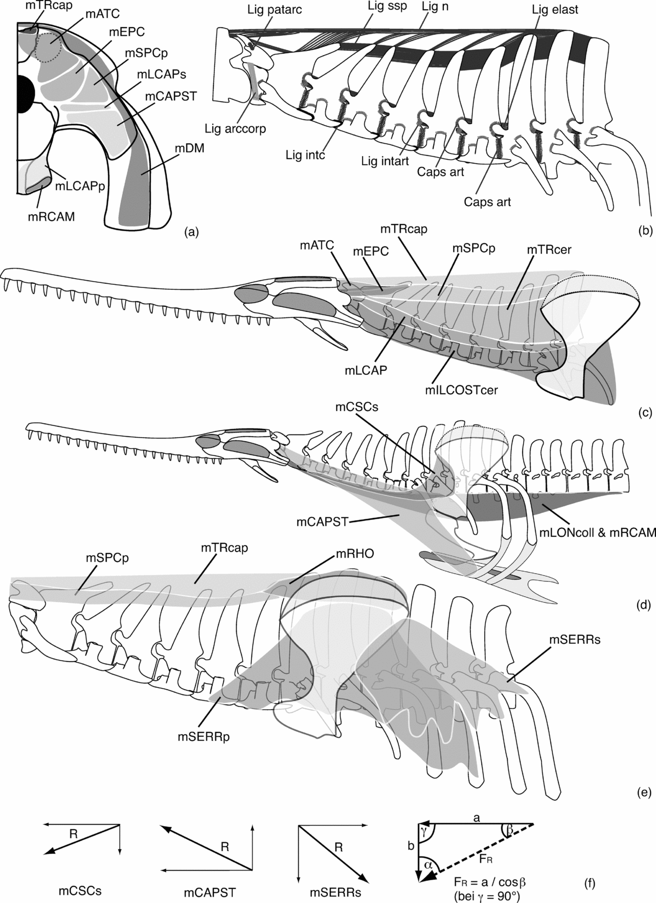
Reconstructed cervical ligaments and muscles of dyrosaurids based on the example of Dyrosaurus. (a) Right side of occiput with presumed insertion areas for cervical muscles; (b) reconstructed cervical ligaments with osteological evidence; (c) reconstructed superficial axial muscles (deeper parts of m. transversospinalis are not depicted); (d) muscles with connection to scapula and neck; (e) m. serratus and connection between m. rhomboideus and m. transversospinalis capitis; (f) direction of the resultant of the force vectors of m. capitisternalis, m. colloscapularis superficialis and m. serratus superficialis, and force triangle with calculation for comparison of the relative sizes of the resultants. Not to scale, for abbreviations see text.

3.b. Relevant soft-tissues of the neck and scapula
3.b.1. Cervical ligaments
The cervical vertebrae of dyrosaurids are amphicoelous with a fossa in the medial two-thirds of the cranial and caudal vertebral articular surfaces (Schwarz, Frey & Martin, Reference Schwarz, Frey and Martin2006). Together with the presence of a rugose rim with lateral striae, this indicates a synovial articulation with a fibrous articular capsule between the cervical vertebrae, similar to other fossil and extant crocodylians (Salisbury & Frey, Reference Salisbury, Frey, Grigg, Seebacher and Franklin2001). In reference to extant Alligator (Frey, Reference Frey1988 b), the atlas-axis complex of dyrosaurids bears osteological evidence for a proatlantoarcual ligament that passed from a craniodorsal circular impression at the atlantic arch to the lateral face of the proatlas (Fig. 4b). The rugose cranial half of the ventral margin of the atlantic arch is the origin of the arcuocorporal atlantic ligament that inserted on the strongly rugose craniolateral half of the atlas centrum (Fig. 4b).
A depression on the lateral face of the axis neural spine forms the attachment surface of the cranialmost branch of the supraspinal ligament (Frey, Reference Frey1988 b). The caudal margin of the third to seventh cervical neural spines of dyrosaurids forms a rugose lamina, which is replaced at the eighth and ninth cervical neural spines by a caudodorsal rugosity. These structures are correlates for the supraspinal ligament (Frey, Reference Frey1988 b), which connected consecutive cervical neural spines with each other in their dorsal fourth (Fig. 4b). The cervical and the prothoracic neural spines of dyrosaurids are significantly higher than in extant crocodylians and in all other crocodylians (Schwarz, Frey & Martin, Reference Schwarz, Frey and Martin2006). The height of the cervical and prothoracic neural spines in dyrosaurids is more similar to that of large artiodactyl mammals, such as Equus and Camelus (Slijper, Reference Slijper1946; Dimery, Alexander & Deyst, Reference Dimery, Alexander and Deyst1985; Gellman, Bertram & Hermanson, Reference Gellman, Bertram and Hermanson2002). Together with a postulated stronger ventral flexibility of the neck in dyrosaurids (see Section 4.a), this suggests a development of the supraspinal ligament into a nuchal ligament similar to that of extant large mammals (Slijper, Reference Slijper1946; Dimery, Alexander & Deyst, Reference Dimery, Alexander and Deyst1985; Gellman, Bertram & Hermanson, Reference Gellman, Bertram and Hermanson2002) and birds (Boas, Reference Boas1929). In dyrosaurids, the nuchal ligament would have expanded from the prothoracic vertebrae to the occipital region of the skull, being connected to every cervical neural spine by a ventral branch (Fig. 4b).
The height of the attachment area for the elastic interlaminar ligament in dyrosaurids reached the height of the postzygapophyses between the axis and the eighth cervical vertebra (Schwarz, Frey & Martin, Reference Schwarz, Frey and Martin2006). From the eighth cervical vertebra onwards, the height of the ligament attachment area increased, which indicates a dorsal expansion of the elastic interlaminar ligament from this vertebra onwards (Fig. 4b). A rugosity around the articular surface of the pre- and postzygapophyses in dyrosaurids suggests the presence of a ligamentous articular capsule enclosing the pre- and postzygapophyses. As in extant crocodylians, the cervical rib corpora of dyrosaurids are strongly rugose in the region where they overlap each other, which indicates the presence of strong intercostal ligaments (Fig. 4b). A lateral rugosity at the prezygapophysis served as the insertion surface for the articulotubercular ligament (Fig. 4b) (Frey, Reference Frey1988 b).
3.b.2. Epaxial cervical muscles
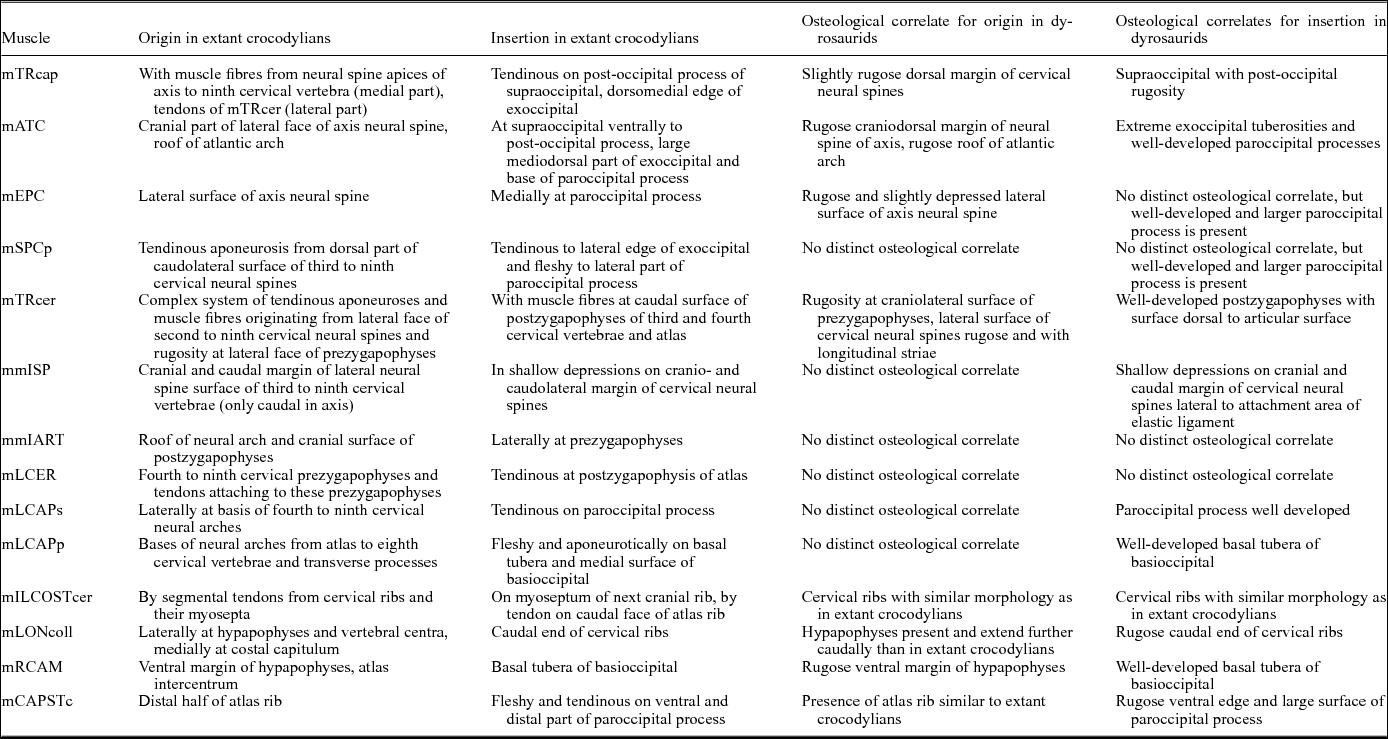
According to the similarities in the morphology of the cervical neural spines of dyrosaurids (Schwarz, Frey & Martin, Reference Schwarz, Frey and Martin2006) and extant crocodylians (Frey, Reference Frey1988 b; Cong et al. Reference Cong, Hou, Wu and Hou1998; Cleuren & De Vree, Reference Cleuren, Vree and Schwenk2000; Salisbury & Frey, Reference Salisbury, Frey, Grigg, Seebacher and Franklin2001; Tsuihiji, Reference Tsuihiji2005, Reference Tsuihiji2007) a similar arrangement of cervical epaxial muscles can be reconstructed for dyrosaurids (Fig. 4; Table 2). In contrast, the occipital region of the skull bears some peculiarities in dyrosaurids that contrast extant crocodylians and indicate differences in the size of some of the epaxial muscles. The exoccipital of dyrosaurids bears well-developed large occipital tuberosities (Denton, Dobie & Parris, Reference Denton, Dobie and Parris1994; Jouve, Reference Jouve2005, Reference Jouve2007; Jouve, Bouya & Amaghzaz, Reference Jouve, Bouya and Amaghzaz2005, Reference Jouve, Bouya and Amaghzaz2008; Jouve et al. Reference Jouve, Bouya and Amaghzaz2005, Reference Jouve, Iarochène, Bouya and Amaghzaz2006), but can vary in size between longirostrine and mesorostrine forms (Jouve et al. Reference Jouve, Bouya and Amaghzaz2005). The occipital tuberosities sit in an area that in extant crocodylians is the insertion surface of the mATC (Fig. 4a) (Tsuihiji, Reference Tsuihiji2005). The mATC connects the atlas neural arches and the axis to the occiput and, according to the large insertion areas at their occiput, in dyrosaurids was most probably larger than in extant crocodylians (Fig. 4c). In extant crocodylians, the paroccipital process is more or less horizontally directed and therefore is in a position dorsal to the foramen magnum (compare for example with Iordansky, Reference Iordansky and Gans1973), whereas in dyrosaurids the process is more ventrolaterally directed and therefore positioned at a level parallel to the occipital condyle and foramen magnum. The more ventral position of the paroccipital process would increase and ventralize the insertion area of the mEPC and mSPCp (Fig. 4a), so that a larger physiological cross-section for both muscles is reconstructed compared to extant crocodylians. In the case of the mEPC, an enlarged and ventralized insertion area at the occiput corresponds to the position of this muscle at the occiput of extant birds (Tsuihiji, Reference Tsuihiji2005; Fig. 5). An enlarged insertion of the mSPCp in dyrosaurids corresponds to their higher cervical neural spines (Schwarz, Frey & Martin, Reference Schwarz, Frey and Martin2006), which provided equally larger origination areas for this muscle (Table 2; Fig. 4a, c). Similarly, higher cervical neural spines also indicate a larger dorsoventral extension and cross-section of the mTRcer in dyrosaurids (Fig. 4c). The rugose lateral face of the cervical neural spines with some vertical crests in dyrosaurids suggests that segmentation of the mTRcer was probably similar to the tendon system of extant crocodylians (Tsuihiji, Reference Tsuihiji2005).
Origin and insertion of most important cervical muscles in extant crocodylians, based mainly on Tsuihiji (Reference Tsuihiji2005, Reference Tsuihiji2007) and Cleuren & De Vree (Reference Cleuren, Vree and Schwenk2000), and osteological correlates for these muscles present in dyrosaurids

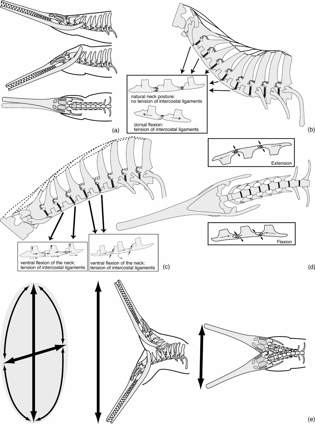
Reconstruction of the flexibility of the skull and neck of dyrosaurids. (a) Flexibility of skull and mandible in dorsal, ventral and lateral direction. (b–c) Flexibility of neck (b) in ventral direction, (c) in dorsal direction and (d) in lateral direction, with boxes showing details of cervical ribs with intercostal ligaments. (e) Feeding envelope of dyrosaurids as reconstructed from the dorsoventral and lateral flexibility of their craniocervical region. Not to scale.

3.b.3. M. longissimus group and m. iliocostalis cervicis
The lateral surface of the cervical neural arches, from their base to the base of the neural spine, was occupied in dyrosaurids by cervical muscles belonging to the m. longissimus complex, similar in origin, insertion and expansion to extant crocodylians (Table 2) (Frey, Reference Frey1988 b; Cleuren & De Vree, Reference Cleuren, Vree and Schwenk2000; Tsuihiji, Reference Tsuihiji2007). The cervical ribs of dyrosaurids indicate an organization of the mILCOSTcer with the myosepta, comparable to extant crocodylians (Frey, Reference Frey1988 b).
3.b.4. M. longus colli and m. rectus capitis anticus major
In the cervical vertebrae, the hypapophyses of dyrosaurids reach only 20% of the height of the hypapophyses in extant crocodylians, which suggests a smaller cross-section of the mLONcoll in dyrosaurids. However, there is some variation among dyrosaurids, as the eighth cervical rib and the hypapophysis of the eighth cervical vertebra in Dyrosaurus is twice as long as in Congosaurus, indicating differences in the cross-section of the mLONcoll between both taxa. The general presence of large hypapophyses in the cranialmost three dorsal vertebra of dyrosaurids, small hypapophyses in the fourth and fifth dorsal vertebrae and a prominent median crest between the sixth and seventh dorsal vertebrae (Schwarz, Frey & Martin, Reference Schwarz, Frey and Martin2006) shows that the mLONcoll of dyrosaurids was large in the prothoracic area and extended caudally until the eighth dorsal vertebra (Fig. 4d). The mRCAM of dyrosaurids was probably similar to that of extant crocodylians (Table 2) (Tsuihiji, Reference Tsuihiji2007).
3.b.5. M. rhomboideus
The scapular blade and suprascapula of dyrosaurids are three times as long as in extant crocodylians (Schwarz, Frey & Martin, Reference Schwarz, Frey and Martin2006), indicating that the mRHO of dyrosaurids was longer than in extant crocodylians (Fig. 4e). It is plausible that the mRHO of dyrosaurids was in close contact cranially with the mTRcap and mSPCp, as in extant crocodylians (Fürbringer, Reference Fürbringer1876; Frey, Reference Frey1988 b; Cong et al. Reference Cong, Hou, Wu and Hou1998).
3.b.6. M. serratus
The m. serratus of dyrosaurids originated as in extant crocodylians (Fürbringer, Reference Fürbringer1876; Frey, Reference Frey1988 b; Cong et al. Reference Cong, Hou, Wu and Hou1998) with its profound portion on the medial surface of the large scapular blade and with its superficial portion on the caudal margin of the scapula (Fig. 4e). The mSERRp in dyrosaurids inserted at the cranial process of the seventh to ninth cervical ribs. The first and second dorsal ribs of dyrosaurids possess cranial processes and therefore plausibly were also in contact with the mSERRp. The mSERRs passed from the scapula caudally to at least the second dorsal vertebra and inserted at the ninth cervical and the first and second dorsal rib ventrally to the mSERRp (Fig. 4e). Most probably, the mSERRs of dyrosaurids inserted also at the fascia lumbodorsalis as in extant crocodylians.
3.b.7. M. colloscapularis superficialis
The mCSCs originated in dyrosaurids from the cranial margin of the scapula, which is much more concave than in extant crocodylians (Fig. 4d). The muscle passed cranioventrally and inserted along the ventral margins of the third to sixth cervical ribs and probably the covering fascia of the mLONcoll, as in extant crocodylians (Fürbringer, Reference Fürbringer1876; Frey, Reference Frey1988 b; Cong et al. Reference Cong, Hou, Wu and Hou1998).
3.b.8. M. capitisternalis
The origin of the cranial part of the mCAPST (‘m. iliocostalis capitis’ M. R. Seidel, unpub. Ph.D. thesis, City Univ. New York, 1978; Cleuren & De Vree, Reference Cleuren, Vree and Schwenk2000; m. atlantimastoideus, Tsuihiji, Reference Tsuihiji2007) in dyrosaurids was from the atlas rib, from which the muscle passed cranially and inserted ventrally and distally at the paroccipital process (Table 2; Fig. 4d). Because of the more ventral position of the paroccipital process in dyrosaurids (see also Sections 3.a.7 and 3.b.2), the mCAPST would equally be positioned slightly more ventrally than in extant crocodylians. The m. capitisternalis pars caudalis (m. sternoatlanticus, Tsuihiji, Reference Tsuihiji2007) in dyrosaurids originated as in extant crocodylians (Frey, Reference Frey1988 b) from the cranial margin of the scapula, the sternocostal segment of the first dorsal rib and the sternum and inserted on the atlas rib. Because of its slightly more ventral origin from the occiput and the long and strongly curved prothoracic ribs, the whole complex of the mCAPST of dyrosaurids reached further ventrally than in extant crocodylians (Fig. 4d).
4. Functional aspects in the skull and neck of dyrosaurids
4.a. Mobility of the skull and neck and feeding envelope
4.a.1. Dorsal flexibility
The occipital condyle of dyrosaurids was more deeply embedded within the bowl-shaped condylar fossa than in extant crocodylians and reached 20% of the total width of the cranial table, whereas the condyle is only 12.5% of the cranial table width in extant crocodylians (Kälin, Reference Kälin1933; Iordansky, Reference Iordansky and Gans1973). Therefore, the occipital condyle of dyrosaurids was more stable during movements of the skull than in extant crocodylians. Dorsally directed motion of the skull in dyrosaurids was limited by the epaxial cervical muscles and the contact between the caudal margin of the cranial table and the proatlas (Fig. 5a), which is comparable to that of extant crocodylians (Virchow, Reference Virchow1914).
During dorsal flexion of the neck of dyrosaurids, the cervical ribs were pulled apart from each other in the region from the axis to the seventh cervical vertebra (Fig. 5b), which stretched the intercostal ligaments. The articular surfaces of the pre- and postzygapophyses approached each other, which stretched their zygapophyseal capsule. The high cervical neural spines of dyrosaurids also approach each other, and would have limited dorsal flexion of the neck at their contact. Even with this limit, an angle of c. 45° to the horizontal plane in dorsal flexion is reconstructed for dyrosaurids (Fig. 5b), similar to extant crocodylians (Ross & Mayer, Reference Ross, Mayer, Rhodin and Miyata1983; S. W. Salisbury, unpub. Ph.D. thesis, Univ. New South Wales, 2001). In extant crocodylids and alligatorids, dorsal neck flexibility is facilitated by the poor segmentation of the epaxial musculature (Frey, Reference Frey1988 b; S. W. Salisbury, unpub. Ph.D. thesis, Univ. New South Wales, 2001), which is reconstructed similarly for dyrosaurids.
4.a.2. Ventral flexibility
During ventral flexion of the skull and mandible, the long retroarticular process of dyrosaurids moved craniodorsally, stretching the dermis of the neck. Assuming a similar large tensibility of the proatlantoarcual ligament and the dermis as in extant crocodylians (Virchow, Reference Virchow1914; S. W. Salisbury, unpub. Ph.D. thesis, Univ. New South Wales, 2001), ventral flexion of the skull of dyrosaurids was possible up to at an angle of 45° to the horizontal plane (Fig. 5a).
The cranial projections of the third to fifth cervical ribs of dyrosaurids were underlapped by the cranially following rib body (Schwarz, Frey & Martin, Reference Schwarz, Frey and Martin2006). During ventral neck flexion, these cervical ribs ‘telescoped' until becoming blocked by the contact to the cranially following costal capitulum and caudally following costal tuberculum. The cranially and caudally directed compressional loads were distributed as in extant crocodylians (Frey, Reference Frey1988 a) to the vertebrae via the costal capitulum and tuberculum, and the intercostal ligaments were stretched during this movement (Fig. 5c). Between the cervical ribs of the atlas and axis and the sixth and seventh cervical ribs, cranial projections of the cervical ribs contacted the subsequently following cervical ribs from a medial direction only with their lateral face. Deflection of the neck resulted in a caudal rotational movement of the ribs, a stretching of the intercostal ligaments, and a lateral and medial compression of the cervical ribs (Fig. 5c). The eighth and ninth cervical ribs were oriented in the vertical plane and did not limit deflection. Ventral neck curvature in dyrosaurids stretched mainly the elastic interlaminar and the supraspinal ligaments. Ventral flexibility in dyrosaurids is reconstructed with an angle of at least 20° below the horizontal plane (Fig. 5c) based on the very reduced bone-by-bone contact limitations between the third to fifth cervical ribs and assuming a similar tensibility of the nuchal ligament of dyrosaurids as in extant artiodactyl mammals and birds (Boas, Reference Boas1929; Dimery, Alexander & Deyst, Reference Dimery, Alexander and Deyst1985; Bennett & Alexander, Reference Bennett and Alexander1987; Dzenski & Christian, Reference Dzemski and Christian2007). In contrast, the neck of extant crocodylians cannot be deflected, as it is stiffened by the rows of cervical ribs.
4.a.3. Lateral flexibility
During lateral bending, medial movement of the long retroarticular process of the flexed side was limited by the cervical musculature. Additionally, the lateral movement of the retroarticular process of the extended side stretched the dermis of the neck, resulting in a limitation of the lateral flexibility of the skull of dyrosaurids to 5–10° (Fig. 5a). Lateral curvature of the neck pulled the axial to seventh cervical ribs on the extended side apart and stretched the intercostal ligaments (Fig. 5d). On the flexed side, cervical ribs glided into one another, which also stretched the intercostal ligaments (Fig. 5d). The movement of the cervical ribs relative to each other led to lateral and medial compression of the costal bodies. Stretching of the intercostal ligaments and compression of the cervical ribs limited lateral flexion of the neck of dyrosaurids, an effect that was increased by the long retroarticular process. Whereas the intrinsic mobility of the neck increased from the eighth cervical vertebra in the caudal direction, the connection with the shoulder girdle in this region completely prevented lateral flexion of the base of the neck (Fig. 4d). As a whole, bone-to-bone contact of the cervical ribs, the ligament stretch, the long retroarticular processes and the shoulder girdle in dyrosaurids limited the lateral flexibility of the neck of dyrosaurids considerably to an angle of c. 15° (Fig. 4d), which is less than in extant crocodylians (Frey, Reference Frey1988 a; Salisbury & Frey, Reference Salisbury, Frey, Grigg, Seebacher and Franklin2001).
4.a.4. Torsion
Torsion of the skull of dyrosaurids against the neck was most probably possible similar to extant crocodylians (Virchow, Reference Virchow1914). Rostroterminal torsion of the neck stretched the intercostal ligaments. The interlocking third to fifth cervical ribs were loaded by transverse shear, a load that was transmitted to the vertebrae via the costal capitulum and tuberculum. In contrast to extant crocodylians (Frey, Reference Frey1988 a), the laterally overlapping rib bodies of the fifth to seventh cervical ribs caused a stretching of the intercostal ligaments during torsion. Stabilization against torsion in the neck of dyrosaurids was achieved by contact between the cervical ribs in the region from the atlas to the fifth cervical vertebra, and by limited ligament stretching between the fifth and seventh cervical vertebra. Rostroterminal torsion in the neck allowed compressional loads to be transmitted via the zygapophyses to the neural arches so that the neck of dyrosaurids was additionally stabilized by the zygapophyseal articulations against rostroterminal torsion, as in extant crocodylians (Frey, Reference Frey1988 a).
4.a.5. Feeding envelope
Combining the reconstructed dorsoventral flexibility of the skull and neck (Fig. 5e), the cervicocranial region of dyrosaurids was flexible with c. 60° of movement dorsally (corresponding to extant crocodylians) and c. 50° ventrally (larger than in extant crocodylians) (Virchow, Reference Virchow1914; S. W. Salisbury, unpub. Ph.D. thesis, Univ. New South Wales, 2001). The combined lateral flexibility of the neck and skull indicates that the cervicocranial region of dyrosaurids was not more than 20° to the horizontal plane, which is considerably less than in extant crocodylians that can reach up to 60° of lateral flexibility (Virchow, Reference Virchow1914; S. W. Salisbury, unpub. Ph.D. thesis, Univ. New South Wales, 2001).
4.b. Functional role of the jaw musculature
4.b.1. Adduction
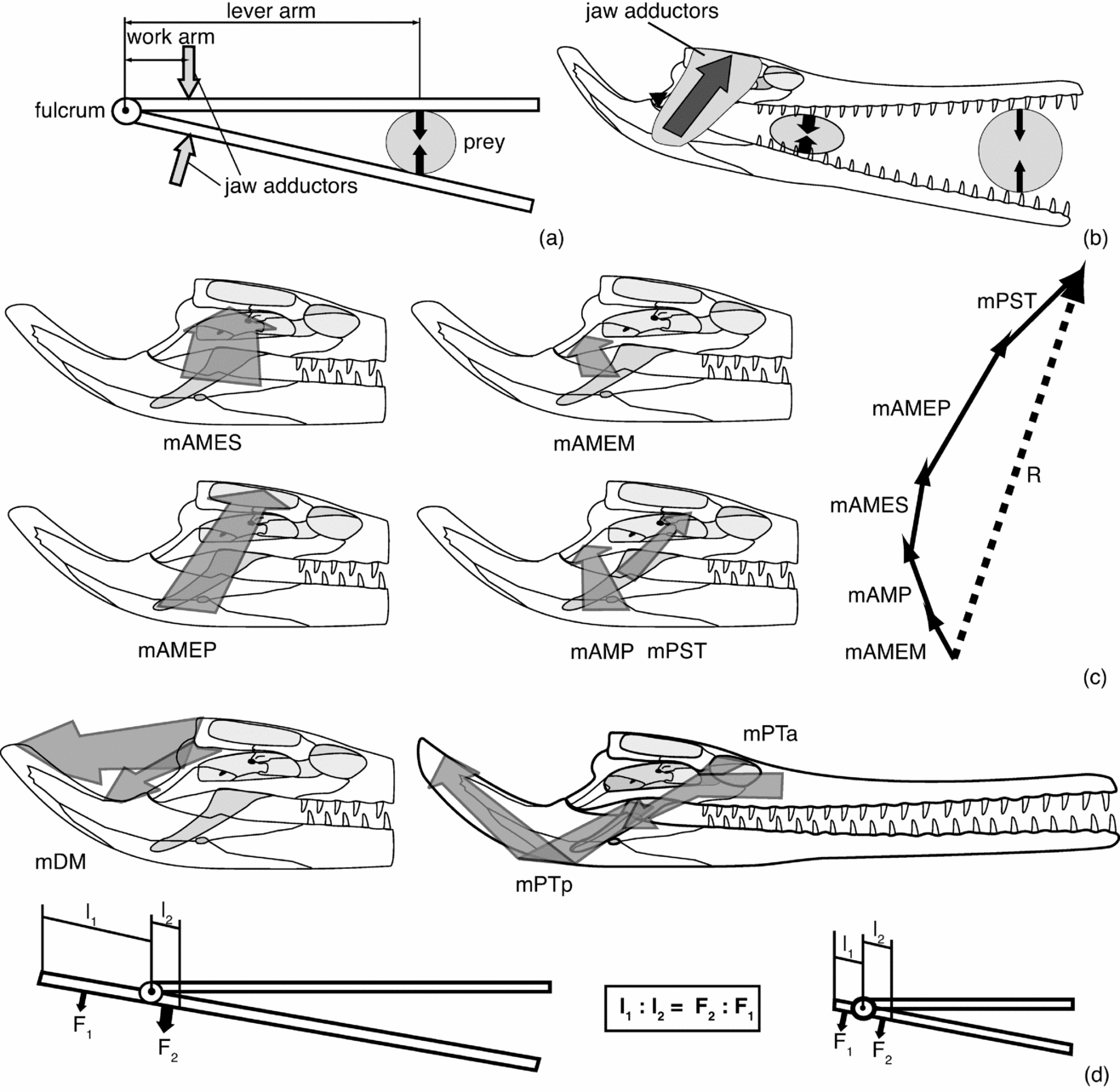
The skull and mandible articulate with each other at the mandibular joint. In the lever model, this joint is the fulcrum and the mandible represents the mobile lever arm (Fig. 6a). The distance between the fulcrum and each reconstructed jaw adductor is the lever arm of a force; the distance between the fulcrum and the prey object between both lever arms is the work arm (Fig. 6a). The resulting force, i.e. the bite force these muscles can exert onto the prey object between the upper and lower jaw, is deduced from the relationship between the lever arm and work arm of the jaw adductors: the longer the work arm is in relation to the lever arm, the smaller is the muscular force. Relative bite force in the jaws of dyrosaurids can be determined from the length of the work arm in relation to the position of the prey object and under the presumption of consistency of the lever arm (Fig. 6b). Owing to the subsequent length increase of the work arm in relation to the force arm, relative bite forces decrease generally in the rostral direction (Sinclair & Alexander, Reference Sinclair and Alexander1987; Pooley, Reference Pooley and Ross1989; Busbey, Reference Busbey and Thomasson1995; Cleuren, Aerts & De Vree, Reference Cleuren, Aerts and Vree1995; Greaves, Reference Greaves and Thomasson1995; Preuschoft & Witzel, Reference Preuschoft and Witzel2002), which is the more dramatic the longer the jaws are.
Functional aspects of the reconstructed jaw musculature in dyrosaurids; skull of Dyrosaurus is used. (a) schematic representation of the unit of skull and mandible as a lever model, (b) skull with position of adductor muscles and different positions of prey objects, which according to the lever model are subject to smaller bite forces in the rostral region of the snout, (c) adductor muscles of the jaws with their main direction of muscle fibres and in a model of force vectors that yield a craniodorsally directed resultant, (d) direction of mDM and mPT and the lever model corresponding to their expansion. Not to scale.

The physiological cross-sections of the mAMES, mAMEP and mAMP of dyrosaurids was larger than in extant crocodylians, suggesting that these jaw adductors could generally exert more force during contraction than in extant crocodylians, which is especially important in the operation of the long rostrum. In extant crocodylians, the mAMEP, mAMP, mPST and mIM are active during the complete feeding process from inertial bites to swallowing (Busbey, Reference Busbey1989; Cleuren & De Vree, Reference Cleuren, Vree and Schwenk2000) and consist mostly of red muscle fibres (Sato et al. Reference Sato, Shimada, Sato and Kitagawa1992), which can be assumed for dyrosaurids. The resultant of the force vectors of the mAME, mAMP and mPST in dyrosaurids was directed from the base of the skull in the rostrodorsal direction (Fig. 6c). The muscle fibres of the mAMES in dyrosaurids were most likely nearly vertically directed, whereas those of the mAMP ran from rostroventrally to caudoventrally (Figs 3, 6c). The position of the muscle fibres of the mAMES and mAMP changed during jaw adduction depending on their orientation at the skull, so that the length of each muscle's lever arm increased. The resulting increase in the forces of these muscles is important, as both muscles are known to produce synchronized tetanic contractions during crushing bites in extant crocodylians (Busbey, Reference Busbey1989; Cleuren & De Vree, Reference Cleuren, Vree and Schwenk2000). In contrast, the muscle fibres of the mAMEP in dyrosaurids were oriented from caudoventrally to rostrodorsally, resulting in a decrease in the length of the lever arm and the resulting force during jaw adduction, probably compensated for by the size increase of this muscle in dyrosaurids.
The position and extension of the mPT and mDM requires the introduction of another lever system with two lever arms, one caudal to the mandibular joint/fulcrum, and one rostral to it (Fig. 6d). According to the law of the lever, the relationship between the lengths of these two lever arms is reciprocally proportional to the relationship between the forces generated on these lever arms (Fig. 6d). In dyrosaurids, the exceptionally long retroarticular process results in a caudal lever arm longer than in extant crocodylians. Thus, in particular the force of the mPTp on the rostral lever arm was larger in dyrosaurids than in extant crocodylians (Fig. 6d), which was also supplemented by the large mPTp cross-section. Because the mPTp of dyrosaurids is reconstructed to have had longer muscle fibres than those of extant crocodylians, it is plausible to also reconstruct a larger speed of contraction for this muscle.
In extant crocodylians, the mAMES, mPTa and mPTp are mostly active when peak activities of the other jaw muscles occur, i.e. at initial capturing and killing or crushing movements (Cleuren & De Vree, Reference Cleuren, Vree and Schwenk2000), and these muscles consist mainly of white muscle fibres (Sato et al. Reference Sato, Shimada, Sato and Kitagawa1992). If the mAMES, mPTa and mPTp in dyrosaurids had similar activities, then they could support the initial stages of feeding quicker and with more muscle force, particularly during prey capturing. Finally, an increase in the muscle force exerted by the mAME, mAMP and mPTp helped to better absorb the joint forces during prey capture (Cleuren, Aerts & De Vree, Reference Cleuren, Aerts and Vree1995; Preuschoft & Witzel, Reference Preuschoft and Witzel2002). In particular, the mAMP with its proximity to the jaw joint and its complex fibre architecture is considered to have had a role in stabilizing the jaw joint during adduction as in extant crocodylians (Iordansky, Reference Iordansky1964; Busbey, Reference Busbey1989), whereas the mIRA and mPTa are considered to form a sort of tension chord to reduce bending moments acting on the mandible (Preuschoft & Witzel, Reference Preuschoft and Witzel2002).
4.b.2. Abduction
As in extant crocodylians (Van Drongelen & Dullemeijer, Reference Van Drongelen and Dullemeijer1982; Busbey, Reference Busbey1989; Cleuren & De Vree, Reference Cleuren, Vree and Schwenk2000), contraction of the mDM in dyrosaurids led to mandibular abduction. The larger physiological cross-section and longer muscle fibres of the mDM of dyrosaurids suggest that this muscle contracted faster and exerted more force than in extant crocodylians (Fig. 6d). Therefore, the long jaws of dyrosaurids could be opened more rapidly and more forcefully than in extant crocodylians.
4.c. Bracing of the neck and suspension from the shoulder girdle
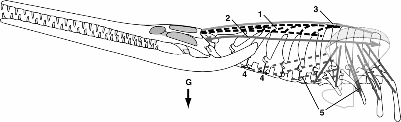
In technical terms, the neck of dyrosaurids can be described as a dorsally tension-braced hydraulically stabilized segmented beam with only rudimentary ventral self-support by the cervical ribs, in contrast to extant crocodylians (Fig. 7) (Frey, Reference Frey1988 a; Salisbury & Frey, Reference Salisbury, Frey, Grigg, Seebacher and Franklin2001). The cervical vertebral column was braced dorsally by the nuchal and supraspinal ligaments in combination with the epaxial musculature. The latter also supported the skull (Fig. 7). In particular, the string of the mRHO and mTRcap/mSPCp connected the skull to the scapula as in extant crocodylians (Frey, Reference Frey1988 b; Salisbury & Frey, Reference Salisbury, Frey, Grigg, Seebacher and Franklin2001) and therefore helped to suspend the skull. Additional suspension of the skull with the long rostrum was provided by the mATC, which was extremely well developed especially in the longirostrine dyrosaurids. The chain of cervical ribs provides ventral stabilization of the vertebral column against ventral flexion by bone-to-bone contact between the third and fifth cervical vertebra (Figs 5, 7). It is unlikely that gravitational forces could be overcome by the stretched ligaments between the sixth and ninth cervical ribs, so that in contrast to extant crocodylians and their strong ventral stabilization component in the neck (Frey, Reference Frey1988 a; Salisbury & Frey, Reference Salisbury, Frey, Grigg, Seebacher and Franklin2001), bracing of this region of the neck of dyrosaurids must have been only dorsally. This corresponds well to the enlarged dorsal epaxial muscles and ligaments in the neck of dyrosaurids. The nuchal ligament was connected to every cervical neural spine in dyrosaurids, which allowed the neck to be suspended dorsally with little muscular support, as in extant large mammals (Dimery, Alexander & Deyst, Reference Dimery, Alexander and Deyst1985; Gellman, Bertram & Hermanson, Reference Gellman, Bertram and Hermanson2002).
Model for the bracing of the neck of dyrosaurids. The self-supported, segmented neck is loaded by gravity and the weight of the skull (1), which leads to dorsal tension and ventral compression of the vertebral column. The supraspinal and nuchal ligaments are tensed and brace the vertebral column dorsally (1); support for the skull is additionally given by the connection to the epaxial musculature (2) and its connection to the shoulder girdle by the string of m. transversospinalis capitis and m. rhomboideus (3). Dorsal bracing of the neck is supported in its cranial half by the paired chain of cervical ribs (4). The neck is also suspended by m. serratus profundus et superficialis and m. colloscapularis superficialis (5).

The scapula of dyrosaurids was connected elastically with the cervical and prothoracic ribs by the m. serratus superficialis et profundus and mCSCs, suspending the cervical vertebral column like tension flanges (Fig. 7). Forces acting on the cervical ribs by these muscles were transmitted to the vertebral corpora and neural arches as in extant crocodylians (Frey, Reference Frey1988 a; Salisbury & Frey, Reference Salisbury, Frey, Grigg, Seebacher and Franklin2001). The direction of the resultant of the mSERRs and mCAPST can be reconstructed from force parallelograms (Fig. 4f). The resultant of the mSERRs was directed from the scapula caudoventrally, that of mCSCs cranioventrally, and that of mCAPST craniodorsally (Fig. 4f). Because of the higher cervical neural spines and long scapula of dyrosaurids (Schwarz, Frey & Martin, Reference Schwarz, Frey and Martin2006), the resultant of these muscles was steeper and had a larger angle to the horizontal plane than in extant crocodylians (Fig. 4f) (Fürbringer, Reference Fürbringer1876; Frey, Reference Frey1988 b; Meers, Reference Meers2003). The relative sizes of the resultants can be compared by using a force triangle (Fig. 4). By the calculation of the resultant FR = a/cos β (a is the horizontal force vector perpendicular to the vertical force vector b; β is the angle to the force vector a) it is demonstrated that owing to its steeper course, the resultant of the three muscles in dyrosaurids was larger than in extant crocodylians (Fig. 4f). This corresponds to the postulated larger vertical flexibility of the cervical vertebral column of dyrosaurids, which requires in compensation for the reduced support by the cervical ribs an enhanced muscular suspension of the neck. Dorsoventral bending loads acting on the cartilaginous suprascapula and the scapula during contraction of the mTRcap – mRHO string and mSERRp were most probably counteracted by the hydraulic pressure of the underlying musculature.
4.d. Functional aspects in the cervical musculature
In extant crocodylians, jaw opening is accomplished by the mDM operating the mandible, but aided also by the bilateral contraction of the epaxial cervical muscles mTRcap, mSPCp and mEPC (Van Drongelen & Dullemeijer, Reference Van Drongelen and Dullemeijer1982; Cleuren & De Vree, Reference Cleuren and Vree1990, Reference Cleuren and De Vree1991, Reference Cleuren, Vree and Schwenk2000). The extremely high development of these muscles in dyrosaurids makes their contribution to the opening of the jaws likely, similar to extant crocodylians. Enlargement of the mSPCp and mEPC in dyrosaurids supported the elevation of the long rostral part of the skull. Cranial elevation in extant crocodylians is usually combined with elevation of the neck, which is accomplished by bilateral contraction of the mTRcer, mLCAPs and mILCOSTcer (Cleuren & De Vree, Reference Cleuren, Vree and Schwenk2000). A development of these muscles in dyrosaurids similar to extant crocodylians indicates a similar function for lifting the neck.
During inertial feeding in extant crocodylians, mostly more complex movements of the skull and neck occur, so that dorsal flexion of the skull and neck is often accompanied by lateral shifting movements of the skull (Cleuren & De Vree, Reference Cleuren and Vree1990, Reference Cleuren, Vree and Schwenk2000). According to electromyographic analyses of the neck muscles of Caiman crocodylus, such lateral movements of the skull and neck require simultaneous activities of several muscles of both sides of the neck (Cleuren & De Vree, Reference Cleuren and De Vree1991, Reference Cleuren, Vree and Schwenk2000). Lateral shifting of the head in extant crocodylians is produced by bilateral contraction of the mTRcap and mILCOSTcap, and ipsilateral contraction of the mSPCp and mLCAPs, and most plausibly by a similar mechanism in dyrosaurids. Unilateral contraction of the mILCOSTcer caused lateral flexion of the neck. Recoil movements were supported by the tensed intercostal ligaments.
Ventral movements of the neck do not occur in extant crocodylians because the chain of cervical ribs effectively prevents neck deflection (S. W. Salisbury, unpub. Ph.D. thesis, Univ. New South Wales, 2001). Ventral neck muscles mainly function as a stabilizer for the occipital joint during inertial feeding (Cleuren & De Vree, Reference Cleuren and De Vree1991, Reference Cleuren, Vree and Schwenk2000). In contrast to extant crocodylians, ventral neck flexibility in dyrosaurids was made possible by the modification of the cervical ribs, and it is likely that there was also muscular support for deflecting the neck. This would most plausibly have been achieved by bilateral contraction of the mILCOSTcer, mCAPST, mCSCs and mLONcoll, all of which are larger in dyrosaurids than in extant crocodylians. During ventral flexion of the neck, the supraspinal, nuchal and elastic ligaments underwent tension and thereby launched elastic recoil of the neck. Because of its connection to the scapula, contraction of the mCSCs required parallel stabilization of the scapula by synchronous contraction of the m. serratus superficialis et profundus. As in extant crocodylians (Cleuren & De Vree, Reference Cleuren, Vree and Schwenk2000; Preuschoft & Witzel, Reference Preuschoft and Witzel2002), it is likely that the occipital joint of dyrosaurids was stabilized during neck and head movements by parallel activity of antagonistic muscles to the firing muscles.
5. Discussion
5.a. Feeding strategy
In the skull of dyrosaurids, jaw adductors and the only jaw abductor were enlarged and possessed longer muscle fibres, which indicates their ability for more forceful and quicker contraction. Iordansky (Reference Iordansky1964) explained the hypertrophied mAMEP of extant longirostrine crocodylians with the special importance of a higher closing velocity of the jaws for fish-catching. Alternatively, the presence of these enlarged jaw adductors can be explained simply by the necessity of operating a longer rostrum in a dense medium such as water (Busbey, Reference Busbey1989) and therefore not necessarily linked with a particular food spectrum.
A most important feature of the feeding apparatus of all dyrosaurids is the increased ventral flexibility of the craniocervical region. This motion is enhanced by the reduction of ventral support of the cervical ribs and the presence of much larger cervical muscles in the neck. Further enhancement comes from a verticalization of the axial neck musculature in combination with a strongly enlarged retroarticular process and associated jaw muscles in the skull. In combination with this, the lateromedial flexibility of the neck was reduced. The increased vertical mobility of the craniocervical apparatus in combination with the mostly longirostrine tubular rostrum of dyrosaurids is very different to all other crocodyliforms and plausibly indicates changes in the mode of prey catching. In particular, behaviours such as occasional poking in the sediment and bottom feeding become likely options for dyrosaurids. The literature lists one record for sediment searching behaviour in extant crocodylians (Pooley, Reference Pooley and Ross1989), stating without further details that longirostrine crocodylians can use their rostrum to search for crabs in subterraneous burrows. A focused poking behaviour in dyrosaurids would be strongly supported by the enlarged jaw musculature that allows a better control of the rapid opening and closing of the long rostrum and overcoming the larger resistance of a muddy substrate and/or the water. The large axial cervical musculature and ligaments would allow control and adjustment of vertical neck movements as well as sustained movements of the neck for poking in the substrate and bottom feeding. Bottom feeding might correspond to extant river dolphins, which in comparison to other cetaceans have a longer and more slender snout and a larger neck flexibility (Kastelein et al. Reference Kastelein, Neurohr, Nieuwstraten and Wiepkema1999; Cassens et al. Reference Cassens, Vicario, Waddell, Balchowsky, Van Belle, Ding, Fan, Lal Mohan, Simőes-Lopes, Bastida, Meyer, Stanhope and Milinkovitch2000; Fish, Reference Fish2002; Geisler et al. Reference Geisler, McGowan, Yang and Gatesy2011) and of which at least the La Plata dolphin Pontoporia blainvillei is known to expand its diet of fish to crustaceans and cephalopods (Ridgway & Harrison, Reference Ridgway and Harrison1989; Rodríguez, Rivero & Bastida, Reference Rodríguez, Rivero and Bastida2002).
A study of the inner ear morphology of Rhabdognathus showed that this dyrosaurid had a very specialized, elaborate vestibular region unlike any other crocodyliform, which has been interpreted as an adaptation of dyrosaurids to walking along the sea floor instead of sustained swimming (Georgi, Reference Georgi2006). Whereas functional morphological studies do not corroborate a reduced swimming ability for dyrosaurids and their swimming abilities were probably even greater than extant crocodylians (Schwarz-Wings, Frey & Martin, Reference Schwarz-Wings, Frey and Martin2009), they do not exclude the animal's ability for expanded walking on the sea floor, and the inner ear morphology would also be in support of a more variable feeding behaviour. Although restricted by the long mandible and the configuration of the cervical ribs, the lateral flexibility of skull and neck in dyrosaurids was still large enough to make the capture of a prey object positioned directly laterally to the rostrum possible. The usual prey-catching strategy of most extant crocodylians of quick lateral skull movements (Thorbjarnarson, Reference Thorbjarnarson1990; McHenry et al. Reference McHenry, Clausen, Daniel, Meers and Pendharkar2006) is therefore likely to have occurred also in dyrosaurids, but probably was expanded by multidirectional and in particular vertical movements of the rostrum for prey capturing, either in the substrate, on the bottom of rivers or the sea floor, or in the open water. The larger vertical mobility of the cervicocranial system could have also aided dyrosaurid crocodyliforms in improved diving, as well as for controlling the long rostrum in turbulent water currents. Dyrosaurid crocodyliforms are known from marginal marine to fluvial environments, which all offer shallow water conditions allowing a large suite of different feeding behaviours. The configuration of the epaxial cervical musculature and the reconstructed dorsal flexibility of the craniocervical region would also allow inertial feeding once the prey object has been grabbed.
5.b. Rostral shape and dentition
The family of Dyrosauridae comprises generally meso- to longirostrine forms with a mostly tubular rostrum (Fig. 2). The cross-section of a tubular rostrum is equal in all potential directions of motion, which means that resistance during movements is smaller than with a narrow or broad platyrostrine rostrum (Busbey, Reference Busbey and Thomasson1995), which can be advantageous in the water. Tubular rostra exhibit better strength against torsion than a platyrostrine rostrum (McHenry et al. Reference McHenry, Clausen, Daniel, Meers and Pendharkar2006), but the smaller cross-section of the tubular rostrum makes it less stable against shear and bending loads (Busbey, Reference Busbey and Thomasson1995; Preuschoft & Witzel, Reference Preuschoft and Witzel2002; McHenry et al. Reference McHenry, Clausen, Daniel, Meers and Pendharkar2006; Pierce, Angielczyk & Rayfield, Reference Pierce, Angielczyk and Rayfield2008), which considerably reduces the chosen prey size available for extant longirostrine crocodylians (Busbey, Reference Busbey and Thomasson1995; McHenry et al. Reference McHenry, Clausen, Daniel, Meers and Pendharkar2006; Erickson et al. Reference Erickson, Gignac, Steppan, Lappin, Vliet, Brueggen, Inouye, Kledzik and Webb2012). It is assumed that the longirostrine and tubular construction of the rostrum covers a larger area for prey catching in a lateral direction, increases the speed of the attack, and results in less-obstructed vision for the hunting crocodile (Thorbjarnarson, Reference Thorbjarnarson1990; McHenry et al, Reference McHenry, Clausen, Daniel, Meers and Pendharkar2006; Erickson et al. Reference Erickson, Gignac, Steppan, Lappin, Vliet, Brueggen, Inouye, Kledzik and Webb2012). In addition to that, the homodont dentition in the elongate jaws results in a long functional mouth gape that is efficient for the catching of small prey items, because it increases the chance of catching the subject during sideward movements of the skull (Gans, Reference Gans1966; Iordansky, Reference Iordansky and Gans1973; P. Vignaud, unpub. Ph.D. thesis, Univ. Poitiers, 1995). A long and uniform tooth row facilitates transport of the food in the direction of the pharynx (Busbey, Reference Busbey and Thomasson1995), which is important for inertial feeding (Cleuren & De Vree, Reference Cleuren, Vree and Schwenk2000). Differences in the rostral shape, as visible in dyrosaurids, are most likely related to differences in the prey spectrum as a reduction in the ‘tubularity’ of the rostrum and a development of a more platyrostrine snout would allow an increase in prey sizes, but does not exclude the food-capturing strategies of sideward movements and poking behaviour as described in Section 5.a.
The dentition of dyrosaurids is homodont, with uniform conical teeth that are slightly labiolingually compressed and curved distolingually with an acute apex, a mesial and a distal carina and labial and lingual striations of the enamel. Only Phosphatosaurus possesses more blunt conical teeth with a broad and rounded apex and a stronger striation of the enamel (Buffetaut, Reference Buffetaut1978c , Reference Buffetaut1979a ). It is plausible that the teeth of dyrosaurids were suited to grabbing and piercing of soft-tissue items, for the separation of part of the soft-tissue items and for crushing of thin shells, or in the case of Phosphatosaurus even of more massive shells (Massare, Reference Massare1987). Together with the morphology of the rostrum of dyrosaurids as discussed above, their dentition allows the reconstruction of the following prey spectrum:
-
(1) Prey objects could not exceed a certain size, because the tubular rostrum was not stable enough for larger, struggling prey. In correspondence to the extant taxa Tomistoma schlegelii and Mecistops cataphractus, which grow to approximately similar sizes as dyrosaurids (Trutnau, Reference Trutnau1994), the size of prey objects could not exceed the size of a dog with a 20 kg body weight.
-
(2) Prey objects other than fish could have thin shells (e.g. crabs, shrimps, benthic gastropods) or could consist of soft-tissues with (e.g. squid) or without (e.g. annelids) a hard endoskeleton.
-
(3) Up to a certain size, prey items were probably taken and swallowed whole.
This reconstructed prey spectrum is in accordance with the occurrence of other vertebrates such as a large variety of osteichthyes and chondrichthyes, aquatic invertebrates and turtles in the same localities (e.g. Hill et al. Reference Hill, McCartney, Roberts, Bouaré, Sissoko and O'Leary2008; Bardet et al. Reference Bardet, Pereda Superbiola, Jouve, Bourdon, Vincent, Houssaye, Rage, Jalil, Bouya and Amaghzaz2010; Hastings et al. Reference Hastings, Bloch, Cadena and Jaramillo2010). Besides fish, benthic animals, in particular crustaceans such as crabs or shrimps, gastropods and annelids, or squid are well within the food spectrum of dyrosaurids. Extant crocodylians feed on everything they can get, and virtually the only restriction to their diet is the size of a prey item (Pooley, Reference Pooley and Ross1989). In comparison with extant crocodylians, there is no evidence that dyrosaurids were specialized for piscivory as previously assumed (Buffetaut, Reference Buffetaut1979 b; Denton, Dobie & Parris, Reference Denton, Dobie, Parris, Callaway and Nicholls1997; Hastings, Bloch & Jaramillo, Reference Hastings, Bloch and Jaramillo2011), and which is known for the extant Gavialis gangeticus (Thorbjarnarson, Reference Thorbjarnarson1990) that has a different shape of the rostrum compared to all dyrosaurids (e.g. Iordansky, Reference Iordansky and Gans1973). Therefore, the prey spectrum of dyrosaurids was certainly supplemented as in extant longirostrine crocodylians (McHenry et al. Reference McHenry, Clausen, Daniel, Meers and Pendharkar2006) by a variety of other prey items, depending mostly on the environment and size. Taxa with a shorter rostrum, i.e. Chenanisuchus and Cerrejonisuchus, were most probably more generalistic feeders (Hastings et al. Reference Hastings, Bloch, Cadena and Jaramillo2010). The occurrence of meso- and longirostrine forms in similar fossil localities (Jouve, Bouya & Amaghzaz, Reference Jouve, Bouya and Amaghzaz2005; Hastings et al. Reference Hastings, Bloch, Cadena and Jaramillo2010; Hastings, Bloch & Jaramillo, Reference Hastings, Bloch and Jaramillo2011) makes it unlikely that they were adapted to a special type of environment or substrate.
The reconstructed enhanced feeding strategy of dyrosaurids, including behaviours such as a poking, would be a new explanation for the long-term evolutionary success of the Dyrosauridae, which survived the mass extinction at the end of the Cretaceous (Denton, Dobie & Parris, Reference Denton, Dobie, Parris, Callaway and Nicholls1997; Hill et al. Reference Hill, McCartney, Roberts, Bouaré, Sissoko and O'Leary2008). Characterization of dyrosaurids as major predators in the marine environment (Barbosa, Kellner & Sales Viana, Reference Barbosa, Kellner and Sales Viana2008) is confirmed by this study, with the restriction of the reconstructed mechanical limitations of the long rostrum to smaller prey items and the reconstructed diet. The postulated replacement of mosasaurs by dyrosaurids in the Paleocene (Denton, Dobie & Parris, Reference Denton, Dobie, Parris, Callaway and Nicholls1997; Barbosa, Kellner & Sales Viana, Reference Barbosa, Kellner and Sales Viana2008) might well be related to their reconstructed feeding adaptations. As the evolutionary success of dyrosaurids seems to be founded at least in part on their specialization for prey capture, these animals might also be important for the reconstruction of transitional Maastrichtian–Paleocene ecosystems and make detailed analyses of the corresponding food webs possible.
Acknowledgements
For their suggestions, discussions and helpful comments I thank cordially my former supervisors Dino Frey and Thomas Martin. I am especially grateful to Stéphane Jouve (MNHN) for providing valuable information (including numerous photographs) on then unpublished material, as well as for fruitful discussions. I am grateful for discussion and helpful comments given by Eric Buffetaut (CNRS) and Christian A. Meyer (NMB) on this topic, and I am in particularly indebted to Hans-Peter Schultze for sharing his thoughts towards the improvement of the initial reconstruction of the feeding apparatus of dyrosaurids. During my visits to different museums and collections, access and information was provided by Sandra Chapman and Angela Milner (both BMNH), Nathalie Bardet (MNHN), Daniel Baudet (MRAC), Daniel Brinkman and Walter Joyce (both YPM). For their efforts in preparing and curating the specimen of Dyrosaurus sp. from the SMNK, I thank Olaf Dülfer and René Kastner. Funding was provided by the FU Berlin (NaFÖG-Program). Financial support for my stay at the NHM was provided by the European Community with the Access to Research Infrastructure action of the Improving Human Potential Programme (SYS-Resource Programme of the NHM) and for the stay at the AMNH and YPM by the Swiss National Science Foundation (SNF No. 200021–101494/1). For the invitation to contribute an article to this special issue I thank Dr Benjamin Kear. The two referees Stéphane Jouve and Alexander Hastings provided valuable comments and suggestions for the improvement of this work, in particular concerning interpretations of the reconstruction of the feeding behaviour of dyrosaurids and the language.