Diaphragmatic paralysis following phrenic nerve injury is a major complication following congenital cardiac surgery. In contrast to unilateral paralysis, patients with bilateral diaphragmatic paralysis present a higher risk group, require different management methods, and have poorer prognosis.Reference Qureshi 1 , Reference de Leeuw, Williams, Freedom, Williams, Shemie and McCrindle 2 Although unilateral palsy may respond to early plication of the affected hemi-diaphragm,Reference Georgiev, Konstantinov, Latcheva, Mitev, Mitev and Lazarov 3 bilateral diaphragmatic paralysis is usually managed with supportive measures, at times resorting to tracheostomyReference Qureshi 1 , Reference Stewart, Alexson and Manning 4 for prolonged respiratory support. In this study, we report the outcome of different management options for bilateral diaphragmatic paralysis following surgery for CHD.
Patient and methods
The Hospital Research Committee approved the study protocol, and consent was waived on account of the retrospective use of anonymous patient data. All the patients who had either closed-heart or open-heart surgery for CHD and diagnosed to have bilateral diaphragmatic paralysis postoperatively from July, 2006 to July, 2014 at our institution were included. In total, seven patients were identified, and hospital records of these patients were retrospectively reviewed. Data collected included age, sex, primary cardiac diagnosis and operation, reason to suspect diaphragmatic paralysis, time to diagnose diaphragmatic paralysis, time to plication if carried out and time to recovery, ventilator days after plication, total ventilator days, and lengths of ICU and hospital stay.
Management strategy
Our approach to bilateral diaphragmatic paralysis comprised mechanical ventilation and immediate plication of the paradoxical hemi-diaphragm upon recovery of the contralateral side. Diaphragmatic excursion was monitored twice a week using ultrasonography. On return of function of one hemi-diaphragm, fluoroscopy was carried out to detect paradoxical movement of the contralateral side to justify its plication. In the absence of paradoxical motion of the unrecovered hemi-diaphragm, weaning from the ventilator was attempted, and when unsuccessful we continued with conservative measures and regular monitoring until either both sides recovered or one side was subjected to plication.
Results
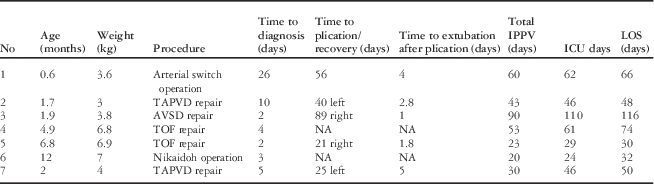
There were seven patients who developed bilateral diaphragmatic paralysis following cardiac surgery from a total of 1020 during the study period with an incidence of 0.68%. The median age was 2 months (0.6–12 months). There was one neonate and six infants with a median weight of 4 kg (3–7 kg), and all the cases developed bilateral diaphragmatic paralysis following open-heart procedures (Table 1). Median time to diagnosis of bilateral diaphragmatic paralysis was 4 days (2–26 days) after the cardiac surgery. Among all, four patients had required re-intubation, and three of them could not tolerate weaning from mechanical ventilation; one patient from the latter group was diagnosed following absence of electrical impulses when assisted with neurally adjusted ventilator assist.
Distribution of bilateral diaphragmatic palsy based on the primary surgical procedure.

AVSD=atrioventricular septal defect; IPPV=intermittent positive-pressure ventilation; LOS=length of hospital stay; TAPVD=total anomalous pulmonary venous drainage; TOF=Tetralogy of Fallot
In total, five children who had unilateral recovery with paradoxical movement of the contralateral hemi-diaphragm underwent plication of the latter at a median interval of 40 days (21–89 days). All five children were extubated at a median of 2.8 days (1.8–5 days) after the plication. The other two patients had a return of function of one hemi-diaphragm at 18 and 49 days after the surgery, respectively, but were not subjected to contralateral diaphragmatic plication due to the absence of paradoxical movement. Both were extubated 2 and 4 days, respectively, (mean 3 days) following unilateral recovery of diaphragmatic excursion. The mean age of these two patients was 8.6 months, compared with 2.6 months for the patients who required plication.
The median ventilation time for the whole group was 48 days (20–90 days). None of the patients required tracheostomy for respiratory support and there were no mortalities; four patients required non-invasive respiratory support with high-flow nasal cannula, continuous positive airway pressure, or bi-level positive airway pressure for 1–3 days following extubation. All except one patient developed ventilator-associated pneumonia. The median length of ICU stay was 46 days (24–110 days), and the median length of hospital stay was 50 days (30–116 days).
Discussion
Cardiac surgery is the most common cause of diaphragmatic palsy that can result in delayed recovery and excess morbidity. The reported incidence of clinically diagnosed diaphragmatic paralysis following congenital heart surgery ranged from 0.28 to 10%.Reference Dagan, Nimri, Katz, Birk and Vidne 5 , Reference Joho-Arreola, Bauersfeld, Stauffer, Baenziger and Bernet 6 The incidence of bilateral diaphragmatic paralysis in our series was 0.68%.
Neonates and infants are diaphragmatic breathers with the intercostal muscles playing a minimal role in respiration. In our study, all the patients who had bilateral diaphragmatic paralysis were <1-year old, and one of them was a neonate. In older children, with higher respiratory capacity and reserves, a higher index of suspicion is required to diagnose bilateral diaphragmatic paralysis as described by de Leeuw et al,Reference de Leeuw, Williams, Freedom, Williams, Shemie and McCrindle 2 who reviewed >10,000 cardiothoracic patients over 12 years of age, and discovered an incidence of bilateral diaphragmatic paralysis of 0.1%.
Clinical diagnosis of diaphragmatic paralysis is difficult when the patient is on the ventilator, and it is usually suspected when there is unexplained difficulty in weaning from the ventilator or following failed extubation. Unilateral paralysis may be evident on a chest X-ray with a notable elevation of the affected hemi-diaphragm; however, diaphragmatic asymmetry is not present in bilateral diaphragmatic paralysis. In our report, four of seven patients were diagnosed with bilateral diaphragmatic paralysis following re-intubation, whereas three patients did not tolerate weaning from the ventilator. Failure to show trigger impulses while on neurally adjusted ventilator assist mode may be useful to help with the diagnosis bilateral diaphragmatic paralysis.Reference Coisel, Chanques and Jung 7
Fluoroscopy is traditionally the gold standard for diagnosing diaphragmatic paralysis. Recent reports have shown that ultrasound is equally reliableReference Miller, Brook and Tacy 8 , Reference Sanchez de Toledo, Munoz and Landsittel 9 and offers the added advantages of bedside availability, avoidance of radiation exposure, and is easily repeatable. In all our patients, diagnosis of bilateral diaphragmatic paralysis was made using bedside ultrasound and confirmation of paradoxical motion by fluoroscopy before surgical plication.
Plication improves the functional residual capacity of the lung facilitating successful extubation in unilateral diaphragmatic paralysis.Reference Dagan, Nimri, Katz, Birk and Vidne 5 , Reference Mickell, Oh, Siewers, Galvis, Fricker and Mathews 10 In our experience, the five patients who underwent plication of the remaining paralysed hemi-diaphragm were subsequently extubated 2.8 days later. In our experience, an aggressive plication strategy was found helpful in the early extubation of patients who developed unilateral diaphragm palsy with a paradoxical movement following cardiac surgery. We adopted the same strategy in patients with bilateral diaphragm palsy once there was unilateral recovery with a paradoxical movement. Alternatively, either weaning from the ventilator or waiting for the contralateral hemi-diaphragm to recover without plication could be considered in older infants and children; however, the recovery time for the paralysed hemi-diaphragm could be unpredictable and variable. Interestingly two patients who had return of function in one hemi-diaphragm, but without paradoxical movement of the other, were also swiftly extubated 3 days after the recovery. It was notable that these two patients were relatively older infants. It is, therefore, difficult to justify an aggressive approach of diaphragmatic plication for patients with bilateral diaphragmatic paralysis, especially in older children. Our results are similar to those of Dagan et alReference Dagan, Nimri, Katz, Birk and Vidne 5 who used a similar conservative approach in nine children with bilateral diaphragmatic paralysis with 100% ventilator-free survival. Bilateral diaphragmatic plications were shown to be beneficial in adults with bilateral diaphragmatic paralysis caused by progressive neurological diseases;Reference Srivastava, Pasha and Knowles 11 , Reference Stolk and Versteegh 12 however, they are not usually considered in children.
Tracheostomy has been advocated in some reports in patients whose phrenic nerve recoveries were significantly prolonged,Reference Qureshi 1 , Reference Stewart, Alexson and Manning 4 , Reference Mickell, Oh, Siewers, Galvis, Fricker and Mathews 10 , Reference Baker, Boulom, Reemtsen, Rollins, Starnes and Wells 13 but it has also been reported that tracheostomy in these patients can be associated with significant mortality.Reference Cotts, Hirsch, Thorne and Gajarski 14 The longest time to extubation in our patients was 90 days, and in spite of this none of the patients were subjected to tracheostomy. The tradeoff was, nevertheless, noteworthy, where all except one of our patients developed ventilator-associated pneumonia – a factor known to increase the risk of mortality in patients with diaphragmatic paralysis.Reference Akay, Ozkan and Gultekin 15 Bearing in mind all these factors, we would recommend considering the use of tracheostomy for bilateral diaphragmatic paralysis in patients who have not recovered after 3 months of conservative management, or in those who have recurrent lung infections. The use of non-invasive positive-pressure ventilation has been reported as an alternative in infants with bilateral diaphragmatic paralysis to avoid tracheostomy or even long-term endotracheal intubation.Reference Kovacikova, Dobos and Zahorec 16 We have limited the use of non-invasive ventilatory assistance to avoid re-intubation in four patients.
Limitations
This study is limited by its retrospective nature and a small number of patients.
Conclusion
Bilateral diaphragmatic paralysis following paediatric cardiac surgery is uncommon but can be a major cause of morbidity and can result in prolonged hospital stay. The recovery time is variable and unpredictable, often necessitating prolonged ventilation, as well as lengthy ICU and hospital stays. The role of diaphragmatic plication for bilateral diaphragmatic paralysis in children is uncertain as it has no impact on the outcome and may not have a fundamental impact on the natural course of recovery. We would recommend that plication should be applied only if the patient remains ventilator-dependent upon recovery of one hemi-diaphragm with paradoxical movement of the unrecovered side.
Acknowledgement
None.
Financial Support
This research received no specific grant from any funding agency, commercial, or not-for-profit sectors.
Conflicts of Interest
None.
Ethical Standards
Institutional review board approval taken and mentioned in patients and methods [L-39,40].