Introductıon
In children with normal cardiac anatomies, 37–50% of supraventricular tachycardia ablations are performed for left-sided arrhythmia foci. The left atrium and mitral valve annulus can be accessed by performing a transseptal puncture after passing through the femoral vein and the right atrium. Transseptal punctures can be performed with low complication rates in children even weighing ≤30 kg. Reference Koca, Akdeniz and Tuzcu1 But paediatric cardiologists can be challenged with different types of congenital cardiac anomalies. Situs inversus totalis-dextrocardia is one of them. Dextrocardia is a congenital heart malformation with a low incidence that occurs in only 1 in 10,000–12,000 people. And Wolff-Parkinson-White exists in 0.1–0.3% of the population. Reference Zhao, Li, Bai, Wang, Zhang and Yang2 Although these two conditions and a Wolff-Parkinson-White with a left posterolateral accessory pathway may be seen rarely. Here, we report a Wolff-Parkinson-White syndrome case in a patient with dextrocardia who was successfully treated with radiofrequency catheter ablation.
Case report
A 10-year-old, 28 kg weighted male patient was admitted to the paediatric emergency room with chest pain. Dextrocardia was found during the examination, and Wolff-Parkinson-White syndrome was detected in his electrocardiogram (ECG). He was referred to our clinic. In the patient’s examinations, a resting heart rate of 105/min, blood pressure of 90/54 mmHg, and an apical pulse on the right chest side were detected; there was no murmur. Twelve-lead ECG (the chest electrodes were placed in a mirror image in accordance with situs inversus) showed manifest preexcitation with positive delta wave in D-I,V5,V6 (Figure 1 A and b).
( a ) Posteroanterior chest radiograph shows dextrocardia. ( b ) Delta pattern is observed in the ECG taken by positioning the electrodes in the mirror-image reverse configuration. ( c ) HV shortening and QRS widening are observed after IV adenosine, proving that it is WPW.

Electrophysiological study and ablation were scheduled for the patient. The procedure was conducted under general anaesthesia. EnSite Precision® Mapping System (St. Jude Medical Inc., St. Paul, MN, USA) was used with limited fluoroscopy. ECG electrodes were positioned in a mirror-image reversed configuration. Similarly, Ensite patches and fluoroscopy views were adjusted to a reversed left–right orientation, enabling familiar imaging perspectives during 3D and fluoroscopic visualisation. Catheters were placed in the high right atrium, right ventricle, and coronary sinus. While accessing the coronary sinus, manoeuvers were performed in a counterclockwise direction, opposite to standard practice.
An accessory pathway risk assessment was performed at baseline electrophysiological study and found a low risk of sudden cardiac death (simultaneous accessory pathway effective refractory period with atrioventricular node effective refractory period was 310 ms, and no pre-excitation during atrial fibrillation). The accessory pathway was unresponsive to intravenous adenosine administration (Figure 1c). Supraventricular tachycardia was induced with basal programmed atrial stimulations. Both with V-pace and during supraventricular tachycardia, the atrial pattern in the coronary sinus was eccentric, and the earliest atrial signal was obtained from the distal part of the coronary sinus catheter; the path was considered left-sided, and it was seen that there was no patent foramen ovale to pass to the left.
The Brockenbrough needle was inserted with the help of an 8F long sheath. Unlike routine cases where the spatial orientation of the transseptal needle is set at 4–5 o’clock, it was adjusted to 7–8 o’clock in this patient due to the mirror-image anatomy. By using the right oblique position instead of the left oblique for septal structures in fluoro and the left oblique position instead of the right oblique for anterior–posterior structures and again by changing the right and left sides in fluoro, the image in the patient with normal heart structure was obtained, in the same way the 3D electroanatomical system was used in a similar way, thus the images were studied as in the normal patient, only during the catheter movements, the manoeuvers normally performed in a clockwise direction were performed in a counterclockwise direction, and in the opposite case, the process was performed by performing it in a clockwise direction. After contrast staining of the septum, the septum was perforated with the stylet, and the left atrium was visualised using contrast. A 0.014” coronary guidewire was advanced into the left upper pulmonary vein (Figure 2). Subsequently, a 4 mm RF Marinr catheter was placed in the left atrium. Intravenous heparin was administered at a dose of 100 IU/Kg.
The coronary guidewire that has passed the septum is observed in the images in ( a ) normal positioning of the fluoroscopy in right oblique position, ( b ) after converting fluoroscopy to a mirror-image view. Three-dimensional anatomy of the atria after ensite patches were adjusted to a reversed left–right orientation is shown in panels C and D. The red triangle indicates the site of the earliest V wave that precedes the delta wave on the surface ECG during delta mapping in sinus rhythm. CS = coronary sinus; IVC = inferior vena cava; LA = left atrium; RA = right atrium; SVC = superior vena cava.

During sinus rhythm, delta mapping revealed the earliest ventricular activation at the distal coronary sinus catheter. At the successful site, the local atrial and ventricular signals overlapped, with a unipolar Q–S complex morphology and ventricular activation preceding the delta wave onset by 25 ms, confirming the left posterolateral accessory pathway location. Radiofrequency energy was delivered at this site using 50 W, a target temperature of 53°C, and an impedance of 112 ohms. During the third test lesion, the delta wave disappeared from the surface ECG by the third second of application. Subsequently, three 30-second lesions were applied to this region and its surroundings to ensure complete pathway elimination. Post-ablation, the rhythm was sinus without preexcitation. With adenosine-induced complete AV block, neither antegrade nor retrograde accessory pathway conduction was observed, confirming successful ablation. A 20-minute observation period followed, during which sinus rhythm was maintained, and no Wolff-Parkinson-White pattern was detected. At the end of the procedure, a repeat administration of 10 mg adenosine confirmed the absence of antegrade and retrograde accessory pathway conduction. The procedure was successfully completed without complications. The total procedure time was 140 minutes, and the total fluoroscopy time was 10.3 min. At 1- and 3-month outpatient follow-up, the patient remained asymptomatic with normal sinus rhythm on ECG and no evidence of preexcitation on Holter monitoring.
Discussion
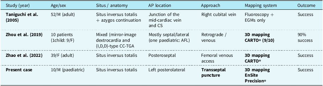
Supraventricular arrhythmias are common in dextrocardia patients and are assumed to be similar to those found in the general population (levocardia). Atrial fibrillation/flutter appears to be the most prevalent of these arrhythmias, but supraventricular tachycardia, including atrioventricular reentrant tachycardia and supraventricular tachycardia associated with Wolff-Parkinson-White syndrome, can also occur. Reference Rao3 Dextrocardia with situs inversus is more common, occurring in around 1 out of every 10,000 live births. Wolff-Parkinson-White syndrome occurred in 0.23% of 427 dextrocardia patients. Mirror image dextrocardia and situs inversus are usually not related with CHD (3%), in contrast to dextrocardia in the situation of situs solitus or situs ambiguus, when CHD incidence can reach 90%. Reference Sternick, Gerken, Max and Vrandecic4 Due to the low occurrence of dextrocardia with situs inversus, there are just a few case reports of catheter ablation and one case series. Reference Zhao, Li, Bai, Wang, Zhang and Yang2,Reference Sternick, Gerken, Max and Vrandecic4–Reference Zhou, Ma and Zhang6 Zhao et al. Reference Zhao, Li, Bai, Wang, Zhang and Yang2 and Taniguchi et al. Reference Taniguchi, Miyauchi and Kobayashi5 reported the AP as posteroseptal. So, catheter ablation could be achieved in the right heart. Zhou et al. Reference Zhou, Ma and Zhang6 reported that in their 10-patient dextrocardia and catheter ablation series, all left atrial accesses were obtained by retrograde approach, and no transseptal puncture was performed.
Table 1 summarises reported paediatric cases of Wolff-Parkinson-White ablation in dextrocardia, highlighting the rarity of our case and the unique approach taken. Notably, our case is among the few reported paediatric cases and, to our knowledge, the only one performed via a transseptal puncture approach, highlighting its feasibility and safety even in mirror-image anatomy.
Comparison of the reported cases of catheter ablation for supraventricular tachycardia in dextrocardia and situs ınversus

AP = accessory pathway; ccTGA = congenitally corrected transposition of the great arteries; AFL = atrial flutter.
In our patient, the AP was left posterolateral. We needed transseptal puncture to reach the left side. The mirror-image inversion of the entire system from the beginning of the procedure allowed us to adapt to the process more easily. First, the ECG electrodes were arranged in a mirror-image inverted pattern. Second, Ensite patches and fluoroscopy views were flipped left–right orientation, during 3D and fluoroscopic visualisation. In fluoroscopy, septal structures were treated in the right oblique position rather than the left oblique position during transseptal puncture. We rotated the catheter counterclockwise when we needed to rotate it clockwise. Intracardiac echocardiography and transesophageal echocardiography were not utilised in this case, as adequate guidance was achieved with fluoroscopy and three-dimensional mapping. Nonetheless, these modalities can be valuable adjuncts in complex paediatric transseptal procedures.
Both transseptal and retrograde aortic approaches have been described for left atrial and left-sided pathway ablation in patients with dextrocardia. The retrograde approach avoids atrial septal puncture but may increase vascular and valvular complications, especially in paediatric patients due to smaller vessel diameters and more fragile aortic valves. Meta-analyses in adults report higher vascular complication rates and longer fluoroscopy times with the retrograde approach compared to transseptal access. In contrast, the transseptal approach allows direct access to the mitral annulus through the left atrium, facilitates better catheter stability and manoeuvrability, and is associated with higher acute success rates in some series. Thus, in experienced hands, the transseptal approach is often preferred even in complex anatomies, including mirror-image configurations. Reference Koca, Akdeniz and Tuzcu1
Performing a transseptal puncture in mirror-image anatomy presents unique challenges due to reversed cardiac orientation and altered fluoroscopic landmarks. Operators must be vigilant to avoid inadvertent puncture of adjacent structures. Careful preprocedural imaging, mirror-adjusted fluoroscopic views, and the use of three-dimensional mapping systems facilitate safer puncture. Paediatric studies confirm that, when performed by experienced operators, transseptal puncture can be safely achieved even in small children and complex anatomies, with low rates of complications such as pericardial effusion. Reference Koca, Akdeniz and Tuzcu1
For situs inversus without additional anatomical anomalies, correcting for projection angulation and catheter manipulation is critical to a successful ablation.
Conclusion
Catheter ablation for tachycardia is effective and safe in patients with dextrocardia. The key to successful ablation in dextrocardia lies in adjusting the projection angles and catheter manipulation differently from standard cases, due to the mirror-image anatomy of the heart.
Acknowledgements
None.
Financial support
This research received no specific grant from any funding agency, commercial, or not-for-profit sectors.
Competing interests
The author(s) declare none.