Introduction
Paediatric heart failure is a complex and heterogeneous clinical syndrome that arises from diverse aetiologies, including CHD, postoperative myocardial dysfunction, and progressive cardiomyopathies such as dilated cardiomyopathy, arrhythmogenic right ventricular cardiomyopathy, and left ventricular non-compaction. Although less prevalent than heart failure in adults, paediatric heart failure is associated with substantial morbidity and mortality, frequently necessitating mechanical circulatory support or heart transplantation, especially in patients with progressive cardiomyopathy.
Given the global shortage of donor organs, optimising medical therapy to delay disease progression remains essential across all healthcare systems. This need is particularly pronounced in developing countries, where access to transplantation is limited or unavailable. In this context, maximising the benefits of available pharmacological treatments—consistent with the principles of guideline-directed medical therapy in adult heart failure—represents an important therapeutic strategy. Guideline-directed medical therapy emphasises the use of all indicated medications at the highest tolerated doses to improve symptoms, reverse ventricular remodelling, and reduce hospitalisation and mortality. Reference Heidenreich, Bozkurt and Aguilar1
Sacubitril/valsartan, an angiotensin receptor–neprilysin inhibitor, has emerged as a cornerstone therapy in adult heart failure with reduced ejection fraction. The pivotal PARADIGM-heart failure trial demonstrated that sacubitril/valsartan significantly reduced cardiovascular mortality and heart failure hospitalisation compared with enalapril (hazard ratio 0.80; 95% confidence interval 0.73–0.87). Reference McMurray, Packer and Desai2 In 2019, based on pharmacokinetic, safety, and extrapolated efficacy data submitted under the Paediatric Written Request, the U.S. Food and Drug Administration approved sacubitril/valsartan for the treatment of symptomatic paediatric heart failure with systemic left ventricular systolic dysfunction in children aged ≥1 year. 3
Despite this regulatory milestone, real-world evidence supporting sacubitril/valsartan use in paediatric cardiomyopathy remains limited. Small observational studies and preliminary trials have suggested potential clinical and hemodynamic benefits; however, robust subgroup analyses—particularly within distinct cardiomyopathy phenotypes—are lacking. Reference Kantor, Abraham and Dipchand4,Reference Fudim and Felker5 Additionally, donor scarcity continues to present a major challenge globally, underscoring the critical need to optimise guideline-directed medical therapy in paediatric heart failure populations. Reference Heidenreich, Bozkurt and Aguilar6 Understanding whether therapeutic responses differ between children with right versus left ventricular dysfunction is particularly important, as ventricular phenotype may influence treatment effectiveness and long-term outcomes.
This study therefore aimed to evaluate the clinical efficacy and tolerability of sacubitril/valsartan in paediatric patients with cardiomyopathy, with specific attention to differences in therapeutic response between those with predominant left versus right ventricular dysfunction. By analysing longitudinal changes in echocardiographic parameters, N-terminal pro B-type natriuretic peptide levels, and functional status, this study sought to provide real-world evidence to guide individualised treatment strategies in paediatric heart failure.
However, data regarding differential ventricular response to sacubitril/valsartan in paediatric cardiomyopathy remain limited. In particular, comparative analyses between right and left ventricular dysfunction are scarce, despite the clinical relevance of ventricular phenotype in paediatric heart failure. Therefore, this study aims not only to evaluate the clinical efficacy and safety of sacubitril/valsartan but also to investigate phenotype-specific treatment responses in a real-world paediatric cohort.
Methods
Study design and setting
This retrospective, single-centre observational study was conducted at the Pediatric Cardiology Department of Istanbul Mehmet Akif Ersoy Thoracic and Cardiovascular Surgery Training and Research Hospital. The study aimed to evaluate the clinical and echocardiographic response to sacubitril/valsartan in paediatric patients with cardiomyopathy and to assess potential differences in therapeutic outcomes between children with predominant left versus right ventricular dysfunction.
Patient identification and selection
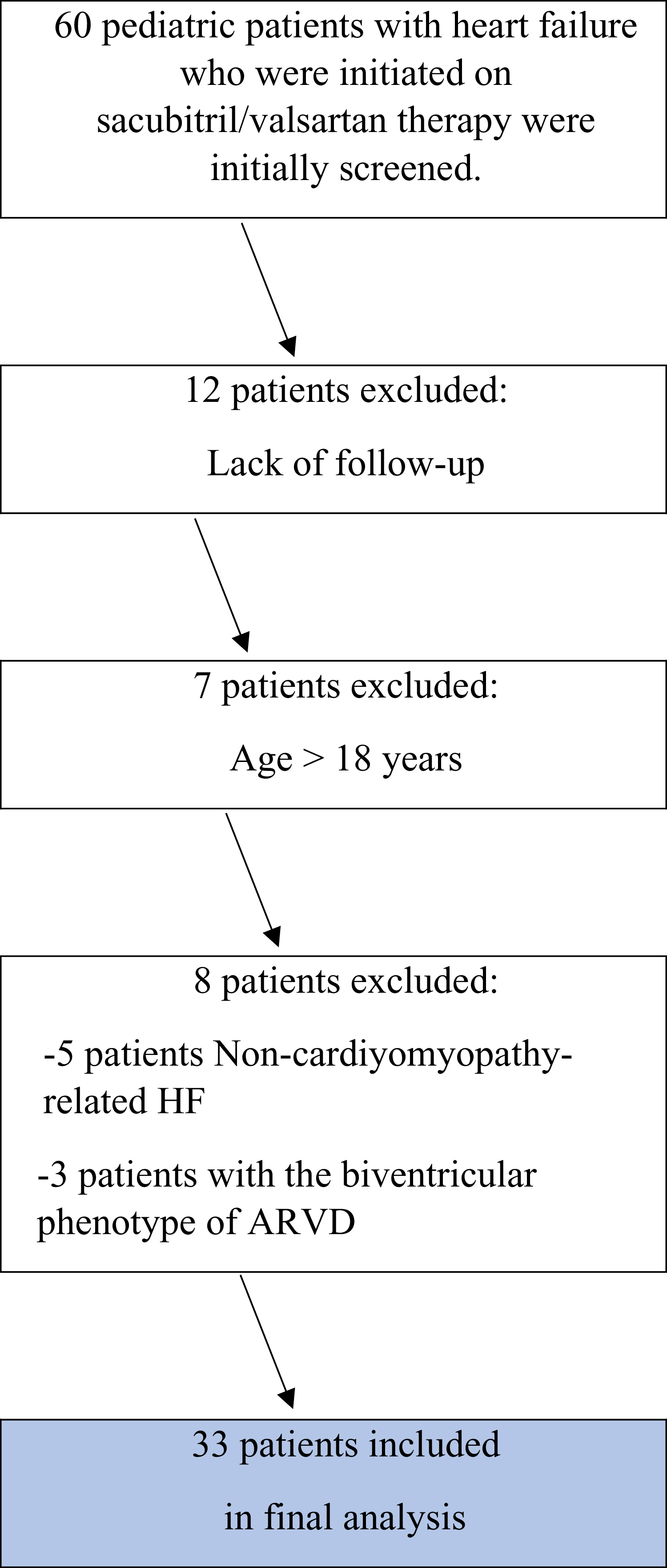
The institutional database was screened to identify paediatric patients diagnosed with cardiomyopathy and treated with sacubitril/valsartan between October 2020 and October 2024. A total of 60 patients initiated on sacubitril/valsartan for heart failure were initially reviewed.
Patients were excluded if they met any of the following criteria:
-
1. Age <1 month or >18 years at the time of treatment initiation.
-
2. Follow-up duration <6 months after starting therapy.
-
3. Incomplete clinical, echocardiographic, or laboratory data at baseline or final assessment.
-
4. Missed ≥2 scheduled follow-up visits or loss to follow-up.
-
5. Heart failure unrelated to cardiomyopathy.
-
6. Presence of biventricular involvement in arrhythmogenic right ventricular cardiomyopathy.
The exclusion of patients with biventricular arrhythmogenic right ventricular cardiomyopathy was performed to ensure a clear distinction between right- and left-ventricular phenotypes, as the primary objective of the study was to compare therapeutic responses based on predominant ventricular dysfunction (Figure 1).
Flowchart of patient selection for sacubitril/valsartan therapy in pediatric heart failure study.

Following the application of these exclusion criteria, a total of 33 patients were eligible and included in the final analysis.
Inclusion criteria
Patients were included in the study if they met all of the following criteria:
-
• Age between 1 month and 18 years at the initiation of sacubitril/valsartan.
-
• A confirmed diagnosis of dilated, arrhythmogenic right ventricular, or left ventricular non-compaction cardiomyopathy (dilated cardiomyopathy, arrhythmogenic right ventricular cardiomyopathy, or left ventricular non-compaction—as a morphological trait rather than a distinct entity) was established based on the 2023 ESC Guidelines for the Management of Cardiomyopathies. Reference Arbelo, Protonotarios and Gimeno7 In the arrhythmogenic right ventricular cardiomyopathy group, only patients with isolated right ventricular dysfunction were included, whereas those with biventricular involvement were excluded.
-
• Evidence of ventricular dysfunction (right or left) as defined by:
-
• Echocardiographic parameters—including left ventricular ejection fraction (left ventricular ejection fraction, <45%), tricuspid annular plane systolic excursion (tricuspid annular plane systolic excursion, <13 mm), right ventricular fractional area change (RVFAC, <35%), and abnormal left ventricular dimension z-scores (based on population normative data) Reference Lopez, Colan and Frommelt8 —were evaluated at baseline and follow-up.
-
• Functional status was assessed using NYHA functional classification for children aged ≥5 years and Ross classification for infants and younger children. A functional class of ≥ Class II was considered indicative of symptomatic heart failure.
-
• Laboratory data included N-terminal pro B-type natriuretic peptide levels, with values above 300 pg/mL considered elevated based on paediatric heart failure references. Reference Cantinotti9
-
-
• A minimum of 6 months of clinical follow-up with complete data at all key time points.
Baseline clinical and echocardiographic assessment
At each visit, blood pressure was measured by an experienced paediatric cardiology nurse, using standardised auscultatory methods, with the average of repeated measurements recorded.
Comprehensive transthoracic echocardiography was performed by a paediatric cardiology specialist, in accordance with the recommendations of the American Society of Echocardiography. Left ventricular ejection fraction was measured using the Simpson biplane method. When feasible, cardiac MRI was performed at baseline, at the 6-month follow-up, and at the final assessment to evaluate right ventricular ejection fraction using standard volumetric methods.
Sacubitril/valsartan administration and titration protocol
Sacubitril/valsartan was initiated during inpatient hospitalisation to ensure close monitoring of vital signs and laboratory parameters, including blood pressure, serum creatinine, and electrolytes. Therapy was initiated according to tolerability and clinical stability, with a target of gradual up-titration to the highest tolerated dose.
In alignment with paediatric protocols and prior studies (e.g. PANORAMA-heart failure), initial dosing was approximately 0.8 mg/kg/day (divided into two doses), with gradual titration up to a maximum of approximately 3.1 mg/kg/day as tolerated. Two available tablet formulations—24/26 mg and 49/51 mg—were used, administered either orally or via nasogastric tube in patients unable to swallow tablets.
Dosing adjustments were individualised based on patient response, blood pressure trends, and renal function. All patients were managed according to a standardised paediatric heart failure protocol implemented at our institution, which includes guideline-directed medical therapy as recommended in current paediatric and adult heart failure guidelines. This protocol emphasises the initiation and up-titration of core heart failure medications—including beta-blockers, renin—angiotensin system inhibitors or ARNIs, and mineralocorticoid receptor antagonists—at age-appropriate maximum tolerated doses, with close monitoring of hemodynamic status and organ function. Outpatient follow-up visits were scheduled at 1, 3, and 6 months, with continued monitoring thereafter. Reference Arbelo, Protonotarios and Gimeno7
Outcome measures
Primary outcome
A composite of:
-
1. All-cause mortality,
-
2. Heart transplantation or listing,
-
3. Unplanned heart failure–related rehospitalisation.
Secondary outcomes
-
• Longitudinal changes in echocardiographic parameters (left ventricular ejection fraction, tricuspid annular plane systolic excursion, fractional area change, right ventricular function, left ventricular dimension z-scores).
-
• Changes in N-terminal pro B-type natriuretic peptide levels.
-
• Changes in NYHA or Ross functional class.
-
• Six-minute walk test distance.
-
• Safety outcomes, including hypotension, renal dysfunction, and hyperkalemia.
Statistical analysis
Descriptive statistics were presented as counts and percentages for categorical variables, and as mean ± standard error for continuous variables. Normality of continuous variables was assessed by skewness and kurtosis coefficients as well as examination of histograms. Since N-terminal pro B-type natriuretic peptide values were not normally distributed, square root transformation was applied for analyses. However, for ease of interpretation, tables include the original N-terminal pro B-type natriuretic peptide means and standard errors.
Longitudinal changes were analysed using repeated measures analysis of variance (repeated measures ANOVA), and mixed-model ANOVA was employed to evaluate the interaction between group and time. The sphericity assumption was tested in repeated measures ANOVA, and Greenhouse-Geisser correction was applied when this assumption was violated. Time-dependent changes in categorical variables were analysed using the McNemar test.
All statistical analyses were performed using IBM SPSS Statistics for Windows, Version 22.0 (IBM Corp., Armonk, NY, USA). A p-Value <0.05 was considered statistically significant.
Ethical approval
The study was approved by the Institutional Ethics Committee of Istanbul Mehmet Akif Ersoy Thoracic and Cardiovascular Surgery Training and Research Hospital (Protocol No: 2025.08-85; Approval Date: 23.09.2025). Owing to the retrospective design of the study, the requirement for informed consent was waived.
Results
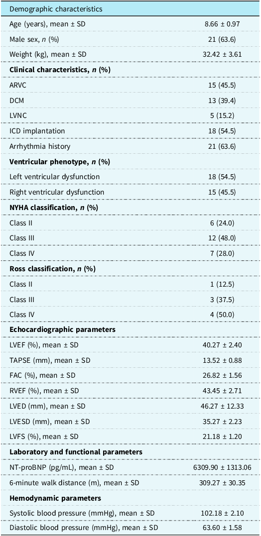
A total of 33 patients were included in the study, with a mean age of 8.66 ± 0.97 years, and 21 were male (63.6%). According to the underlying diagnosis, 15 patients (45.4%) had arrhythmogenic right ventricular cardiomyopathy, 13 (39.4%) had dilated cardiomyopathy, and 5 (15.2%) had left ventricular non-compaction. While arrhythmogenic right ventricular cardiomyopathy is less commonly observed as the predominant diagnosis in paediatric heart failure compared to dilated cardiomyopathy, in our cohort, the higher representation of arrhythmogenic right ventricular cardiomyopathy may reflect a tendency to initiate sacubitril/valsartan in patients presenting with more advanced clinical symptoms despite its lower overall prevalence. This likely reflects a selection bias inherent to the retrospective design. Based on predominant ventricular involvement, 18 patients (54.5%) had left ventricular involvement (dilated cardiomyopathy or left ventricular non-compaction), and 15 (45.5%) had right ventricular involvement.
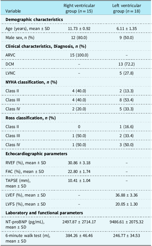
In patients with right heart failure, the mean tricuspid annular plane systolic excursion was 10.41 mm (SD ± 1.04), fractional area change was 22.80% (SD ± 1.74), right ventricular ejection fraction was 30.86% (SD ± 3.18), and the six-minute walk distance was 384.26 m (SD ± 46.46). In patients with right heart failure, the mean N-terminal pro B-type natriuretic peptide level was 2,498 pg/mL (SD ± 2,714), reflecting substantial variability within the arrhythmogenic right ventricular cardiomyopathy cohort. In patients with left heart failure, the mean left ventricular ejection fraction was 36.88% (SD ± 3.36), FS was 20.05% (SD ± 1.30), N-terminal pro B-type natriuretic peptide level was 9,486.61 pg/mL (SD ± 2,075.32), and the six-minute walk distance was 246.77 m (SD ± 34.53). The demographic and baseline clinical characteristics of the study population are summarised in Tables 1 and 2.
Demographic and baseline clinical profiles of the study population (n = 33)

ARVC = arrhythmogenic right ventricular cardiomyopathy; DCM = dilated cardiomyopathy; FAC = fractional area change; ICD = implantable cardioverter defibrillator; LVED = left ventricular end-diastolic; LVESDs = left ventricular end-systolic diameter; LVEF = left ventricular ejection fraction; LVFS = left ventricular fractional shortening; LVNC = left ventricular non-compaction cardiomyopathy; NT-proBNP = N-terminal pro B-type natriuretic peptide; RVEF = right ventricular ejection fraction; TAPSE = tricuspid annular plane systolic excursion; SE = standard error.
Descriptive baseline clinical characteristics of patients diagnosed with right heart disease and left heart disease

Table 2. Long description
The table compares demographic and clinical characteristics of patients with right and left ventricular groups. It has 15 rows and 4 columns. The columns are labeled ‘Variable’, ‘Right ventricular group (n = 15)’, and ‘Left ventricular group (n = 18)’. The rows are grouped into sections: Demographic characteristics, Clinical characteristics, Diagnosis, NYHA classification, Ross classification, Echocardiographic parameters, and Laboratory and functional parameters. Row 1: Variable, Right ventricular group (n = 15), Left ventricular group (n = 18). Row 2: Demographic characteristics,, . Row 3: Age (years), mean ± SD, 11.73 ± 0.92, 6.11 ± 1.35. Row 4: Male sex, n (%), 12 (80.0), 9 (50.0). Row 5: Clinical characteristics, Diagnosis, n (%),, . Row 6: ARVC, 15 (100.0), -. Row 7: DCM, -, 13 (72.2). Row 8: LVNC, -, 5 (27.8). Row 9: NYHA classification, n (%),, . Row 10: Class II, 4 (40.0), 2 (13.3). Row 11: Class III, 4 (40.0), 8 (53.4). Row 12: Class IV, 2 (20.0), 5 (33.3). Row 13: Ross classification, n (%),, . Row 14: Class II, 0, 1 (16.6). Row 15: Class III, 1 (50.0), 2 (33.4). Row 16: Class IV, 1 (50.0), 3 (50.0). Row 17: Echocardiographic parameters,, . Row 18: RVEF (%), mean ± SD, 30.86 ± 3.18, -. Row 19: FAC (%), mean ± SD, 22.80 ± 1.74, -. Row 20: TAPSE (mm), mean ± SD, 10.41 ± 1.04, -. Row 21: LVEF (%), mean ± SD, -, 36.88 ± 3.36. Row 22: LVFS (%), mean ± SD, -, 20.05 ± 1.30. Row 23: Laboratory and functional parameters,, . Row 24: NT-proBNP (pg/mL), mean ± SD, 2497.87 ± 2714.17, 9486.61 ± 2075.32. Row 25: 6-minute walk test (m), mean ± SD, 384.26 ± 46.46, 246.77 ± 34.53.
Values are presented as mean ± SD or n (%).
ARVC = arrhythmogenic right ventricular cardiomyopathy; DCM = dilated cardiomyopathy; FAC = fractional area change; LVEF = left ventricular ejection fraction; LVFS = left ventricular fractional shortening; LVNC = left ventricular non-compaction cardiomyopathy; NT-proBNP = N-terminal pro B-type natriuretic peptide; RVEF = right ventricular ejection fraction; TAPSE = tricuspid annular plane systolic excursion; SD = standard deviation.
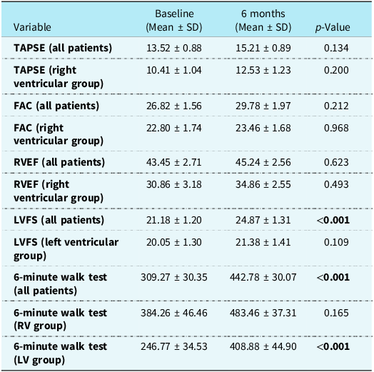
The participants’ tricuspid annular plane systolic excursion, fractional area change, right ventricular ejection fraction, LVFS, N-terminal pro B-type natriuretic peptide levels, and six-minute walk test results were evaluated at five follow-up points: baseline, 1st month, 3rd month, 6th month, and final assessment.
No statistically significant changes over time were observed in tricuspid annular plane systolic excursion, fractional area change, and right ventricular ejection fraction parameters in the overall patient group (p = 0.134; p = 0.212; p = 0.623, respectively). Similarly, no significant differences were found in these parameters among patients with right heart disease (tricuspid annular plane systolic excursion p = 0.200; fractional area change p = 0.968; right ventricular ejection fraction p = 0.493).
Left ventricular fractional shortening (LVFS) showed a significant increase in the overall patient group (p < 0.001). Post- hoc analyses revealed that baseline values were significantly lower compared to the 3rd month (p = 0.033), 6th month (p = 0.009), and final assessment (p = 0.011). However, the increase in LVFS was not statistically significant in patients with left heart disease (p = 0.109).
The six-minute walk test distances demonstrated a significant increase over time in the entire cohort, with post hoc analyses indicating significant differences between baseline and 6th month (p < 0.001) and between baseline and final assessment (p < 0.001). While patients with left heart disease showed a significant increase in walking distance, the increase in patients with right heart disease was not statistically significant (p = 0.165). A progressive improvement trend was observed across follow-up visits, with the most pronounced changes at 6 months and sustained at final follow-up. These findings are presented in detail in Table 3.
Changes in echocardiographic and functional parameters from baseline to 6 months

Values are presented as mean ± SD.
p-Values were derived from repeated measures analysis.
FAC = fractional area change; LVFS = left ventricular fractional shortening; RVEF = right ventricular ejection fraction; TAPSE = tricuspid annular plane systolic excursion; SD = standard deviation.
*Repeated measures one-way ANOVA.
The boldface indicates the significance of the p-value.
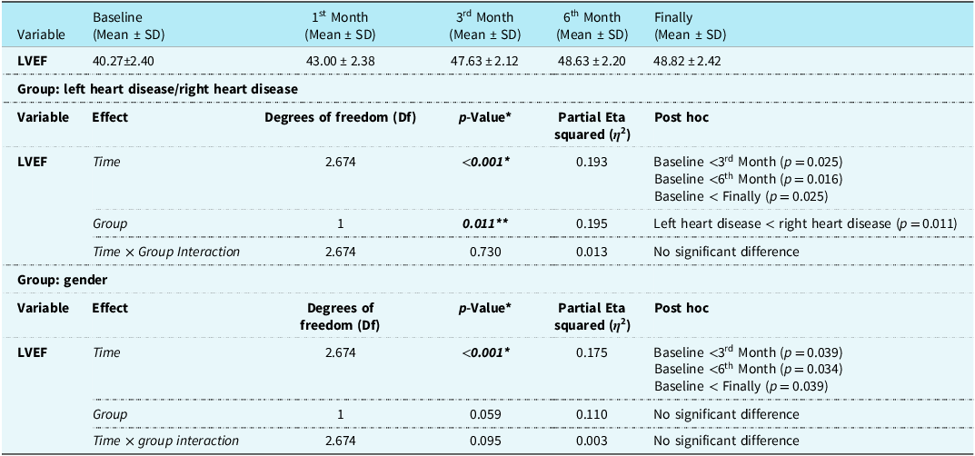
Changes in left ventricular ejection fraction from baseline to the final follow-up, as well as differences across diagnostic and sex-based subgroups, were examined (Table 4).
Group comparisons and longitudinal changes for left ventricular ejection fraction

LVEF = Left ventricular ejection fraction.
*Repeated measures mixed ANOVA.
Over time, left ventricular ejection fraction values showed a statistically significant increase in the overall cohort (p < 0.001). Post hoc analyses indicated that baseline values were significantly lower than those at the 3rd month (p = 0.025), 6th month (p = 0.016), and final follow-up (p = 0.025).
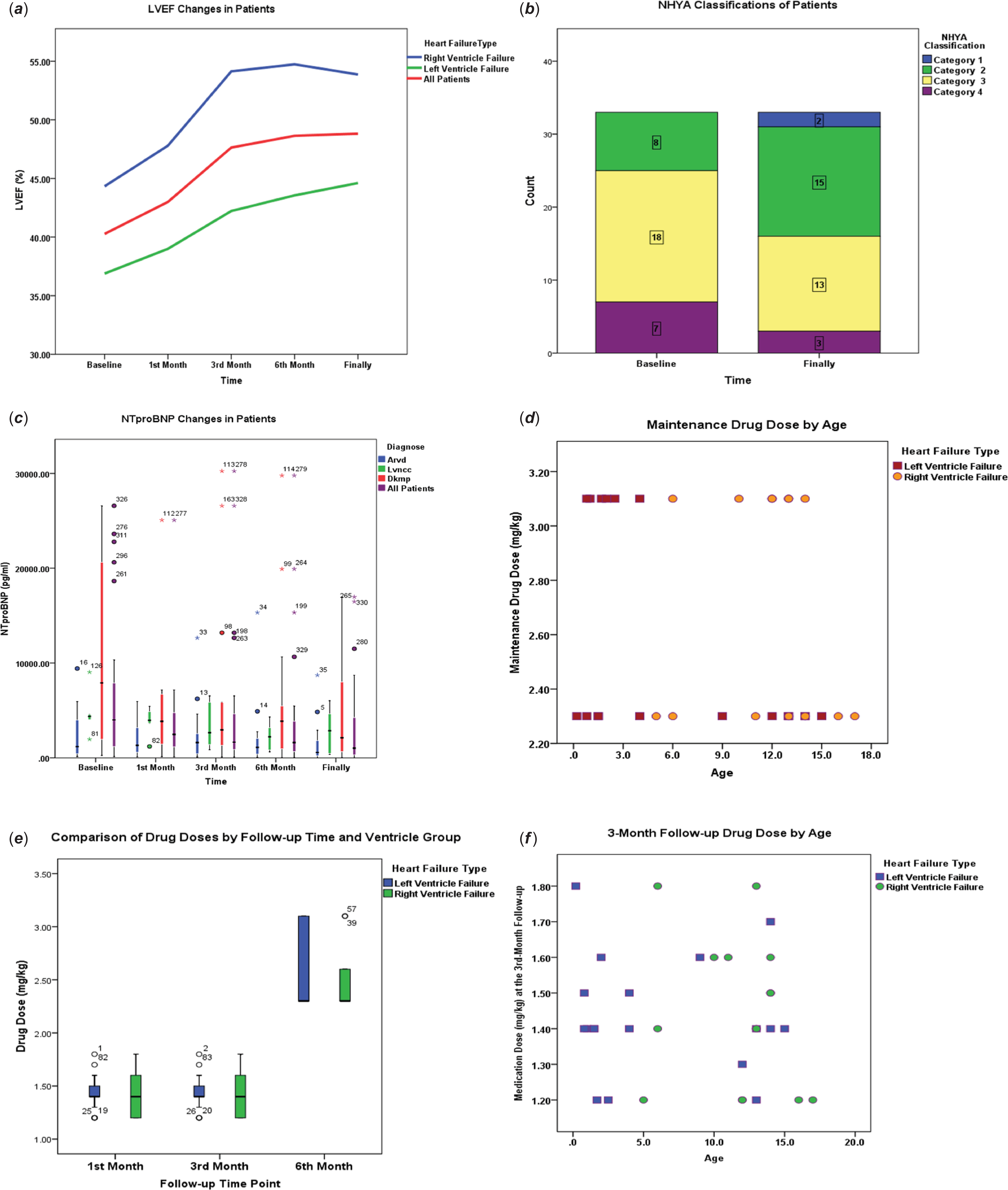
At baseline, patients with left heart disease had significantly lower left ventricular ejection fraction values compared to those with right heart disease (p = 0.011). However, the interaction between time and group was not statistically significant (p = 0.730), indicating a similar pattern of left ventricular ejection fraction increase over time in both groups (Figure 2).
Mean left ventricular ejection fraction (LVEF, %) values over time, stratified by type of heart failure ( a ). Distribution of patients according to NYHA functional classification at baseline and at the final follow-up ( b ). Distribution of NT-proBNP levels at five different time points (Baseline, 1st month, 3rd month, 6th month, and final) for diagnostic subgroups (arrhythmogenic right ventricular cardiomyopathy, left ventricular non-compaction cardiomyopathy, DCM) and the total patient cohort. Boxplots display the median, interquartile range (IQR), and overall distribution. Outliers are shown as individual points with corresponding patient identifiers( c ). Distribution of maintenance drug dose (mg/kg) by patient age, stratified by type of heart failure. Each point represents an individual patient, with groups classified as left or right ventricular failure ( d ). Boxplots showing the distribution of drug dose (mg/kg) at the 1st, 3rd, and 6th months in patients with left and right ventricular failure. Boxes represent the interquartile range (IQR), and the central line indicates the median value. Outliers are shown as individual points ( e ). Scatterplot showing the distribution of follow-up drug dose (mg/kg) at the 3rd month by patient age, stratified by heart failure type. Each point represents an individual patient ( f ).

Figure 2. Long description
Panel A: A line graph shows LVEF (%) changes in patients over time, stratified by type of heart failure. The x-axis represents time points (Baseline, 1st Month, 3rd Month, 6th Month, Finally), and the y-axis represents LVEF (%). Three lines represent different heart failure types: Right Ventricle Failure, Left Ventricle Failure, and All Patients. Panel B: A stacked bar chart displays the distribution of patients according to NYHA functional classification at baseline and at the final follow-up. The x-axis represents time points (Baseline, Finally), and the y-axis represents the count of patients. Different colors represent different NYHA categories (Category 1, Category 2, Category 3, Category 4). Panel C: A scatter plot shows NTproBNP levels at five different time points (Baseline, 1st month, 3rd month, 6th month, and final) for diagnostic subgroups (Arrhythmogenic right ventricular cardiomyopathy, Left ventricular non-compaction cardiomyopathy, DCM) and the total patient cohort. The x-axis represents time points, and the y-axis represents NTproBNP (pg/ml). Different colors and symbols represent different diagnostic subgroups. Panel D: A scatter plot displays the distribution of maintenance drug dose (mg/kg) by patient age, stratified by type of heart failure. The x-axis represents age, and the y-axis represents maintenance drug dose (mg/kg). Different colors represent different heart failure types (Left Ventricle Failure, Right Ventricle Failure). Panel E: Box plots show the distribution of drug dose (mg/kg) at the 1st, 3rd, and 6th months in patients with left and right ventricular failure. The x-axis represents follow-up time points (1st Month, 3rd Month, 6th Month), and the y-axis represents drug dose (mg/kg). Different colors represent different heart failure types (Left Ventricle Failure, Right Ventricle Failure). Panel F: A scatter plot shows the distribution of follow-up drug dose (mg/kg) at the 3rd month by patient age, stratified by heart failure type. The x-axis represents age, and the y-axis represents medication dose (mg/kg). Different colors represent different heart failure types (Left Ventricle Failure, Right Ventricle Failure).
In sex-based comparisons, a significant increase in left ventricular ejection fraction was observed over time in both male and female patients (p < 0.001). No statistically significant difference was detected in left ventricular ejection fraction values between sexes throughout the study period (p = 0.059), and the time × sex interaction was also not significant (p = 0.095), suggesting a parallel trend of left ventricular ejection fraction improvement across sexes.
These findings indicate a significant improvement in left ventricular ejection fraction during follow-up, regardless of sex or heart failure subtype, although patients with left heart involvement exhibited lower left ventricular ejection fraction values at baseline.
Hypotension was observed in six patients (18.2%); however, all episodes were transient and manageable, and no patient required treatment discontinuation. During the follow-up period 14 patients (42.4%) experienced heart failure–related rehospitalisation. Additionally, three patients died and two required heart transplantation, reflecting the severity of underlying disease in this cohort.
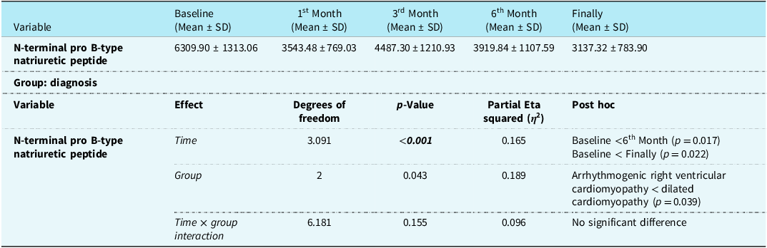
Longitudinal changes in N-terminal pro B-type natriuretic peptide levels were evaluated across all patients and compared between diagnostic groups (Table 5). A significant decrease in N-terminal pro B-type natriuretic peptide levels over time was observed in the overall cohort (p < 0.001). Post hoc analyses revealed that baseline N-terminal pro B-type natriuretic peptide levels were significantly higher than those at the 6th month (p = 0.017) and at final assessment (p = 0.022) (Figure 2).
Group comparisons and longitudinal changes in N-terminal pro B-type natriuretic peptide levels among all patients

At baseline, N-terminal pro B-type natriuretic peptide levels were significantly lower in the arrhythmogenic right ventricular cardiomyopathy group compared with the dilated cardiomyopathy group (p = 0.039). However, no significant time × group interaction was found (p = 0.155).
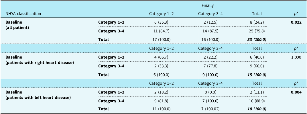
In this study, the distribution of NYHA functional classification at baseline and at the final assessment was evaluated. In the overall patient group, the proportion of patients classified as NYHA class I–II increased from 24.2% at baseline to 48.5% at the final assessment, which was found to be statistically significant (p = 0.022). Among patients with right heart disease, the proportion of those in NYHA class I–II was 40.0% at baseline and 44.4% at the final assessment; this change was not statistically significant (p = 1.000). In contrast, among patients with left heart disease, the proportion of patients in NYHA class I–II increased from 11.1% at baseline to 44.4% at the final assessment, and this improvement was statistically significant (p = 0.004). These findings are present in detail in Table 6 and Figure 2.
Distribution of NYHA functional classification at baseline and final assessment

* McNemar’s test.
Discussion
The introduction of sacubitril/valsartan has marked an important advancement in the management of paediatric heart failure, with regulatory approval extending its use to younger patient populations. Despite this regulatory milestone, real-world evidence supporting the use of sacubitril/valsartan in paediatric cardiomyopathy remains limited. Small observational studies and preliminary trials have suggested potential clinical and hemodynamic benefits; however, robust subgroup analyses across distinct cardiomyopathy phenotypes are lacking. In particular, comparative data on treatment response in patients with predominant left versus right ventricular dysfunction remain scarce, despite the potential influence of ventricular phenotype on therapeutic effectiveness and long-term outcomes. Accordingly, this study aimed to evaluate the clinical efficacy and tolerability of sacubitril/valsartan in paediatric patients with cardiomyopathy, with particular emphasis on phenotype-specific differences in treatment response. This study provides real-world evidence on the use of sacubitril/valsartan in a heterogeneous cohort of paediatric patients with cardiomyopathy and represents one of the few analyses to directly compare treatment response between children with predominant left versus right ventricular dysfunction. The key findings of this investigation are: (1) sacubitril/valsartan was generally well tolerated across all age groups, with no cases requiring permanent drug discontinuation; (2) significant improvements were observed in left ventricular systolic function, N-terminal pro B-type natriuretic peptide levels, and functional capacity; (3) patients with right ventricular dysfunction demonstrated parallel but non-significant trends toward improvement; and (4) clinically meaningful outcomes, including NYHA class improvement and enhanced exercise tolerance, were achieved in a substantial proportion of patients. Taken together, these findings suggest that sacubitril/valsartan may offer clinical benefits across diverse paediatric cardiomyopathy subtypes, although the magnitude of improvement varies according to ventricular phenotype.
In adults with heart failure with reduced ejection fraction, sacubitril/valsartan has consistently demonstrated superiority over conventional renin–angiotensin system inhibition, leading to improved survival, reduced hospitalisations, and reverse remodelling. Reference McMurray, Packer and Desai2,Reference Arbelo, Protonotarios and Gimeno7,Reference Lopez, Colan and Frommelt8 Paediatric data, however, remain limited. Early experience from the PANORAMA-heart failure trial and subsequent observational studies has supported the safety and feasibility of sacubitril/valsartan in children with systemic left ventricular dysfunction. Reference McMurray, Packer and Desai2,Reference Bruns, Slaverio-Witt, Pahl, Huh and Auerbach13,Reference Tan, Fang, Gong, Chen and Wu14 Our findings align with this emerging evidence, demonstrating significant increases in left ventricular ejection fraction and substantial reductions in N-terminal pro B-type natriuretic peptide. Improvements in LV dimensions and systolic indices further support the drug’s beneficial effects on reverse remodelling in paediatric dilated cardiomyopathy and left ventricular non-compaction.
A unique contribution of this study is the ventricular-stratified analysis, which revealed notable differences in treatment response between patients with left and right ventricular involvement. Children with left ventricular dysfunction exhibited clear improvements in left ventricular ejection fraction, NYHA class, and exercise capacity, consistent with adult and paediatric angiotensin receptor–neprilysin inhibitor data. In contrast, while modest increases in tricuspid annular plane systolic excursion, fractional area change, and right ventricular ejection fraction were observed in the right ventricular dysfunction group, these changes did not reach statistical significance. This divergence likely reflects the distinct pathophysiology of arrhythmogenic right ventricular cardiomyopathy, characterised by fibrofatty myocardial replacement, arrhythmogenic substrate formation, and impaired electromechanical coupling. As such, the extent of functional reversibility achievable through neurohormonal modulation may be inherently limited. Nevertheless, the observed trends toward improvement in right ventricular parameters suggest a potential benefit that warrants investigation in larger, phenotype-specific cohorts.
Functional improvement was further supported by parallel changes in NYHA and Ross classifications. NYHA class improvement was evident in older children capable of symptom-based assessment. Additionally, the Ross classification was used in eight younger patients who did not meet the developmental threshold for NYHA evaluation. Improvements in Ross class mirrored those observed in NYHA, indicating that sacubitril/valsartan provided consistent functional benefit across different paediatric age groups. The concordance between Ross and NYHA trajectories reinforces the robustness of the functional improvement observed and highlights the importance of age-appropriate functional assessment tools in paediatric heart failure.
The significant decline in N-terminal pro B-type natriuretic peptide across the entire cohort underscores the hemodynamic benefits of sacubitril/valsartan. As a biomarker closely correlated with ventricular wall stress and clinical outcomes, reductions in N-terminal pro B-type natriuretic peptide reflect improvements in global cardiac loading conditions. Reference Chen, Wu and Zhu12,Reference Docherty, Vaduganathan, Solomon and McMurray15–Reference Volpe17 The dual mechanism of action—enhancing natriuretic peptide activity while suppressing RAAS—likely contributed to symptomatic improvement and enhanced exercise capacity, as reflected in six-minute walk test results. Improvements were particularly prominent in left ventricular dysfunction, but even numerically positive changes in the right ventricular group suggest systemic benefit.
Safety and tolerability are essential considerations in paediatric heart failure management. In our cohort, hypotension occurred in 18.2% of patients but was manageable with dose adjustments, and importantly, no child required drug discontinuation. All patients—including those who later died—successfully achieved target doses during titration. These findings are notable, as previous paediatric studies have reported hypotension as a limiting factor in angiotensin receptor–neprilysin inhibitor up-titration. Reference McMurray, Packer and Desai2,Reference Bruns, Slaverio-Witt, Pahl, Huh and Auerbach13,Reference Tan, Fang, Gong, Chen and Wu14 Achieving target doses in every patient in this cohort demonstrates both the medication’s tolerability and the effectiveness of our standardised titration protocols.
Despite these benefits, a proportion of patients experienced adverse clinical outcomes, including rehospitalisation (42.4%), death (n = 3), and heart transplantation or listing (n = 2). These findings are not unexpected given the severity of disease in paediatric cardiomyopathy populations. The Pediatric Cardiomyopathy Registry has shown that approximately 40% of symptomatic children with dilated cardiomyopathy reach an endpoint of death or transplantation within two years. Reference Docherty, Vaduganathan, Solomon and McMurray15 Thus, while sacubitril/valsartan may slow disease progression or improve functional status, some patients may still require timely escalation to mechanical circulatory support or transplantation.
Although formal anthropometric measurements such as height-for-age and weight-for-age z-scores were not systematically analysed, clinicians observed that many children demonstrated visible improvements in appetite, weight gain, and overall growth trajectory during follow-up. These clinical impressions suggest that hemodynamic stabilisation with sacubitril/valsartan may contribute to improved nutritional status and daily functional well-being, consistent with prior paediatric heart failure experience. However, these observations remain descriptive and require confirmation in future studies incorporating standardised growth assessments.
This study has several strengths, including ventricular phenotype-based subgroup analysis, cardiac MRI-supported right ventricular evaluation in eligible patients, achievement of target angiotensin receptor–neprilysin inhibitor doses in all participants, and comprehensive longitudinal assessment of functional, biochemical, and echocardiographic parameters. Nonetheless, certain limitations must be acknowledged. The retrospective, single-centre design introduces selection bias and limits generalisability. The sample size was relatively small—particularly in the right ventricular subgroup—reducing statistical power for subgroup analyses. Cardiac MRI could not be performed in all patients, leading to reliance on echocardiographic right ventricular parameters in some cases. Additionally, heterogeneity in age, underlying diagnosis, and duration of prior therapy may have influenced treatment response.
Future studies should incorporate multicentre prospective designs with larger sample sizes and standardised imaging and functional assessment protocols. Particular emphasis should be placed on right ventricular cardiomyopathies, including arrhythmogenic right ventricular cardiomyopathy, to better characterise sacubitril/valsartan’s therapeutic role in this underserved population. Studies examining biomarkers, myocardial tissue characteristics, and genetic predictors of treatment response may also aid in developing personalised therapeutic strategies for paediatric heart failure.
Overall, our findings support the safety and potential efficacy of sacubitril/valsartan in paediatric cardiomyopathy and highlight the importance of individualised guideline-directed medical therapy optimisation. While treatment responses varied based on ventricular phenotype, consistent improvements across multiple domains underscore sacubitril/valsartan’s clinical utility in paediatric heart failure management.
Conclusion
Sacubitril/valsartan therapy resulted in improvements in functional, biochemical, and echocardiographic parameters in paediatric cardiomyopathy, with the magnitude of benefit varying by ventricular phenotype. These findings underscore the importance of individualised treatment strategies and support the need for larger, multicentre prospective studies to clarify its role in diverse paediatric populations.