Transcatheter closure of muscular ventricular septal defects is considered a first-line therapy when anatomically appropriate, while closure of perimembranous ventricular septal defects have historically been considered to be best addressed surgically given the potential complications of complete atrioventricular block or aortic valve regurgitation.Reference Warnes, Williams and Bashore1, Reference Baumgartner and Taylor2 Advancements in device design, however, have allowed for significant improvements in deployment techniques and overall safety.Reference Tanidir, Baspinar, Saygi, Kervancioglu, Guzeltas and Odemis3 Here we describe the first use of the Lifetech Konar-MF™ (multi-functional) occluder (Lifetech, Shenzhen, China) in North America, and our specific use of this device to close complex post-operative muscular and a perimembranous ventricular septal defects in the same patient.
Case description
The patient is a 15 kg, 4-year-old female born full term with trisomy 21 and associated issues of hypotonia, obstructive sleep apnea, hypothyroidism, and global developmental delay. She was diagnosed with a balanced Rastelli Type C atrioventricular canal defect. She underwent modified single-patch repair of her defect at 6 months of age with post-operative echocardiogram demonstrating moderate-severe left atrioventricular valve regurgitation (mean inflow gradient 4 mmHg) and mild right atrioventricular valve regurgitation (mean inflow gradient 3 mmHg). There was also a small to moderate residual ventricular septal defect and normal biventricular systolic function with right ventricle hypertrophy.
Over the course of the following 3.5 years, the patient was followed regularly by pediatric cardiology with follow-up echocardiograms demonstrating post-operative ventricular septal defect in the perimembranous location and a muscular ventricular septal defect, persistent moderate-severe left atrioventricular valve regurgitation and left atrial enlargement. She had normal left ventricular systolic function and diastolic indices. She was continued on enalapril, but clinically was becoming more symptomatic with increasing work of breathing and reduced exercise tolerance.
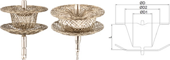
At 4 years old, the patient underwent diagnostic cardiac catheterisation demonstrating normal end diastolic pressures of both right and left ventricle (6 mmHg). Her Qp was calculated at 5.46 L/min/m2 by Fick with mean pulmonary pressure of 18 mmHg and pulmonary vascular resistance of 1.83 indexed Woods units. Significant shunting was noted through two separate ventricular septal defects with a Qp:Qs of 1.63:1. Two ventricular septal defects were defined by angiography and transesophageal echocardiography: (1) muscular defect in the inlet septum 5 mm below the level of the atrioventricular valves with an left orifice measuring 8 mm and right ventricular opening of 4 mm; (2) long tubular-shaped perimembranous defect 4.5 mm below the aortic valve with left ventricular orifice of 4.5 mm and right ventricular opening of 3.5 mm. Due to significant shunt, associated left chamber enlargement and severe left atrioventricular valve regurgitation, a multidisciplinary decision was made to pursue a cardiac catheterisation procedure to close the residual ventricular septal defects. We anticipated that reducing the left-to-right shunt would allow for reversal of left chamber enlargement and subsequent improvement in left atrioventrcicular valve regurgitation without need for repeat cardiac surgery. A Lifetech Konar-MF™ occluder ventricular septal defect device (Fig 1) was chosen to close the defects due to the complex position and shape of the defects.
Lifetech MF™ Occluder Device.

To acquire compassionate use FDA approval, documents were prepared (cover letter, prescription letter explaining the need to manufacturer, physicians curriculum vitae, IRB consent document, independent letter for case review, device documents and a letter from manufacturer that they can provide the device) and then submitted to the FDA for review.
-
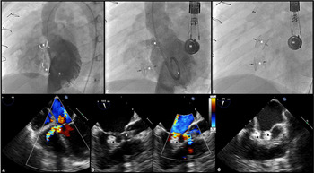
(1) Closure of muscular ventricular septal defect: The defect was crossed with a JR catheter over an 0.035” wire in retrograde fashion and then exchanged for a long 5 French delivery sheath. A 6/4 mm Lifetech Konar-MF™ Occluder was chosen, prepared in the usual fashion, was then advanced through the sheath and retrograde across the defect. Under transesophageal echocardiography guidance the right ventricular disc was deployed followed by the left ventricular disc. Further selective angiography and transesophageal echocardiography images were used to ensure adequate device position with a disc on either side of the interventricular septum and no interference with adjacent structures including free movement of right atrioventricular valve leaflets (Fig 2). The rhythm was constantly analysed and there was no change in the QRS waveform and no signs of conduction delays. Once positioning was felt to be adequate, the device was released. Repeat transesophageal echocardiography images showed a well-positioned device with mild increase in right atrioventricular valve regurgitation.
Figure 2.Angiographic and Transesophageal imaging during case: (1) LV angiography (60° LAO, 20° Cranial) using pigtail catheter demonstrating post-operative perimembranous VSD with RV orifice of 3.2 mm (a) and LV orifice of 4.3 mm (b), additional muscular VSD with RV orifice of 3.1 mm (c) and LV orifice of 5.1 mm (d). (2) LV angiography after placement of device in muscular VSD showing shunt through perimembranous VSD. (3) Fluoroscopy showing placement of both closure devices in muscular and perimembranous VSDs.
(x) post-operative residual perimembranous VSD. (star) muscular VSD. (ii) pigtail catheter in LV chamber. (iii) transesophageal echocardiography probe.

-
(2) Closure of post-surgical perimembranous ventricular septal defect: The defect was crossed in retrograde fashion with a 0.014” BMW wire which was snared from the venous side in order to create a rail. This was exchanged for a glide catheter to safely cross the defect. A 5/3 mm Lifetech Konar-MF™ occluder was then prepared and deployed in a similar retrograde manner – first deploying the right ventricular disc and then the left ventricular disc while using transesophageal echocardiography guidance and selective angiography to ensure adequate device position with a disc on either side of the interventricular septum and no conduction/rhythm issues or interference with adjacent structures including free movement of right atrioventrcicular valve leaflets. (Fig 2) Once positioning was felt to be adequate, the device was released. Repeat transesophageal echocardiography images showed a well-positioned device and improved right atrioventrcicular valve regurgitation.

At follow-up at 1 and 6 months post-procedure, the patients symptoms had improved with no concerns of fatigue or decreased energy. Echo showed no shunting through the devices with a trivial to small residual shunt through a defect just lateral to post-surgical peri-membranous device. Left atrial and ventricular measurements, and left atrioventrcicular valve regurgitation were similar to previous.
Discussion
The Lifetech Konar-MF™ occluder is a self-expandable, double-disc device made from 144 individually woven nitinol threads, and discs linked together by an articulated and expanding cone-shaped waist. (Fig 1) The flexible waist is designed to reduce the risk of heart block and can be adjusted to a variety of ventricular septal defects. Its design of a microscrew on each surface allows for placement from either the left ventricle or the right ventricle (antegrade or retrograde) through a 4–7 French sheath. Soft woven mesh provides high conformability with septal defects making it particularly well suited for residual postoperative defects. Safe and effective use of this device has been demonstrated for closure of perimembranous and muscular ventricular septal defects as well as coronary artery fistula.Reference Tanidir, Baspinar, Saygi, Kervancioglu, Guzeltas and Odemis3–Reference Egorova, Ewert, Hadamitzky and Eicken6
The device was first implanted in 2013 and since this time has been implanted in over 2000 patients throughout Asia and Europe. Compared to other devices that have been designed and used for catheter closure of ventricular septal defect the Konar MF™ is considered ideal, as it has a low profile, making implantation more successful (>97%) as well as safer with low risk of major complications (only 1 reported case of congenital heart block).Reference Tanidir, Baspinar, Saygi, Kervancioglu, Guzeltas and Odemis3, Reference Schubert, Kelm, Koneti and Berger4 This device, like all devices when first used, becomes easier to implant with less complications after experience with the device. Embolisations were seen early on in studies but after experience was gained device embolisation has not been documented.
The group from Istanbul in Turkey published a sizing guideline for ventricular septal defectsReference Tanidir, Baspinar, Saygi, Kervancioglu, Guzeltas and Odemis3 that suggests muscular defects you should add 2 mm to the defect size for the right ventricular disc size, and for perimembranous -ventricular septal defect you add 2 mm to the defect size for the right ventricular disc unless the aneurysm won’t accommodate the size of the device or the aortic valve may be compromised in which case you only add 1 mm to the defect size. This method was used in our patient and provided for appropriately sized devices with no evidence of aortic or tricuspid valve involvement.
After surgical closure of ventricular septal defects residual defects can persist. These defects can be haemodynamically significant and cause symptoms to the patient. It is often more difficult to close these defects than native defects. In our patient she had one defect that was muscular and one defect that was described as perimembranous-ventricular septal defect that was a residual post operation defect. This case shows that with the Konar-MF™ occluder it is not only possible to close residual ventricular septal defects but to do so easily and safely in the catheterisation lab. In this patient, closure significantly reduced the left to right shunt. The previously reported safety of this device, with only one reported case of heart block, helped us to choose it for use in the post-surgical perimembranous defect. We also believed the anterior location of this defect limited this risk for heart block as the conduction system in complete atrioventricular canal defects run posteriorly.
A great aspect of this device is its ability to be deployed from either antegrade or retrograde approach. One complication that has been described over the years with transcatheter ventricular septal defect closure is damage to the tricuspid valve. By allowing the delivery cable to be attached to the left ventricular disc, a rail is no longer needed for deployment. The wire can be placed from the left ventricle into the right ventricle or pulmonary artery and the delivery sheath advanced over the wire into the right ventricle. The right ventricular disc is then deployed and with it in place on the right ventricular septal wall, the tricuspid valve can be analysed. If there is no evidence of regurgitation, then the device can be deployed safely knowing that the tricuspid valve is not involved. This has reduced the major complications with this device.
Conclusion
The Konar MF™ Occluder device is a safe and effective device for the closure or perimembranous-ventricular septal defects, Muscular ventricular septal defects and post-operative residual ventricular septal defects.
Acknowledgements
Transesophageal imaging was obtained by Dr. Natasha Gonzalez-Estevez and Dr. Bijoy Thattaliyath. FDA approval and additional logistic support for the case facilitated by Jennifer Maldonado.