Introduction
Patients with substance use disorders are often characterized by high impulsivity, with impaired inhibitory control and impulsive decision making (Goldstein & Volkow, Reference Goldstein and Volkow2002; Baler & Volkow, Reference Baler and Volkow2006; Perry & Carroll, Reference Perry and Carroll2008; van Holst & Schilt, Reference van Holst and Schilt2011). Dual-process models of addictive behaviors posit the importance of sensitized reward processes in combination with impaired inhibitory control (Jentsch & Taylor, Reference Jentsch and Taylor1999; Robinson & Berridge, Reference Robinson and Berridge2003; Lubman et al. Reference Lubman, Yucel and Pantelis2004; Vandermeeren & Hebbrecht, Reference Vandermeeren and Hebbrecht2012), together leading to a vicious cycle of craving, bingeing, intoxication and withdrawal (Goldstein & Volkow, Reference Goldstein and Volkow2002), which in turn contributes to the chronic character of substance use. However, findings on the role of inhibitory control in alcohol dependence (AD) are inconsistent (Kirby & Petry, Reference Kirby and Petry2004; Lipszyc & Schachar, Reference Lipszyc and Schachar2010), with some studies reporting impaired (Lawrence et al. Reference Lawrence, Luty, Bogdan, Sahakian and Clark2009; Noel et al. Reference Noel, Van der Linden, Brevers, Campanella, Verbanck, Hanak, Kornreich and Verbruggen2012) and others unimpaired inhibitory control (Li et al. Reference Li, Luo, Yan, Bergquist and Sinha2009; de Ruiter et al. Reference de Ruiter, Oosterlaan, Veltman, van den Brink and Goudriaan2012; Schmaal et al. Reference Schmaal, Joos, Koeleman, Veltman, van den Brink and Goudriaan2012). Therefore, the exact nature of the relationship between inhibition and AD, and especially its neural correlates, needs further study.
These inconsistent findings with regard to inhibitory control in AD could be due to the heterogeneous character of AD. Different typologies have been proposed to define more homogeneous subgroups (e.g. Cloninger et al. Reference Cloninger, Bohman and Sigvardsson1981; Babor et al. Reference Babor, Hofmann, DelBoca, Hesselbrock, Meyer, Dolinsky and Rounsaville1992; Dom et al. Reference Dom, D'haene, Hulstijn and Sabbe2006), including typologies based on the presence/absence of co-morbid psychiatric disorders. This is important because psychiatric co-morbidity may complicate the presentation and the course of AD, especially when it comes to impulsivity and behavioral inhibition, and its neural correlates. For example, AD is highly co-morbid with depression and/or anxiety disorders (de Graaf et al. Reference de Graaf, Bijl, Smit, Vollebergh and Spijker2002; Boschloo et al. Reference Boschloo, Vogelzangs, Smit, van den Brink, Veltman, Beekman and Penninx2011). However, most behavioral and neuroimaging studies in AD either exclude AD patients with co-morbid depressive and/or anxiety disorders, thus obtaining a clinically not very relevant sample with low ecological validity, or fail to consider the presence of co-morbid depression and/or anxiety as a potential confounder of the (lack of) observed associations between AD and impulsivity or behavioral inhibition. This research bias fails to address a key issue because depression and anxiety are considered ‘internalizing’ disorders, which are presumed to be associated with decreased impulsivity, whereas substance use disorders such as AD are presumed to belong to an ‘externalizing’ dimension (Achenbach, Reference Achenbach1966; Krueger, Reference Krueger1999). As a result of these high co-morbidity rates between AD and internalizing psychopathology, AD patients with co-morbid mood disorders may resemble patients with mood disorders only, and therefore may be less impulsive than patients with other substance use disorders.
However, studies on response inhibition and related impulsivity assessments in depression or anxiety disorders are scarce, and have moreover shown heterogeneous results. For example, some studies have found impaired, rather than enhanced, inhibition in patients with mood and anxiety disorders in response to affective stimuli (Gray & McNaughton, Reference Gray and McNaughton2000; Lau et al. Reference Lau, Christensen, Hawley, Gemar and Segal2007; Ngo et al. Reference Ngo, Street and Hulse2011), which may underlie rumination and failed regulation of negative mood states (Joormann et al. Reference Joormann, Dkane and Gotlib2006; Joormann & Gotlib, Reference Joormann and Gotlib2010). In addition, motor impulsivity has been related to depression (Peluso et al. Reference Peluso, Hatch, Glahn, Monkul, Sanches, Najt, Bowden, Barratt and Soares2007; Hur & Kim, Reference Hur and Kim2009) whereas co-morbid anxiety has been shown to increase impulsivity in patients with major depressive disorder (MDD; Bellani et al. Reference Bellani, Hatch, Nicoletti, Ertola, Zunta-Soares, Swann, Brambilla and Soares2012), indicating that depression/anxiety may also be associated with increased impulsivity and related inhibition impairments, which is therefore at odds with decreased impulsivity intuitively associated with internalizing psychopathology. It should be noted, however, that these studies were performed with either affective paradigms, assessing inhibition in a negative emotional context, or with the Barratt Impulsiveness Scale Version 11 (BIS-11; Patton et al. Reference Patton, Stanford and Barratt1995), a subjective impulsivity questionnaire. Moreover, two studies in AD patients showing that depression (Jakubczyk et al. Reference Jakubczyk, Klimkiewicz, Topolewska-Wochowska, Serafin, Sadowska-Mazuryk, Pupek-Pyziol, Brower and Wojnar2012) and anxiety (Karch et al. Reference Karch, Jager, Karamatskos, Graz, Stammel, Flatz, Lutz, Holtschmidt-Taschner, Genius, Leicht, Pogarell, Born, Moller, Hegerl, Reiser, Soyka and Mulert2008) symptom severity was associated with increased impulsivity excluded AD patients with an established co-morbid depression/anxiety DSM-IV diagnosis. Thus, it remains unclear how depression/anxiety is related to (non-affective) response inhibition, and especially whether AD patients with co-morbid depression/anxiety disorders resemble patients with depression/anxiety only.
Therefore, in the current study, we compared a large sample of AD patients with co-morbid depression/anxiety disorders with a group of depression/anxiety only (D/A) patients and a healthy control (HC) group on response inhibition during functional magnetic resonance imaging (fMRI). Response inhibition, as one of the core features of impulsivity, can be defined as the ability to inhibit a prepotent response (Evenden, Reference Evenden1999) and was measured with the Stop Signal Task (SST; Logan, Reference Logan, Dagenbach and Carr1994). A ‘hyperdirect’ cortico-subthalamic-pallidal pathway (Nambu et al. Reference Nambu, Tokuno and Takada2002) is thought to underlie response inhibition during the SST. This pathway projects from motor and premotor areas such as the pre-supplementary motor area (pre-SMA; Lambert et al. Reference Lambert, Zrinzo, Nagy, Lutti, Hariz, Foltynie, Draganski, Ashburner and Frackowiak2012) and the inferior frontal gyrus (IFG) via the subthalamic nucleus and striatum to the globus pallidus and back to the motor cortex via the thalamus. Neuroimaging studies using the SST in healthy subjects have shown involvement of the basal ganglia, the thalamic area, the IFG/insula and the medial/lateral dorsal prefrontal cortex including motor areas such as the (pre-)SMA during response inhibition (Liddle et al. Reference Liddle, Kiehl and Smith2001; Rubia et al. Reference Rubia, Smith, Brammer and Taylor2003; Aron & Poldrack, Reference Aron and Poldrack2006; Li et al. Reference Li, Huang, Constable and Sinha2006, Reference Li, Yan, Sinha and Lee2008b ; Zandbelt & Vink, Reference Zandbelt and Vink2010; Cai & Leung, Reference Cai and Leung2011). Therefore, we focused on these brain regions in the current study.
To the best of our knowledge, this is the first study to directly compare (non-affective) response inhibition and its neural correlates between an ecologically valid group of AD patients with co-morbid depression/anxiety and a group of patients with depression/anxiety only. We aimed to distinguish AD-associated inhibition deficits from inhibition deficits associated with depression/anxiety, and to delineate the neural substrate of response inhibition in these groups. We therefore tested the following hypotheses: (1) patients with AD show increased impulsivity and altered brain activity of response inhibition-related regions compared to HCs and D/A patients; and (2) impairments in SST performance and brain activations are positively associated with AD duration and severity, but negatively with depression/anxiety severity.
Method
Participants
Forty-two AD patients were included with a current (<6 months) DSM-IV diagnosis of AD and no co-morbid lifetime Axis I diagnosis other than major depressive disorder (MDD) or anxiety disorder (generalized anxiety disorder and/or panic disorder and/or social anxiety disorder). In addition, a comparison group of 22 patients with a current (<1 month) DSM-IV diagnosis of MDD and/or anxiety disorder (D/A patients) but no other lifetime diagnosis of Axis I disorders was included. Finally, a group of 20 HCs, without any lifetime Axis I disorders, was included.
To study a sample of AD subjects with a wide variety of disease duration and severity, AD patients were recruited from two sources. One half of the group was recruited from local addiction treatment clinics and the other AD patients were recruited from the Netherlands Study for Depression and Anxiety (NESDA; Penninx et al. Reference Penninx, Beekman, Smit, Zitman, Nolen, Spinhoven, Cuijpers, de Jong, van Marwijk, Assendelft, van der Meer, Verhaak, Wensing, de Graaf, Hoogendijk, Ormel and van Dyck2008), a large cohort study with patients from primary care and out-patient mental health services. The comparison group of D/A patients and the HC subjects were also recruited through the NESDA.
The VU University Medical Center Ethical Review Board approved this study, and written informed consent according to the Declaration of Helsinki was obtained from all participants prior to the study.
Procedure and clinical assessments
Participants were screened for current and lifetime psychiatric diagnoses using the Composite International Diagnostic Interview (CIDI; Robins et al. Reference Robins, Wing, Wittchen, Helzer, Babor, Burke, Farmer, Jablenski, Pickens, Regier, Sartorius and Towle1988). All participants were free of major internal or neurological disorders and MRI contraindications, did not use psychotropic medication other than the stable use of selective serotonin reuptake inhibitors (SSRIs) or infrequent benzodiazepine use, and were free of current substance abuse or dependence (other than alcohol abuse or dependence in the AD group, and/or smoking). On the day of scanning, a urine test was performed to check for unreported recent use of benzodiazepines and drugs of abuse. Exclusions based on this check are reported in the Results section. All participants were asked to abstain from alcohol at least 24 h and from caffeine a few hours prior to the study.
During the scanning day, participants had a confirmed breath alcohol level of 0.0% (Alcoscan Daisy-AL7000; Sentech, Korea). No participants scored more than 8 points on the revised Clinical Institute Withdrawal Assessment for Alcohol scale (CIWA-Ar; Sullivan et al. Reference Sullivan, Sykora, Schneiderman, Naranjo and Sellers1989) and were therefore considered withdrawal free.
Self-report questionnaires were filled out for assessment of demographic and clinical characteristics. Handedness was measured by the Hand Preference Questionnaire (van Strien, Reference van Strien1992). Addiction severity was assessed by the Alcohol Use Disorder Identification Test (AUDIT) for alcohol use disorders (Babor et al. Reference Babor, Kranzler and Lauerman1989), and the Fagerstrom questionnaire (Heatherton et al. Reference Heatherton, Kozlowski, Frecker and Fagerstrom1991) for cigarette use. Self-reported rates of impulsivity were assessed with the BIS-11 (Patton et al. Reference Patton, Stanford and Barratt1995), yielding a total impulsivity score and subscale scores for attentional, motor and non-planning impulsivity. Anxiety severity was assessed by the Beck Anxiety Inventory (BAI; Beck et al. Reference Beck, Epstein, Brown and Steer1988) and the Spielberger State/Trait Anxiety Inventory (STAI; Spielberger et al. Reference Spielberger, Gorsuch and Lushene1970), and depression severity by the Inventory of Depressive Symptomatology (IDS; Rush et al. Reference Rush, Gullion, Basco, Jarrett and Trivedi1996). Disease durations in years were calculated by subtracting the year of (alcohol use/depression/anxiety) disorder onset from the most recent year disorder symptoms contributed to a DSM-IV diagnosis, both as obtained by the CIDI (Robins et al. Reference Robins, Wing, Wittchen, Helzer, Babor, Burke, Farmer, Jablenski, Pickens, Regier, Sartorius and Towle1988).
Task paradigm
Participants performed an SST (Heslenfeld & Oosterlaan, Reference Heslenfeld and Oosterlaan2003) while fMRI images were obtained. For a detailed description of the task see the online supplementary material (text and Supplementary Figs S1 and S2). In brief, during the SST, participants had to respond as rapidly and accurately as possible by pressing a button with their left or right index finger in response to an airplane (Go stimulus) facing either to the left or to the right. Occasionally, a Stop signal, a white cross, was superimposed over the Go stimulus after a small delay, prompting an attempt to inhibit the (already initiated) Go response. The difficulty of stopping varied by adjusting the interval between the onset of the Go stimulus and the Stop signal, the stop signal delay (SSD), using a staircase tracking algorithm (de Jong et al. Reference de Jong, Coles, Logan and Gratton1990), which approached a critical SSD representing the delay required for participants to successfully stop their response on approximately 50% of the stop trials. The time required for the Stop signal to be successfully processed, the stop signal reaction time (SSRT), was calculated by subtracting the SSD from the mean Go reaction time. A longer SSRT indicates poorer response inhibition and thus higher impulsivity.
Imaging data acquisition and preprocessing
MRI scanning was performed at the Academic Medical Centre (AMC) in Amsterdam using a 3-T Philips Intera full-body MR system (Philips Medical Systems, The Netherlands) with a phased array SENSE RF eight-channel head coil. Functional blood oxygen level-dependent (BOLD) signals were acquired sequentially with a T2*-weighted, gradient echo planar imaging (EPI) sequence and an estimated time-course series of approximately 370 volumes (self-paced) per session [repetition time (TR) = 2300 ms, echo time (TE) = 20 ms, matrix size = 96 × 95, voxel size = 2.29 × 2.29 × 2.50 mm, 45 slices]. Imaging parameters were optimized according to the methods of Deichmann et al. (Reference Deichmann, Gottfried, Hutton and Turner2003): to minimize susceptibility and distortion artifacts in the orbitofrontal cortex, each volume was scanned with an orientation of 30° from the anterior commissure–posterior commissure (AC-PC) line. Three-dimensional T1-weighted images were obtained using a gradient echo sequence for anatomical reference with the EPI data (TR = 9 ms, TE = 3.6 ms, matrix size = 256 × 231, voxel size = 1 × 1×1 mm, 170 slices).
Imaging data were preprocessed and analyzed using SPM8 (Wellcome Trust Centre for Neuroimaging, London, UK). Images were manually reoriented to the AC-PC line, slice timed, realigned, warped to Montreal Neurological Institute (MNI) space, and smoothed using a Gaussian kernel of 8 mm at full-width at half-maximum (FWHM).
Statistical analyses
Sample characteristics and task performance data were analyzed using IBM SPSS Statistics 20 (IBM, USA). Group differences were examined using a one-way ANOVA design and non-parametric Kruskal–Wallis or χ 2 tests where necessary. In the case of significant group effects, additional analyses were conducted to evaluate pairwise differences between the three groups, controlling for Type I error across tests by using the Bonferroni approach. Correlation analyses between task performance parameters, substance use characteristics (AD duration and severity of alcohol and smoking), depression/anxiety severity and brain activity parameter estimates were performed using Spearman's ρ. The significance level for ANOVA and correlation analyses was set to p < 0.05.
Statistical analysis of individual imaging data was performed using a first-level fixed-effects analysis, in the context of the General Linear Model (Friston et al. Reference Friston, Holmes, Poline, Grasby, Williams, Frackowiak and Turner1995), with the onsets of the Go stimulus convolved with a canonical hemodynamic response function to model each outcome type. To remove low-frequency signal drift, a high-pass filter (128 Hz) was applied. Four main trial types were distinguished: Go success (Go), Stop success (SS), Stop error (SE), and Go trial omissions. For each individual subject, a contrast image SS>Go was constructed to examine regional brain activation specifically related to response inhibition. These contrast images were entered into a second-level random-effects analysis using a one-way ANOVA design to investigate between-group effects, and a two-samples t test with the two control groups pooled (D/A+HC) to further investigate activated areas specific to the AD group.
We focused on activated brain areas within regions of interest (ROIs) that have been implicated in the hyperdirect–indirect model of response inhibition (Nambu et al. Reference Nambu, Tokuno and Takada2002), including the IFG, a motor region including the primary motor cortex and the (pre-)SMA, the striatum covering the caudate and the nucleus lentiformis (the putamen and globus pallidus), and a thalamic region including the thalamus and subthalamic nucleus. Bilateral masks for the subcortical ROIs in the caudate and nucleus lentiformis and the thalamic region were derived from the automated anatomical labeling (AAL) atlas (Tzourio-Mazoyer et al. Reference Tzourio-Mazoyer, Landeau, Papathanassiou, Crivello, Etard, Delcroix, Mazoyer and Joliot2002) incorporated in the WFU PickAtlas Tool version 2.5.2 (Maldjian et al. Reference Maldjian, Laurienti, Kraft and Burdette2003). As a separate SMA mask is not provided in the WFU PickAtlas, bilateral cortical masks for the IFG and motor region were defined from probabilistic volumes of interest (Nielsen & Hansen, Reference Nielsen and Hansen2002) based on the BrainMap database (Fox & Lancaster, Reference Fox and Lancaster1994).
We report significant brain activations that survived family-wise error correction for multiple comparisons on the voxel-level within the ROIs using a small volume correction (p FWE-SVC) (Worsley et al. Reference Worsley, Marrett, Neelin, Vandal, Friston and Evans1996) or whole-brain correction for regions not of a priori interest (p FWE). To investigate whether activity in the identified regions was associated with performance parameters or clinical characteristics, we calculated the effect size of activity change within the identified regions during group comparisons for each subject using the MarsBaR toolbox (Brett et al. Reference Brett, Anton, Valabreque and Poline2002) and entered these in SPSS for correlation analyses.
Results
Sample characteristics
After data collection we excluded the following participants: (1) subjects with imaging data of poor quality (three AD, one D/A, two HC); (2) subjects showing poor task involvement with either extremely long reaction times (>3 s.d. from the group mean) or with more than 6% omissions (two AD, one D/A, two HC); and (3) subjects urine-testing positive for cocaine or benzodiazepines on the day of scanning (seven AD, two D/A, no HC). The final dataset included 31 AD, 18 D/A and 16 HC participants for task performance and imaging data analyses.
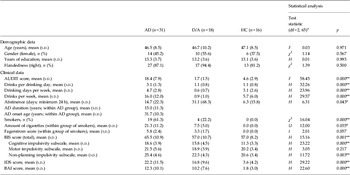
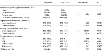
Table 1 presents detailed information on sample characteristics and Table 2 compares the two patient groups on depression/anxiety characteristics. All groups were matched for age, gender, years of education and handedness. Groups differed on alcohol use characteristics (F 2,62 = 58.445, p < 0.001) because, as expected, the AD group scored significantly higher than the HC and D/A groups (both p < 0.001) whereas the HC and D/A groups did not differ (p = 0.438). On the BIS-11 questionnaire, AD patients had higher total (F 2,62 = 15.16, p = 0.001), cognitive (F 2,62 = 23.22, p < 0.001) and non-planning (F 2,62 = 11.72, p = 0.003) impulsivity scores but did not differ from the D/A and HC groups on motor impulsivity (p = 0.217). Both AD and D/A patient groups had more depression symptoms than the HC group (U 1,47 = 21.5, p < 0.001 and U 1,34 = 25.5, p < 0.001 respectively) and also more anxiety symptoms (U 1,47 = 58.5, p < 0.001 and U 1,34 = 23.0, p < 0.001 respectively). The AD and D/A groups did not differ significantly on depression or anxiety severity (U 1,49 = 208, p = 0.140 and U 1,49 = 254.5, p = 0.611 respectively). The groups differed in the percentage of smokers (χ 2 2,65 = 19.225, p < 0.001) and number of cigarettes smoked (H 2,65 = 20.62, p < 0.001): none of the HC participants smoked whereas 19 of the 31 (61%) AD patients and four of the 18 (22%) D/A participants smoked. Post-hoc analyses with smoking as a covariate did not affect the results. The findings of these post-hoc analyses are described in the online supplementary material.
Demographic, clinical and task performance data for patients with alcohol dependence (AD), patients with depression/anxiety (D/A) and healthy controls (HC)

AUDIT, Alcohol Use Disorder Identification Test; BAI, Beck Anxiety Inventory; BIS, Barratt Impulsiveness Scale; df, degrees of freedom; F, Fisher test statistic for ANOVA; H, non-parametric Kruskal-Wallis one-way ANOVA; IDS, Inventory of Depressive Symptomatology; s.d., standard deviation; U, Mann–Whitney test statistic; χ2, chi-square distribution test statistic.
a df = 1, 23 for amount of cigarettes and total Fagerstrom score within smoker groups of AD and D/A; df = 2, 58 for abstinence period (non-drinking participants could not report a valid abstinence period); df = 2, 62 for the BIS-11 questionnaire.
* Significant effect p < 0.05. ** Significant effect p < 0.01.
Comparison of depression and anxiety characteristics between patients with alcohol dependence (AD) and patients with depression/anxiety (D/A)

BAI, Beck Anxiety Inventory; IDS, Inventory of Depressive Symptomatology; s.d., standard deviation; SSRI, selective serotonin reuptake inhibitor; t, Student's t test statistic; U, Mann–Whitney non-parametric test statistic; χ 2, chi-square distribution test statistic.
a Statistical tests performed between AD and D/A only, total n = 49. Except for: b n = 40, c n = 38, d n = 47.
Within the AD group, the mean age of AD onset was 31.68 years (s.d. = 10.31, range 16–51) and AD duration was 14.98 years (s.d. = 11.23, range 1–37). On average, the AD patients were abstinent for 2 weeks [range 24 h (required minimum) to 3 months]. Most AD patients had developed lifetime co-morbid depression/anxiety before the onset of AD (n = 27) and can thus be considered secondary AD patients. However, because of the subjective and retrospective character of the onset data obtained by a clinical interview, the reported temporal sequence should be considered with caution. Therefore, we did not take this information into account in our analyses.
Task performance
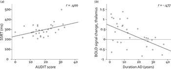
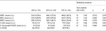
Groups did not differ on mean Go reaction time (H 2,65 = 3.093, p = 0.213), SSD (H 2,65 = 1.621, p = 0.445) or SSRT (H 2,65 = 0.542, p = 0.762) (Table 3). All groups approached 50% successful stop trials of all stop trials, without group differences, indicating the efficacy of the tracking algorithm, and therefore accounting for the degree of attentional monitoring. Within the AD group, SSRT was positively correlated with the severity of alcohol problems according to the AUDIT (r 29 = 0.400, p = 0.026) (see Fig. 1a ), and also with the number of cigarettes smoked (r 29 = 0.43, p = 0.017). The correlation between AUDIT and SSRT scores remained significantly positive after correction for smoking (r 29 = 0.379, p = 0.039). Depression (IDS) and anxiety severity (BAI) were not associated with SSRT scores over all groups or in each group separately.
Scatterplots of correlations within the alcohol dependence (AD) group. (a) Positive correlation between stop signal reaction time (SSRT) and alcohol problem severity (measured with the Alcohol Use Disorder Identification Test, AUDIT), indicating higher impulsivity with more severe alcohol use problems. (b) Negative correlation between blood oxygen level-dependent (BOLD) signal change in the thalamus during response inhibition and duration of AD, indicating decreased engagement of the thalamus with a longer drinking history.

Task performance data for patients with alcohol dependence (AD), patients with depression/anxiety (D/A) and healthy controls (HCs)

df, Degrees of freedom; H, Kruskal–Wallis test statistic for non-parametric one-way ANOVA; MRT, mean reaction time; s.d., standard deviation; SE, stop error; SS, stop success; SSD, stop signal delay; SSRT stop signal reaction time.
a
Effect size for effects between three or more groups: d = (
 $\bar x$
largest –
 $\bar x$
smallest)/√(mean standard error).
Imaging data
Main effect of inhibition
The main effect of inhibition (SS>Go) is reported in the online supplementary material (supplementary results, Table S1 and Fig. S3), largely replicating brain activation patterns associated with response inhibition in previous imaging studies (Liddle et al. Reference Liddle, Kiehl and Smith2001; Rubia et al. Reference Rubia, Smith, Brammer and Taylor2003; Aron & Poldrack, Reference Aron and Poldrack2006; Li et al. Reference Li, Huang, Constable and Sinha2006, Reference Li, Yan, Sinha and Lee2008b ; Zandbelt & Vink, Reference Zandbelt and Vink2010; Cai & Leung, Reference Cai and Leung2011).
Group comparisons
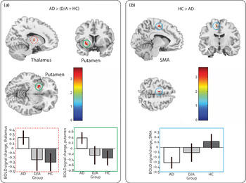
Compared to the D/A and HC groups, the AD group showed a significantly higher engagement of the ventral lateral nucleus in the right thalamus (Z = 3.58, p FWE-SVC < 0.05) and a borderline significant hyperactivation of the left putamen (Z = 3.57, p FWE-SVC = 0.06), which was significant with a unilateral mask (p FWE-SVC < 0.05) (see Fig. 2 a). In addition, the AD group showed hypoactivation of the left SMA (Z = 3.48) compared to the D/A and HC groups, but this difference was only significant between the AD and the HC groups (p FWE-SVC < 0.05) (see Fig. 2 b). No differences in brain activation during inhibition were seen between the D/A patients and HCs during ROI analyses. Whole-brain group comparisons did not yield results corrected for multiple comparisons.
Group differences during inhibition (SS>GO). (a) The thalamus and putamen were activated more in patients with alcohol dependence (AD) than in both depression/anxiety (D/A) patients and healthy controls (HCs); (b) the supplementary motor area (SMA) was activated more in HCs than in AD patients; D/A patients did not differ from the two other groups on SMA activation. Activated areas displayed at p < 0.005. Colored bars: Z values of activated voxels. Plots of parameter estimates per group; error bars: 90% confidence interval. BOLD, blood oxygen level-dependent.

Correlations between brain activations and performance/clinical characteristics
We extracted cluster parameter estimates from the putamen (peak MNI coordinates: x = –28, y = 8, z = –8), thalamus (peak MNI coordinates: x = 14, y = –9, z = 5) and SMA (peak MNI coordinates: x = –11, y = –11, z = 55). Correlation of these cluster parameter estimates with clinical characteristics showed that, within the AD group, activation in the right thalamus was negatively correlated with duration of alcohol dependence (r 29 = –0.477, p = 0.007). The SMA activation in HCs was negatively correlated with severity of depression symptoms (r 14 = –0.515, p = 0.041), whereas depression and anxiety severity were not correlated with regional brain activation in the two patient groups. Within the AD group, putamen and SMA activation were significantly inversely correlated (r 29 = –0.440, p = 0.013).
To test for the influence of smoking on the results obtained, a post-hoc analysis was performed with the number of cigarettes as a covariate in the ANOVA group comparisons for performance and imaging data. No influence of smoking was observed (data reported in the online supplementary material).
Discussion
In the current study there were no differences in response inhibition between AD patients, D/A patients and HCs based on the SST using the SSRT as the main performance parameter. However, alcohol problem severity (AUDIT) was positively associated with SSRT in AD patients, indicating that more severe AD was associated with more severe response inhibition impairments, and thus higher motor impulsivity, whereas the severity of depression and anxiety was not associated with behavioral impulsivity. These findings also indicate that depression/anxiety is not related to decreased (or increased) response inhibition and that depression/anxiety co-morbidity in AD patients is presumably not responsible for the inconsistent findings in the literature regarding response inhibition deficits in patients with AD. Of note, AD patients reported higher levels of subjective impulsivity compared with D/A patients and HCs on the cognitive and non-planning subscales of the BIS but not on the motor impulsivity subscale. The latter is probably the closest to the concept of impaired response inhibition measured by the SST, and the lack of differences between the three groups on BIS motor impulsivity is therefore in agreement with the observed absence of group differences on the SST.
Importantly, although we did not find clear behavioral group differences in our study, our imaging data were more sensitive to detecting impairments in the AD group. We showed increased engagement of the ventral lateral thalamus and putamen in AD patients compared with both D/A patients and HCs. In combination with similar SST performances in all three groups, this suggests the presence of either a compensatory mechanism or an alternative strategy of subcortical engagement in AD patients to achieve a similar level of performance compared with D/A patients and HCs. The thalamus and putamen are both part of the pathway involved in motor response and inhibition (Nambu et al. Reference Nambu, Tokuno and Takada2002; Aron, Reference Aron2011). The thalamus is an important node and output nucleus to the motor cortex in the ‘direct pathway’, associated with motor responsiveness, and also the ‘hyperdirect’ pathway, a fast circuit for global stopping of motor responses (Nambu et al. Reference Nambu, Tokuno and Takada2002; Aron, Reference Aron2011). The thalamus is thought to have a primarily excitatory connection through glutamate pathways to the motor cortex, and activation of the thalamus is therefore thought to initiate a motor response. However, the activity of the thalamus has also been associated with inhibition of motor responses (Li et al. 2008b), and increased activation of the ventral lateral nucleus of the thalamus was recently found to be associated with improved inhibitory control in AD patients treated with the cognitive enhancer modafinil compared with placebo (Schmaal et al. Reference Schmaal, Joos, Koeleman, Veltman, van den Brink and Goudriaan2012). Based on these findings, we propose that AD patients in the current study are still able to activate the thalamus to maintain their inhibition performance at a level comparable to the D/A patients and HCs, which AD patients with impaired SST performance may only achieve by administration of a cognitive enhancer (Schmaal et al. Reference Schmaal, Joos, Koeleman, Veltman, van den Brink and Goudriaan2012).
Increased activity in the putamen in the AD group is of particular interest, considering the involvement of the putamen in motor habits and habit formation in chronic addiction (Everitt & Robbins, Reference Everitt and Robbins2005; Everitt et al. Reference Everitt, Belin, Economidou, Pelloux, Dalley and Robbins2008; Tricomi et al. Reference Tricomi, Balleine and O'Doherty2009). Activation of the putamen in AD patients during a motor task that, over time, could be associated with more automated, habit-based motor behavior matches this addiction-related shift towards habit pathways in the brain. However, the area we found to be overactivated in this study is part of the anterior putamen (y = 8), whereas the posterior putamen (posterior to y = 0) is assumed to be mainly involved in habit formation (Tricomi et al. 2009). Moreover, we did not find an association between putamen activation with AD duration or severity. We therefore consider the interpretation of our findings in terms of increasing habit formation premature and in need of further study.
Within the AD group, activity in the thalamus was negatively correlated with duration of AD, implying that AD patients with a longer drinking history were less able to invoke this compensatory or alternative strategy during inhibition. It should be noted, however, that we found no direct association between AD duration or thalamus activity and SSRT score, and therefore these data do not provide direct evidence that decreased recruitment of the thalamus with a longer drinking history is related to response inhibition deficits. However, AD duration was, as might be expected, positively correlated with AD severity (AUDIT score), which in turn was correlated with SSRT in the AD group. Therefore, we suggest that a longer drinking history, associated with more severe alcohol use problems, underlies an increasing disability to compensate for inhibition impairments by recruitment of the thalamus, leading to higher impulsivity.
In addition to increased engagement by AD patients of subcortical areas, we found hypoactivity in AD patients in the SMA compared with HCs during the response inhibition task, which was negatively correlated with putamen activity within this group. Together, these findings suggest that cortical hypoactivity in AD patients is compensated for by subcortical hyperactivity during response inhibition leading to performance levels equal to comparison groups. Engagement of the SMA was lower in AD and D/A patients compared to HCs, but significant only between the AD patients and HCs. The lack of a difference between the AD and D/A groups in SMA activity may suggest that hypoactivity in the SMA during response inhibition is not specific to addiction-related disorders, but could also be associated with internalizing psychopathology and might thus be expressed with co-morbid depression/anxiety in an AD group as well. A possible association between decreased inhibition-related SMA activity and depression/anxiety is further indicated by the fact that, in the HC group, and hence within participants that do show engagement of the SMA, a negative correlation was seen between activity in this area and severity of depressive symptoms. However, it should be noted that a negative correlation between SMA activation and depression/anxiety symptoms in a healthy group has very little clinical relevance, and should therefore be considered with caution. Hypoactivity of frontal midline structures, including the (pre-)SMA and anterior cingulate cortex (ACC), during both successful and failed response inhibition has been fairly consistently reported in substance use disorders (Kaufman et al. Reference Kaufman, Ross, Stein and Garavan2003; Forman et al. Reference Forman, Dougherty, Casey, Siegle, Braver, Barch, Stenger, Wick-Hull, Pisarov and Lorensen2004; Hester & Garavan, Reference Hester and Garavan2004; Fu et al. Reference Fu, Bi, Zou, Wang, Ye, Ma, Ming and Yang 2008 ; Li et al. Reference Li, Huang, Yan, Bhagwagar, Milivojevic and Sinha2008a ; de Ruiter et al. Reference de Ruiter, Oosterlaan, Veltman, van den Brink and Goudriaan2012; Schmaal et al. Reference Schmaal, Joos, Koeleman, Veltman, van den Brink and Goudriaan2012). Additionally, decreased activation of these areas, especially the rostral ACC, has been observed in depression/anxiety during inhibition of neutral (Matthews et al. Reference Matthews, Simmons, Strigo, Gianaros, Yang and Paulus2009; Katz et al. Reference Katz, De Sanctis, Mahoney, Sehatpour, Murphy, Gomez-Ramirez, Alexopoulos and Foxe2010) and positive (Eugene et al. Reference Eugene, Joormann, Cooney, Atlas and Gotlib2010) stimuli, and during other neutral cognitive control paradigms such as error processing and emotion regulation paradigms (Etkin et al. Reference Etkin, Prater, Hoeft, Menon and Schatzberg2010; Diener et al. Reference Diener, Kuehner, Brusniak, Ubl, Wessa and Flor2012). Although increased activation of the ACC during response inhibition has also been reported in adolescent MDD (Yang et al. Reference Yang, Simmons, Matthews, Tapert, Frank, Bischoff-Grethe, Lansing, Wu, Brown and Paulus2009), and in late-life depression (Bobb et al. Reference Bobb, Adinoff, Laken, McClintock, Rubia, Huang, Husain, Mapes, Tamminga, Cullum, Gopinath, Trivedi and Kozel2012), the current findings provide additional evidence for decreased engagement of frontal midline areas in both substance use disorders and depression/anxiety during inhibitory control. This hypoactivation can therefore be thought of as a sign of general psychopathology, instead of a pattern seen in AD patients only.
The findings of this study should be considered in the light of some limitations. First, many of our patients were recruited from primary care, and many were not receiving any (current) treatment for AD and/or depression/anxiety, despite their established DSM-IV-TR diagnoses. Therefore, our sample could represent a less severe group of AD and/or D/A patients (see also Table 1), and this could (partly) explain the lack of behavioral differences and the ability of the AD patients to compensate for any (subclinical) inhibition impairments. Moreover, the data that were available on treatment showed considerable heterogeneity within and between groups with regard to treatment types (pharmacotherapy, psychotherapy, or both). Therefore, reliable group comparisons regarding treatment could not be made. Second, most AD patients in the current study developed AD after the onset of depression/anxiety symptoms and can therefore be considered to be secondary AD patients, which may (partly) explain why these patients were very similar to D/A patients in terms of their response inhibition. Unfortunately, we were unable to study the effect of the order of onset of AD in the current sample because there were only four patients with primary AD. To further elaborate on response inhibition in co-morbid AD, prospective data are necessary, looking at primary versus secondary AD. Third, we chose to use the SST, which is a neutral and objective assessment of response inhibition. However, it only measures one aspect of impulsivity, and the use of additional fMRI paradigms, such as a delay discounting task, measuring impulsive decision making, along with self-report questionnaires, may aid in covering a broader range of the impulsivity domain. As cognitive and neural processes underlying these different aspects of impulsivity show minimal overlap (Broos et al. Reference Broos, Schmaal, Wiskerke, Kostelijk, Lam, Stoop, Weierink, Ham, de Geus, Schoffelmeer, van den Brink, Veltman, de Vries, Pattij and Goudriaan2012), and even show heterogeneous impairments between various addictive and impulse control disorders (Lawrence et al. Reference Lawrence, Luty, Bogdan, Sahakian and Clark2009), future studies should further elucidate the specificity of impulsivity impairments in AD compared with other disorders using additional paradigms and impulsivity measurements.
The aim of the current study was to investigate AD patients with co-morbid mood disorders and to compare them with patients with mood disorders only, so as to test whether an ecologically valid group of AD patients resembles a group of D/A patients in terms of having a relatively low level of impulsivity. Intuitively, it might be desirable to include a third patient group, namely a group of AD patients without any lifetime internalizing psychopathology. However, pure AD patients are rare and including such a group in the present study was not considered feasible. Therefore, although we were able to distinguish AD-specific impairments in inhibitory control in the current study, inclusion of a pure AD group in future studies could further clarify the specificity of our findings.
To the best of our knowledge, the current study is the first to directly compare response inhibition in AD patients and D/A patients, that is a comparison of a supposedly externalizing group (with internalizing co-morbidity) with a purely internalizing group. We conclude that, on a performance level, AD patients with co-morbid D/A show a very similar pattern of response inhibition to a matched group of D/A patients and HCs. Therefore, response inhibition as measured by an SST does not seem to be impaired in (less severe) AD patients, and internalizing co-morbidity does not seem to be the explanation for this lack of response inhibition deficits in AD. However, at a neural level, AD patients seem to rely on an alternative strategy to maintain normal performance levels, thereby differing from D/A patients, and indicating the presence of an AD- or addiction-specific correlate of impaired response inhibition.
Supplementary material
For supplementary material accompanying this paper visit http://dx.doi.org/10.1017/S0033291713002274.
Acknowledgments
We thank the local treatment clinics Jellinek, De Maliebaan and the Home Clinic, along with the NESDA, for the recruitment of alcohol-dependent patients, MDD and anxiety patients and healthy controls. Funding for this study was provided by grant 31160004 from the Netherlands Organization for Health Research and Development (Zon-Mw), commissioned by the Netherlands Organization for Scientific Research (NWO); the NWO had no further role in the study design; in the collection, analysis and interpretation of data; in the writing of the report; or in the decision to submit the paper for publication.
Declaration of Interest
None.