At the end of 2019, the first cases of coronavirus disease 2019 have emerged from Wuhan, China, with rapid spread worldwide, it became a global pandemic. 1 Although COVID-19 was initially thought to be a disease characterised mainly by respiratory tract involvement, subsequent data have highlighted extrapulmonary manifestations. Reference Guan, Ni and Liang2 There is increased recognition of cardiac involvement in patients with COVID-19. The most common cardiac manifestations are myocardial infarction, myocarditis, arrhythmias, ventricular dysfunction, and heart failure. Reference Hendren, Grodin and Drazner3 Reports of cardiac tamponade secondary to COVID-19 infection in an otherwise healthy patient are rare in the literature.
Case report
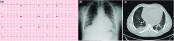
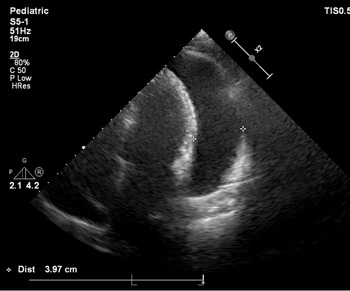
An otherwise healthy 17-year-old adolescent male was admitted to the emergency department with cough and low-grade fever for a few days. The patient’s past medical history was unremarkable. His presenting blood pressure was 111/57 mmHg, heart rate was 123 beats/minute, respiratory rate was 28/minute, body temperature was 37.1°C, and oxygen saturation on room air was 90%. Physical examination revealed distant heart sounds, diminished respiratory sounds, and rales at the lung bases. There was no finding consistent with haemorrhage, including petechiae, purpura, or haemoptysis. An electrocardiogram on admission showed normal sinus rhythm without significant pathological findings (Fig 1a). Chest X-ray demonstrated bilateral pulmonary infiltrates, right basal atelectasis with minimal pleural effusion, and cardiomegaly (Fig 1b). Chest CT showed bilateral peribronchial pulmonary infiltrates, peripheral ground-glass opacities, and pleural effusion predominantly on the right side was consistent with COVID-19 infection (Fig 1c). Transthoracic echocardiography revealed very large pericardial effusion (maximum, 39 mm) with signs of early right ventricular diastolic collapse and preserved biventricular cardiac functions (Fig 2, Supplementary videos 1 and 2). Laboratory tests included normal leukocyte count with low lymphocyte count (5590/µL versus 690/µL), haemoglobin level 11.1 g/dl, platelet count 352×109/L, blood urea nitrogen level 11 mg/dl. Inflammatory markers included elevated CRP (189 mg/dl) and ESR (31 mm/hour), as well as procalcitonin (0.12 ng/ml), and ferritin (410.9 ng/ml). Fibrinogen and D-dimer were 440 mg/dl and 1.81µg/ml, respectively. BNP was mildly elevated (166 ng/L), while high-sensitivity troponin T (6.86 ng/L) and CK-MB (0.62 ng/ml) were normal. Serological tests for hepatitis viruses, cytomegalovirus, human immune deficiency virus were negative. Serum autoantibody workup for rheumatological diseases was negative. Lower respiratory specimens were negative for influenza A and B, respiratory syncytial virus, human coronavirus (OC43, 229 E, and NL 63), metapneumovirus, parainfluenza virus, rhinovirus, Mycoplasma pneumonia, Chlamydia pneumonia, and bacteria. Although he had previously received a BCG vaccine, a tuberculin skin test was negative. Reverse transcription–polymerase chain reaction of a nasopharyngeal swab was positive for severe acute respiratory syndrome coronavirus 2.
(a) An electrocardiogram on admission showed normal sinus rhythm without significant pathological findings. (b) Chest X-ray showing an enlarged cardiac silhouette, bilateral pulmonary infiltrates, and pleural effusion on right basal lung fields. (c) Chest CT demonstrating pericardial effusion, bilateral peribronchial pulmonary infiltrates, peripheral ground-glass opacities, and bilateral pleural effusion predominantly on the right side consistent with COVID-19.

Apical four-chamber view on transthoracic echocardiography showing large circumferential pericardial effusion.

The patient was haemodynamically stable without inotropic support. We elected to proceed with pericardiocentesis to prevent evolving cardiac tamponade. The procedure was performed under conscious sedation without intubation in order to reduce the risk of COVID-19 contamination, and 1000 ml of haemorrhagic fluid were aspirated. Pericardial drainage was continued for 48 hours, yielding an extra 990 ml of fluid. The pericardial fluid analysis showed polymorphonuclear predominance, glucose of 74 mg/dl, lactate dehydrogenase of 974 U/L, and protein of 44.2 g/L. The fluid-to-serum protein ratio was 0.66, and the fluid-to-serum lactate dehydrogenase ratio was 3.37. Microbiological and cytological investigation for typical causes of haemorrhagic pericarditis revealed an exudative process with no evidence of malignancy, tuberculosis, or bacterial or fungal infection. For severe acute respiratory syndrome coronavirus 2, polymerase chain reaction of pericardial fluid was negative. In the absence of positive findings for other causative agents, we concluded the haemorrhagic effusion was secondary to COVID-19. Treatment with ibuprofen and hydroxychloroquine was added to the ongoing intravenous antibiotic regimen. Serial echocardiography demonstrated no recurrence of pericardial fluid. For the first few days of hospitalisation, the patient received supplemental oxygen via nasal cannula (4–6 L/minute) to maintain the target oxygen saturation of 90% was given. The patient improved without further decompensation, and he was discharged from the hospital 15 days after the first admission.
Discussion
Herein, we describe a previously healthy adolescent male who presented with mild fever, cough, and chest pain, and was diagnosed with COVID-19 infection. The most striking feature of his presentation finding was a very large haemorrhagic pericardial effusion, while his respiratory symptoms were relatively mild.
Children seem to be less severely affected by COVID-19 infection, with most manifesting asymptomatic. Most children appear to have an asymptomatic, mild, or moderate disease. They are more likely than adults to present with extra-respiratory symptoms. Reference Iguoro, Pilotto and Bonanni4 Recently, a presentation in children similar to incomplete Kawasaki disease or toxic shock syndrome has been described. Reference Feldstein, Rose and Horwitz5 The condition has been termed multisystem inflammatory syndrome in children. Some affected children appear to have coronary artery involvement accompanying severe cardiac dysfunction requiring inotropic support. Pericardial effusion has been reported in 26–32 % of these patients. Reference Feldstein, Rose and Horwitz5,Reference Matsubara, Kauffman and Wang6 The exact mechanism for multisystem involvement, and whether organ damage is mediated by ongoing viral replication in the affected tissues, a hyperinflammatory host response, or both, is a mystery for now.
Pericardial effusion leading to tamponade appears to be rare in COVID-19, with only a few cases in adults having been reported thus far. Reference Dabbagh, Aurora and D’Souza7,Reference Singh, Fuentes and Ellison8 To our knowledge, this is the youngest patient reported to have a large haemorrhagic pericardial effusion associated with COVID-19 infection.
Viral infections are the most common cause of pericarditis in children. Occasionally, such patients may present with cardiac tamponade. COVID-19 is known to provoke an inflammatory response that in some patients results in pericarditis and pericardial effusion; however, the exact mechanism is unclear. Negative high-sensitivity troponin and CK-MB were evidence against myocardial involvement. As there is limited number of reported cases with cardiac tamponade in COVID-19 patients, describing characteristics of pericardial fluid is important. In the literature, only two patients with haemorrhagic pericardial effusion were defined. Reference Dabbagh, Aurora and D’Souza7,Reference Singh, Fuentes and Ellison8 In our case, the appearance of pericardial fluid was obviously haemorrhagic without any distinguishing features from other etiologies. Haemorrhagic characteristics of the pericardial fluid might be related to an autoinflammatory response instead of a direct viral invasion of the cardiovascular system. Although definitive diagnosis requires molecular testing of pericardial fluid, usually, it is an exclusion diagnosis when no other causes of the pericardial effusion can be identified. Our patient’s pericardial fluid reverse transcriptase–polymerase chain reaction for SARS-CoV-2 was negative. Among reported cases, only one patient’s pericardial fluid was found positive for a single target gene of COVID-19. No amplification was detected for the other two SARS-CoV-2 genes in this patient, thus suggesting a low viral load. Reference Farina, Uccello and Spreafico9
Despite large pericardial effusion, our patient’s clinical condition was stable during follow-up. Since intubation and positive pressure ventilation may exaggerate the haemodynamic consequences of pericardial effusion, we preferred early pericardiocentesis. Also, non-invasive ventilation with high flow of oxygen from the beginning of the disease course may help to prevent further compromising of cardiac filling.
High-dose aspirin, non-steroid anti-inflammatory drugs, or colchicine are the mainstay of therapy for acute pericarditis. Nevertheless, the treatment protocol for COVID-19 related pericardial effusion has not been developed. Concerns have been raised that non-steroid anti-inflammatory drugs may be associated with an increased risk of adverse effects when used in patients with acute viral respiratory infections, including COVID-19. However, there have been no clinical or population-based data that directly address this risk. 10 We have not seen an adverse effect related to ibuprofen in our case.
Conclusion
This report highlights that large pericardial effusion without severe respiratory tract involvement might be seen in COVID-19 infection. Proper management with early pericardiocentesis can be life-saving.
Acknowledgements
None.
Financial support
This research received no specific grant from any funding agency, commercial, or not-for-profit sectors.
Conflicts of interest
None.
Ethical standards
All procedures performed in this case were in accordance with the ethical standards of the institutional and national research committee and with the 1975 Helsinki declaration and its later amendments or comparable ethical standards.
Supplementary material
To view supplementary material for this article, please visit https://doi.org/10.1017/S1047951120004850.