Introduction
The habitat of single-cell bacteria is very complex and widespread in nature. Intensive studies of the biological effect of nanomaterials have found that nanoparticles have an adverse effect on many bacteria (Zhao and Chai, Reference Zhao and Chai2005; Zhao et al., Reference Zhao, Zhao and Ye2005). Adapting to this harsh environment is a challenge to bacteria.
With the rapid development of materials science, nanotechnology is widely used in many different fields of scientific research and production. For example, on exposing a nanoparticle to bright light, it can accept strong optical electrons, resulting in excessive free radical toxicity, which produces a bactericidal action. There are many nanoparticles in the air and on the surface of plants, and it is necessary for microorganisms to overcome these negative effects in order to survive on the plant leaf surface. Since 2003, scientists have been discussing the effects of nanomaterials and nanotechnology on the environment, including the potential negative impact of nanomaterials on human health (Brumfiel, Reference Brumfiel2003; Service, Reference Service2003, Reference Service2004), the living environment (Oberdörster, Reference Oberdörster2004) and social security (Jia et al., Reference Jia, Wang and Zhao2005).
To date, most research on the biological effects of an environment containing nanoparticles has been conducted in animal experiments, which are preliminary in nature, such as toxicity analysis on DNA (Hirakawa et al., Reference Hirakawa, Mori, Yoshida, Oikawa and Kawanishi2004) and at a cellular level (Rahman et al., Reference Rahman, Lohani and Dopp2002). Research regarding the impact of nanoparticles on plants is rare. Nano-TiO2 P25 (Degussa Co.) is an international standard sample of bacteriostatic materials (Mills and Lee, Reference Mills and Lee2002). In this work P25 was chosen as the nanomaterial used to study the impact on phyllosphere bacteria, and a system was established to research the impact of nanoparticles on microorganisms on plant surfaces. The benefit of this research was not only to reveal the biological effects of an environment containing nanoparticles, but also to provide materials for the study of useful phyllosphere microbial populations and the screening of beneficial organisms.
Material and methods
Plant materials
The cucumber seed (Cucumis sativus cv. QIUPENG ERHAO) was used in this study (YUNONG Institute of Vegetable and Horticulture, Beijing, China). The trials were conducted in a greenhouse located at Shangzhuang Agricultural Experimental Station, China Agricultural University, Beijing, China. Plants in the early period of florescence were prepared for follow-up work.
Nanoparticles
AEROXIDE® TiO2 P25 (Degussa, Germany) was used for this research. It was treated with high-pressure sterilization and kept in the dark. TiO2 in phosphate buffered saline (PBS; 0.1 mol/l, pH 7.0) was sonicated for 30 min before the experiment.
Sample treatment
Leaves were sprayed with various concentrations of the TiO2 suspensions (0, 0.002, 0.02, 0.2, 2 and 20 mg/ml) using an artist's airbrush (HD-470, Yehong Air Tools Co., China). An airbrush was selected that did not wet the leaf surface in order to avoid spatial displacement of the microbes (Monier and Lindow, Reference Monier and Lindow2005). After being treated, the cucumber then grew in a greenhouse for 5 h, with irradiation of sunlight.
Recovery of phyllosphere bacteria
To collect leaf samples, every fourth ful expanded leaf (30 g per replication) was chosen randomly from the plant canopy, using sterile scissors to nip individual leaves into sterile plastic bags containing 50 ml washing buffer (0.1 M PBS, pH 7.0, containing 0.1% bacto-peptone (Difco Laboratories, Detroit)) and sonicated for 7 min in an ultrasonic cleaning bath to recover bacterial cells. Appropriate dilutions of leaf washings were plated on tryptic soy agar (TSA) containing trypton (15 mg/ml), soya peptone (5 mg/ml), sodium chloride (5 mg/ml) and agar (16 mg/ml) pH 7.0. The population sizes were estimated from plate counts made after 7 to 14 days of incubation at 30°C. The results were analysed using SAS 8.0 statistical software. All of the experiments were repeated in triplicate.
Genomic DNA extraction
Leaf washings were harvested by centrifugation for 15 min at 4°C and 18 000 rpm with a 50 ml centrifuge tube. The method of genomic DNA extraction was carried out as in Sambrook and Russel (Reference Sambrook and Russell2001).
PCR-DGGE analyses
The V3 region of 16S rDNA of the phyllosphere community was amplified by polymerase chain reaction (PCR) with primer PRBA338f and PRUN518r (Øvreås et al., Reference Øvreås, Forney, Daae and Torsvik1997). After amplification, a 236-bp product was obtained. Denaturing gradient gel electrophoresis (DGGE) was performed with a Dcode Universal Mutation Detection System (Bio-Rad, USA). PCR samples (25 μl) were loaded onto an 8% (wt/vol) polyacrylamide gel, 1.0-mm thick, with a linear chemical gradient ranging from 30 to 60% denaturant (100% denaturing solution contained 40% (vol/vol) formamide and 7 M urea). After running for 7 h at 130 V and 60°C in 1× Tris-acetate-EDTA (TAE) electrophoresis buffer, the gels were stained for 10–15 min with ethidium bromide and photographed with an AlphaImager (Alpha Innotech, USA).
Sequencing of DGGE fragments
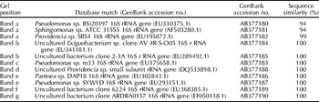
The dominant fragments of the V3 region of 16S rDNA from the cucumber phyllosphere community were excised. DNA fragments were cut from the gel and placed in sterilized water at 4°C overnight to allow the DNA to diffuse passively into the water. The eluate was then used as template DNA in a PCR with the primers PRBA338f and PRUN518r (Øvreås et al., Reference Øvreås, Forney, Daae and Torsvik1997). The PCR amplified products were ligated into pEASY-T3, and were further cloned into competent cell TOP10. The products were sequenced by SinoGenoMax Co. The sequences were aligned to 16S rRNA sequences obtained from the National Center for Biotechnology Information database using a BLAST (basic local alignment search tool) search (Table 1).
BLAST results of predominant 16S rDNA DGGE bands obtained from the phyllosphere of cucumber leaves

Results
Plant count analysis of TiO2 on foliar bacterial community of cucumber
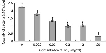
After two weeks of growth, cucumber leaves were sprayed with different concentrations of TiO2 suspensions in the greenhouse. After 5 h of solar heat, the plant count analysis revealed that TiO2 inoculation significantly decreased the biomass accumulation of the foliar bacterial community. Total culturable leaf surface populations in the phyllosphere communities were gradually reduced, corresponding to an increase of TiO2 concentrations (Fig. 1). Culturable leaf surface populations in the phyllosphere communities were reduced from 2.2×10⁷ to 3.1×10⁶ compared with the control group. Culturable populations of phyllosphere bacteria treated with TiO2 were all significantly different from the control (P=0.05). Total culturable bacterial populations were markedly diminished with every tenfold increase in TiO2 concentration. However, there was no significant difference between 0.2 and 2 mg/ml. These results demonstrate that the higher the concentration of nano-TiO2 suspension, the greater the impact on the culturable bacterial ability to survive in the phyllosphere.
Effect of different concentrations of nano-TiO2 on the bacterial community of the cucumber phyllosphere (P=0.05). Symbols indicate significant differences (P<0.05) based on Tukey's significantly different test using SPSS (SPSS Inc., Chicago, IL, USA).

16S rDNA DGGE analysis of TiO2 on foliar bacterial community of cucumber
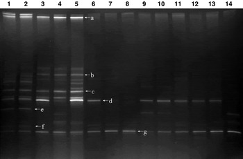
After two weeks of growth, cucumber leaves were sprayed with different concentrations of TiO2 suspensions in the greenhouse. After 5 h of solar illumination, the DNA of phyllosphere bacteria was extracted. The genome was amplified with specificity primer of 16S rDNA. We then acquired same-length DNA fragments with different nucleotide sequences. When the concentration of nano-TiO2 suspension was 20 mg/ml, 16S rDNA fragments of phyllosphere bacteria were not amplified. DGGE was used to separate 16S rDNA fragments of the same length but with differing nucleotide sequences. Species abundance was based on the number and intensity of bands in the DGGE profile. More bands indicated a greater abundance of microbial diversity. The light intensity of bands positively correlated with the number of microbial communities. As shown in Fig. 2, the concentration of TiO2 had a significant impact on community structures. Only band d, e, f and g could be monitored after TiO2 concentrations reached 0.02 mg/ml. Band g was the only band that had the same light intensity for all concentrations. This result revealed that the resistances of different phyllosphere bacteria to high concentrations of TiO2 varied. Most bacteria could not tolerate high TiO2 doses. Only the bacterial community of band g demonstrated resistance to high concentrations of TiO2.
PCR-DGGE 16S rDNA banding profiles of microorganisms from the cucumber phyllosphere sprayed with different concentrations of TiO2. Lanes: 1–2, 0 mg/ml TiO2 treatments; lanes 3–5, 0.002 mg/ml TiO2 treatments; lanes 6–8, 0.02 mg/ml TiO2 treatments; lanes 9–11, 0.2 mg/ml TiO2 treatments; lanes 12–14, 2 mg/ml TiO2 treatments. Bands a–g correspond to bands in Table 1.

Sequence analysis of TiO2 on foliar bacterial dominant species of cucumber
Sections of the special bands in the DGGE profiles were sequenced. The results indicated that at least seven species of bacteria survived on cucumber leaf surfaces. One of the uncultured bacteria was not influenced by nano-TiO2, but the other species, Pseudomonas sp., Providencia sp., uncultured bacterium, Sphingomonas sp., uncultured Exiguobacterium sp., Proteobacterium sp., uncultured Providencia sp., Pantoea sp. and Exiguobacterium sp. were affected by nano-TiO2.
Discussion
Phyllosphere microbes are very vulnerable to environmental effects. With the wide use and release of nanomaterials into the environment, the population and the structure of phyllosphere microbes may be affected by nanoparticles. At present, research on the biological effects of nanoparticles is still lacking. Most research focuses predominantly on animals, in particular for effects on DNA (Hirakawa et al., Reference Hirakawa, Mori, Yoshida, Oikawa and Kawanishi2004) and toxicity assays at the cellular level (Rahman et al., Reference Rahman, Lohani and Dopp2002). This study explored the effect of nano-TiO2 nanoparticles on the composition of phyllosphere microbes of cucumber, as a powerful supplement to the study of environmental factors and guidance on the application of nano-TiO2.
In this experiment, different concentrations of nano-TiO2 suspension were used on the surfaces of cucumber leaves. The results of bacterial plates recovery showed that the quantity of culturable phyllosphere bacteria was significantly reduced compared to control (P=0.05). Furthermore, the reduction was more significant with increasing concentration of nano-TiO2. These results coincide with those of other studies (Amézaga-Madrid et al., Reference Amézaga-Madrid, Silveyra-Morales and Córdoba-Fierro2003; Ye, Reference Ye, Liu and He2003; Cho et al., Reference Cho, Chung and Choi2005). At present, there is very little research on the mechanism of bacterial resistance to nanoparticles. This study provides some biological resources to be applied in research about microbial resistance for the photocatalysis of nanoparticles.
Analysing cucumber phyllosphere bacteria by PCR-DGGE revealed that different phyllosphere bacteria have different sensitivities to different TiO2 concentrations. We observed, from the DGGE fingerprint, that culturability bacterial species was very low at TiO2 concentrations above 0.02 mg/ml. This suggests that most bacteria were sensitive to nano-TiO2, and only a few bacteria have high resistance to TiO2. This may be related to their own bacterial structural features and the mechanisms for their resistance to TiO2 photocatalysis. The sequencing results of DGGE bands showed that at least seven kinds of dominant bacteria existed on cucumber leaf surfaces. Only one (Fig. 2, band g) displayed high resistance to nano-TiO2; the others were affected by nano-TiO2 to varying degrees. This reflects that various bacteria have different sensitivities to the oxidative toxicity produced by TiO2.
Before the onset of nanometre technology, humans neglected the damage cause by nanoparticles to people and the environment. With the increasing presence of nanoparticles, further study is needed to determine their negative impact and the far-reaching consequences for their safe use. With the industrialization of nano-coated fertilizers, soil treatments or photosynthesis accelerators that use TiO2, regulations should be set out to standardize the use of nanomaterials. In the future, we will research the minimum doses of nano-TiO2 that vary the diversity of phyllosphere microbes in the cucumber and identify or screen for the beneficial microorganisms from phyllosphere microbes as recovered from resistance assays.
Acknowledgements
We are indebted to our referees for valuable comments and suggestions. This project was supported by grants from the National Natural Science Foundation of China (award nos 30871665 and 30640044) and Beijing Natural Science Foundation (award no. 6073028).