With the continuous advances in the methods of imaging in medicine, and particularly in the recognition of gross anatomy, concerns have emerged with regard to the role to be played by the pathologist in the final macroscopic diagnosis. In the field of the congenitally malformed heart in particular, the richness of detail now provided by cross-sectional echocardiography, magnetic resonance, and computerised tomographic imaging, not to mention the promise of three-dimensional echocardiography, is truly amazing.
Alongside these developments, tertiary medial centres around the World have been experiencing, over recent decades, a vertiginous decrease in the number of autopsies performed. Data from a teaching hospital in Sheffield, United Kingdom, revealed a decline in the rate of post-mortem examination from around one-quarter of hospital deaths in the late 1970s, to less than one-twentieth in 2001.Reference Burton and Underwood1 In Australia, a national survey revealed an autopsy rate in hospitals of one-fifth in the period from 1992 through 1993, and one-eighth 10 years later.2 At the Heart Institute of São Paulo University in Brazil, a big tertiary centre for the treatment of cardiovascular disease, with more than 13,000 hospital admissions a year, the rate of autopsies declined from just over half the total number of deaths in the early nineties, to less than one-fifth last year. The impact of this decline on the process of educating clinicians and pathologists has yet to be fully evaluated, but the possible reasons are deserving of debate. Possible causes would include the technological progress in medical imagingReference O’Grady3 and laboratory diagnosis, but the extent of the influence of the scandal concerning retention of organs in the United Kingdom is not known. An immediate consequence of the inappropriate retention of organs, nonetheless, was a relative lack of confidence of families in the process of use of tissues for research, and certainly could explain a proportion of the current denials of consent for either autopsy or the subsequent retention of organs.
The impact of retention of organs
As is stated in the report of the Inquiry into retention of organs at Royal Liverpool Children’s Hospital,Reference Redfern, Keeling and Powell4 the need for the inquiry arose from evidence given by one of us (RHA) to the Bristol Royal Infirmary Inquiry.Reference Kennedy5 Paradoxically, as stated in the background to the Liverpool inquiry “He spoke of the benefits of retaining hearts for the purpose of study and teaching.” When this evidence was given, all of those practising in the United Kingdom, and almost certainly many practising elsewhere in the World, were unaware that many parents themselves had not given “proper consent” for the removal, examination, and subsequent retention of the organs of their loved ones. The knock-on effects of the evidence given in Bristol have been considerable, not least because the subsequent inquiry of happenings in Liverpool revealed that, in addition to the retention of hearts, there had been wholesale removal and retention of bodily organs explicitly against the wishes of parents. The inquiryReference Redfern, Keeling and Powell4 refers to this “systematic full-scale removal of organs” as the “van Velzen years”. As is stated in the pages of the inquiry,Reference Redfern, Keeling and Powell4 the discovery of these organs seriously “compounded the problems”, since “the large majority of organs remained untouched throughout the period.” Removal of organs in this latter fashion is unforgiveable. The earlier difficulties reflected problems of communication, rather than any wish to compromise parental consent. In most instances, the hearts had been removed for the purposes of coronial autopsies, and the legal position was unclear as to the ownership of the organs at the conclusion of the autopsy. When hearts had been removed for hospital, rather than coronial, autopsies, many thought that parents had granted permission, but in retrospect it proved that wordings in the forms used to obtain consent had been obscure, often referring to retention of “tissues” rather than giving explicit indications that the entirety of the heart, and often the lungs, trachea, oesophagus, and tongue, would be retained. As can be judged from the content of the inquiry,Reference Redfern, Keeling and Powell4 there was appreciable outrage. In the aftermath, there was unequivocally a significant reduction in the willingness of parents to agree to autopsies, and even greater reluctance to grant permission for retention of organs. As news of these happenings spread, similar outrage emerged in many other countries, notably Australia and Canada. The situation was not nearly as bad in the United States of America, since most hearts in the numerous archives in this country had been removed with full consent for subsequent use for the purposes of education and research. The end result, quite rightly, is that forms have now been totally re-written in the United Kingdom, and the other countries that suffered most subsequent to the revelations of previously inadequate consent. Those dealing with the increasingly small number of families losing children with congenitally malformed hearts continue to recognise the value of the autopsy, but now also recognise the need fully to take these grieving relatives into their full confidence, explaining the benefits that unequivocally do emerge from the opportunity to study and retain the heart in these tragic circumstances. We hope that, with time, the number of autopsies will again increase. In the meantime, nonetheless, it is essential that we retain, and make full use, of the existing archives.
The role of the autopsy in education
Despite the remarkable changes in the undergraduate medical education over the last decades, most educators still consider the autopsy as an important tool for teaching specific skills to their medical students.Reference Charlton6, Reference Burton7 Of the advantages to be accrued from the autopsy, perhaps the matter of greatest importance is the development of an analytical attitude in overall clinical practice. But many would argue that autopsies are expensive and unpleasant, and that there are several alternative methods of training, such as video demonstrations, and analysis of specimens in museums. The immediacy of the autopsy in providing clinicopathological correlation, however, will never be replaced by such alternative resources. Moreover, a recent metanalysis study demonstrated that, although the number of misdiagnoses detected by autopsy has decreased over time, there is still a great chance that important unsuspected diagnoses are revealed by the post mortem study.Reference Shojania, Burton, McDonald and Goldman8 At the same time, the importance of live demonstrations of cardiac specimens cannot be neglected in the process of education of cardiologists, irrespective of whether or not they are intimately involved with new methods of imaging. Since the late and great Maude Abbott began to organize the McGill Medical Museum at the end of the 19th Century, specimens have been extensively used, and proved to be of outstanding educational value for students. From the 1950s, with the advent of cardiac surgery, they have also proved their worth for cardiologists and cardiac surgeons.
The autopsy as a guide to improvements in treatment
In this respect, morphological studies, both gross and microscopic, have been highly significant over recent decades in the development of the modern cardiology and new surgical strategies. Numerous seminal papers demonstrated the echo-morphological correlations in congenitally malformed hearts,Reference Sutherland, Godman, Smallhorn, Guiterras, Anderson and Hunter9, Reference Silverman, Zuberbuhler and Anderson10 clarifying the significant anatomic features and providing better understanding, at that time, of a new method of diagnosis. The attitude of the morphologist, working side-by-side with the clinician or surgeon, was fundamental at this stage of knowledge, as it still is, and as it always should be. The technique of dissecting and sectioning the cardiac specimens changed considerably over the years, in order to facilitate such correlations with the anatomy of the living heart. Moreover, the knowledge acquired about the course of the atrioventricular conduction tissue in malformed hearts, again derived from studies of autopsied hearts, was instrumental in refining the optimal surgical strategies for these groups of patients.Reference Bharati and Lev11, Reference Anderson, Ho and Becker12
The potential contributions of microscopic analysis of congenitally malformed hearts, however, are far from complete. Myocardial remodelling shows distinct features related to the distorted gross anatomy present since morphogenesis that cannot be compared directly with the usual responses to dilation, hypertrophy, and valvar dysfunction found in hearts with normal anatomy.Reference Akiba and Becker13–Reference Salih, McCarthy and Ho15 Inherent abnormalities in the density of the capillaries in the setting of hypoplastic left ventricles, for example, may have implications for the success of surgery and postnatal ventricular development.Reference Salih, McCarthy and Ho15 Many other aspects require further investigation. Patients with palliated or corrected defects are now reaching adulthood, and there is much to be learnt about the ongoing evolution of the surgically repaired but congenitally malformed heart.
Financial considerations
For any specific institution, the advantages of autopsy must be judged in terms of cost and benefits. The medical staff, as well as the administrators, must be convinced that information obtained from autopsies improves the quality of health care. This is fundamentally linked to the quality of the data obtained. In this regard, therefore, the cardiac autopsy should ideally be performed by trained cardiovascular pathologists, in order to respond conveniently to the increasingly complex strategies used in the diagnosis and treatment of cardiac diseases. In an American and Canadian survey, two-fifths of responding Centres of Pathology indicated that Institutional funding was available for the hire of a cardiovascular pathologist.Reference McManus, Wilson and Struve16 The question must also be posed, nonetheless, as to whether the cardiovascular pathologist also has the necessary expertise in examination of the congenitally malformed heart. Certainly in the United Kingdom, and arguably elsewhere, those training in pathology rarely have the opportunity, nor the time, adequately to study the range of abnormalities seen when the heart is congenitally malformed. The best solution, therefore, could be to have a fully trained pathologist working in harness with an experienced cardiac morphologist, or alternatively with a pathologist’s assistant who has been properly trained in the examination of the congenitally malformed heart.
Describing the congenitally malformed heart
It is, perhaps, surprising that so few of those who have written in recent years about the congenitally malformed heart have been “card-carrying” pathologists. It is equally surprising that many highly skilled pathologists, who have relatively few problems in distinguishing malignant from non-malignant cells when looking down the microscope, still consider congenital cardiac disease to represent a difficult topic. To those of us familiar with examination of congenitally malformed hearts, it does not seem that difficult to reach the correct diagnosis, providing that attention is paid to a few basic rules. Recognition of the relationship of holes between the atrial chambers to the oval fossa serves to distinguish those simple interatrial connections within, and the more complicated lesions outside, this important component of the fetal circulation. Examination of the borders of holes between the ventricles as viewed from the morphologically right side permits them to be classified into ones with exclusively muscular borders, ones characterised by fibrous continuity between the leaflets of the atrioventricular and arterial valves, and ones with fibrous continuity between the leaflets of the arterial valves themselves. All interventricular communications can then be placed into one of these three groups, making allowance for necessary modifications when both arterial trunks arise from one ventricle, or alternatively when there is a common arterial valve. In similar fashion, it is an easy matter to distinguish the common atrioventricular junction that characterises almost all hearts having an atrioventricular septal defect, not least because of the trifoliate nature of the left component of the common atrioventricular valve. It was, however, the introduction of the so-called “morphologic method”Reference Van Praagh, David, Wright and Van Praagh17 that did most to defuse the multiple controversies that surrounded the description of congenitally malformed hearts in the latter half of the 20th century. This crucial concept was first advanced as justifiable criticism of the attempt by a group of European investigators, including one of the current authors (RHA), to disqualify a chamber from ventricular state in the setting of double inlet right ventricle.Reference Keeton, Macartney and Hunter18 The philosophy thus established has now proved its worth in several contentious areas. In essence, the method states that one feature of a given part of the heart, which is itself variable, should not be used as the defining feature for that particular cardiac component. Instead, the component should be defined on the basis of its most constant feature. This now serves as the underlying philosophy for the latest versions of sequential segmental analysis,Reference Anderson and Ho19, Reference Anderson, Moller and Hoffman20 themselves developed from the segmental approach that revolutionised the diagnosis of congenital cardiac disease.Reference Van Praagh and Bergsma21
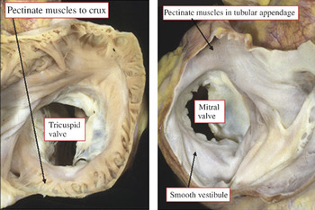
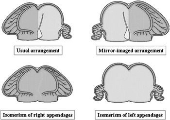
Thus, if we consider the distinction of the morphologically right and left atrial chambers, we know that the venous connections are themselves markedly variable, not least in the setting of visceral heterotaxy, which in the past was often considered to represent an ambiguous arrangement.Reference Van Mierop, Gessner, Schlieber and Bergsma22 Attention to the structure of the appendages, the most constant atrial component, and defined on the basis of the extent of the pectinate muscles relative to the atrioventricular vestibules, permits all appendages to be recognised as being either morphologically right or morphologically left (Fig. 1). Once the appendages have been distinguished in this fashion, it is then an easy matter to appreciate that the paired appendages can only be arranged in the usual fashion, in mirror-imaged arrangement, or in the patterns of right and left isomerism (Fig. 2). Any additional ambiguity in the setting of visceral heterotaxy can then be dispelled by describing separately the arrangement of the various thoraco-abdominal organs. In similar fashion, it is possible for the pathologist to distinguish the nature of ventricles according to the structure of their apical trabecular components, and arterial trunks on the basis of their patterns of branching. Using these basic rules, pathologists should then be able to describe the entire spectrum of congenital cardiac disease, even if a specific combination of segmental unions and associated malformations has never previously been encountered or described.
The panels show how the atrial chambers in the normal heart can be distinguished on the basis of the extent of the pectinate muslces relative to the atrioventricular vestibules. In the morphologically right atrium (left hand panel, the pectinate muscles encircle the entirety of the vestibule, whereas in the morphologically left atrium (right hand panel, the pectinate muscles are confined within the tubular appendage, leaving a smooth vestibule that contains the coronary sinus.

On the basis of the extent of the pectinate muscles relative to the atrioventricular junctions, and recognising that all hearts have two atral appendages, there are only four possible arrangements of the appendages. These are the usual pattern, the mirror-imaged variant, and the variants of right and left isomerism. When analysed in this fashion, the stage is set for subsequent sequential segmental analysis, taking care to assess venoatrial connections as a separate feature of any individual heart.

How can we increase the number of autopsies?
We suggest that actions can be taken by both the medical and the general communities so as to increase the number of autopsies in a given Institution. From the stance of the medical community, clinico-morphological correlations, if properly organised, can stimulate young doctors and junior students to become critical of the methods of diagnosis and treatment, and are to be encouraged. Case-based learning exercises have already been shown to improve the academic performance of medical students,Reference O’Grady23 and could be of equal value in the setting of training of paediatric cardiologists and paediatric cardiac surgeons. The autopsy will be key to such advances. From the stance of the community, conferences with families after the autopsy will go a long way to restoring their confidence in the process, emphasising the importance of the morphological diagnosis for genetic counselling, and the increasing need to retain tissue for the purposes of research and teaching. Having made initial inroads, it should then be hoped that the community itself will promulgate the value of the autopsy.
New methods for the autopsy
Recently, post-mortem imaging by magnetic resonance and computed tomography has been considered as an alternative to the autopsy. The term Virtopsy® was coined for this procedure, and its potential utilization for the study of the heart was addressed by Jackowski et al.Reference Jackowski, Schweitzer and Thali24 A learning curve is under way now, because the interpretation of the images has to be adapted to post-mortem conditions, such as clotting, loss of circulation, and internal lividity. Although its validation may take time, the new methodology is a promising substitute for the autopsy if combined with minimal sampling of tissue, and will represent a new frontier for the pathologists.
The fetal, perinatal and paediatric autopsy
The autopsy of those unfortunately dying during the fetal, perinatal and childhood periods is an important tool that should be utilized by all clinicians involved. With the advent of high resolution, targeted, echocardiographic studies we know more now than ever about the status of the patient, including the fetus well before birth. In this respect, the significant advances in fetal, maternal, and neonatal medicine and surgery have allowed many premature infants born quite early to survive. The fetal echocardiographer, obstetrician, neonatologist, surgeon and perinatal or paediatric pathologist must now work together to sort out what went right or wrong when there is a fetal or neonatal death. At centers where there is an active programme involving fetal echocardiographers and paediatric cardiac surgeons, a surgical repair or interventional procedure is often planned prior to birth. The baby is then born at a center where the chosen treatment can immediately be implemented with well informed medical personnel, and with the full support of the parents.
The indications for fetal ultrasound are many and diverse, and can be used in unison with multiple other studies, such as amniocentesis, to gain crucial knowledge about an ongoing pregnancy. Many major and minor structural anomalies can be reliably detected during fetal life, but it is unrealistic to expect total accuracy in all cases. The autopsy can be used as a valuable tool to fill in the gaps left by the many variables that come into play during ultrasonic evaluation, such as gestational age at time of scan, the experience of the sonographer, and the types of equipment used. Even with all of the new molecular and genetic advances, the autopsy still can give us the best and most complete answer.
A properly trained paediatric or perinatal pathologist, or a suitably knowledgeable pathologists’ assistant, can add significantly to the study at autopsy of an intrauterine fetal death, a neonatal death, or a death subsequent to surgical intervention. The knowledge gained from the properly performed autopsy will almost always benefit the clinicians involved, and in many cases will give the parents a sense of peace when certain questions are answered. In some cases, the parents may also feel that the death of their infant was not a total loss, since in the future the results of the autopsy can serve to help another family faced with a similar situation. In one study,Reference Elder and Zuccollo25 reports of autopsies carried out on extremely premature infants showing that new findings were discovered in four-fifths of cases. The new findings resulted in a significant change in the diagnosis in over one-quarter. In two-fifths of the cases, iatrogenic lesions were identified, and were the main cause of death in one-sixth. Many questions are answered by, and future treatments and surgical procedures can be refined from, these lessons. Autopsy can improve the quality of care in this population of patients, as well as provide necessary genetic and morphological analysis for the medical history of both the patient and the relatives. Future pregnancies can be evaluated earlier in gestation, and informed decisions concerning termination, surgery, and so on, can be made in the best interest of the mother and fetus. Siblings of these fetuses will also benefit by having a well documented history when they reach child-bearing age.
Just as cardiac morphology has been broken down into the morphological method, and sequential segmental analysis, the perinatal and paediatric autopsy needs to be performed in systematic fashion. With good powers of observation, looking at the body, and each system of organs, in a systematic manner will allow for identification of minor variations of normal, as well as the more obvious complex anomalies or malformations. These, in turn, can be compared with those recognized clinically. If approached in a practical manner the procedure is not as complex or as daunting as it seems. A good gross description, along with photographs, can then be sent to a consultant, often with a quite complete diagnosis being rendered. This allows for a more cost-effective situation for the institution, and less of a strain on the family when the fetus or infant does not have to be transported to another institution for autopsy. Efforts have already been made to publish the necessary information to assist those pathologists in community hospitals in the performance of the perinatal or paediatric autopsy, in hopes of adding to their skills of observation, practical knowledge and communication with clinicians.26, Reference Gilbert-Barness and Debich-Spicer27
In many community hospitals, the examination of a fetus or child is considered a complex and time-consuming procedure, or else is not considered important at all. The latter is a great disservice to the family and the public in general. In the experience of one of the authors (DDS), many institutions will gladly employ the services of a well trained perinatal or paediatric pathologists’ assistant, as apposed to the supervising pathologist doing the examination. Offering autopsy services to multiple institutions in one given area allows for each institution to give the very best results to the families and clinicians in a cost-effective manner, without the time constraints on the pathologist, or the “in house” pathologists’ assistants.
Echomorphological and autopsy correlation can yield more information than ever for all of the clinicians involved in the care of a fetus or infant. A close collaboration between clinician and pathologist has the potential to bring the autopsy back to the forefront of teaching our medical professionals, especially in the perinatal and paediatric arenas. Over time, the relationship between cardiologists, cardiac surgeons, and the pathologist or morphologist have proved very beneficial. The sharing of information, answering and formulating questions about the care of this population of patients, can only serve to improve outcomes. The perinatal or paediatric pathologist, along with their assistants, must continue to play an integral part in the correlation of the developing imaging techniques and autopsy findings.
The role of the pathologist’s assistant
In the field of perinatal or paediatric pathology, the autopsy is one of the best teaching tools, not only for the clinicians, but especially for the pathologist, his or her assistant, or the pathology resident. Many of the diseases, syndromes, associations, and so on encountered in this population of patients are not seen in older children, let alone adults. Most syndromes have a constellation of abnormalities associated with them, but within that group there is a high degree of variability. It can take years of training to become proficient in performing a perinatal or paediatric autopsy. It is the practice of doing them that is the best teaching tool. There are many well qualified paediatric pathologists, most working at paediatric hospitals. At many of those institutions, pathologists’ assistants have been trained by the pathologist to perform the autopsy with the same degree of precision.Reference Gilbert-Barness and Debich-Spicer28 This allows the pathologist to supervise the procedure, and to free up his or her time for other duties. As well, this is cost effective for the institution. Some of the same pathologists’ assistants have become proficient with cardiac morphology, and interact closely with the clinicians at their respective institutions. Recognising their prowess as cardiac morphologists, this small group is often sought out by other institutions for their expertise. Pathologists’ assistants in general are highly trained and skilled providers of health care, no matter what branch of pathology they are trained in. The profession has grown significantly over the recent past within the United States of America and Canada, with a wide range of opportunities for employment. It may be the wave of the future to train pathologists’ assistants to do these very interesting, but sometimes very time-consuming, duties associated with the perinatal and paediatric autopsy, along with the morphological study of the congenitally malformed heart.