Gap junctions are expressed throughout the body and take part in many processes that are necessary for normal physiological function. Gap junction channels are formed by connexin protein subunits, six of which combine to produce a hemichannel (Ref. Reference Goodenough and Paul1). Two hemichannels from the plasma membranes of adjacent cells align in the extracellular space to form a complete gap junctional channel (Ref. Reference Harris2), which allows ions and other small molecules to pass between the connected cells. In addition, connexin hemichannels can be functional in nonjunctional membranes, allowing the exchange of solutes with the extracellular space (Refs Reference Jiang and Gu3, Reference Saez4). There are more than 20 connexin genes that are expressed throughout the body in overlapping and unique patterns (Ref. Reference Sohl5). Gap junction channels may be assembled from the same connexin, or a combination of different connexins, depending on the cell type (Fig. 1). Each connexin protein has four transmembrane domains (TM1–TM4), connected by two extracellular loops (EC1 and EC2) and a cytoplasmic loop (CL). The N- and C-termini of the protein extend into the cytoplasm of the cell. Two organs where connexins and gap junctions are necessary for normal function are the skin and the inner ear. Mutations in connexin genes have been linked to human hereditary diseases affecting both the epidermis and cochlea.
Connexin channel formation. Diagram shows how connexons (hemichannels) form at cell membranes and the terminology used to describe the different types of combinations. Connexin proteins assemble into hemichannels (connexons) and intercellular channels (gap junctions). Connexons may be homomeric or heteromeric, and gap junction channels may be homotypic or heterotypic.

The role of connexins in the auditory process
Hearing is a complex process through which sound is converted from a physical pressure wave into neuronal signals. The critical step takes place in the sensory hair cells of the cochlea, where an array of proteins work together to convert the pressure wave sensed by the basilar membrane into an electrical signal propagated by the auditory nerve. Gap junctions do not have a direct role in signal conversion in the inner ear or vestibular system, and are not found between hair cells and the supporting cells that separate them (Refs Reference Santos-Sacchi and Dallos6, Reference Zhao7). However, connexins and gap junctional communication have been well documented in other cochlear cells (Refs Reference Jagger and Forge8, Reference Kikuchi9). Cells interconnected by gap junctions fall into two groups: nonsensory epithelial cells among which hair cells are dispersed, and connective tissue cells at more distal locations to the hair cells. Connexin-26 (Cx26; also known as gap junction β-2 protein and encoded by GJB2) is present in almost all locations in which cochlear gap junctions have been found (Ref. Reference Kikuchi10).
The mammalian cochlea has an unusual arrangement of extracellular fluid-filled spaces, which provides a unique environment for the sensory cells. Basolateral hair cell surfaces are bathed in perilymph, which has an ionic composition similar to that of other extracellular fluids. The apices of hair cells, however, are bathed in endolymph, which has high K+ and low Na+ concentrations, similarly to intracellular fluids. When hair cells are activated by sound, receptor potentials are generated from the flow of K+ ions from endolymph into the hair cell. The K+ ions are then released from hair cells into interstitial space within the organ of Corti (Ref. Reference Johnstone11), where K+ channels in cochlear supporting cells permit uptake of these ions (Ref. Reference Oesterle and Dallos12). Normal hearing requires this K+ movement, and it is possible that junctional communication influences the ionic environment of inner ear sensory epithelia. It has been proposed that serially arranged gap junctions of epithelial and connective tissue cells serve as a mechanism for recycling endolymphatic K+ ions that pass through sensory cells during auditory transduction by providing an intercellular pathway for conveyance and distal release of K+ ions.
There are models explaining how gap junctions could achieve this task. In the original model, it was speculated that passive diffusion through gap junctions may be required for K+ recycling from the perilymph back to the endolymph after sound transduction (Refs Reference Kikuchi13, Reference Wangemann14, Reference Hibino and Kurachi15). In this model, after uptake by the supporting cells, K+ moves through epithelial cell gap junctions laterally toward the lower part of the spiral ligament. There, K+ ions are released into the extracellular space within the spiral ligament by root cells, and are taken up by the Na+/K+-ATPase and Na+/K+/Cl– cotransporter (NKCC1) in the plasma membrane of type II fibrocytes. This uptake incorporates K+ into the connective tissue gap junction system, where it can pass through the tight junctional barrier at the basal cell layer of the stria vascularis and then be released into the extracellular space. The marginal cells of the stria vascularis then take up the K+ ions using the Na+/K+-ATPase and NKCC1, and release the K+ into the endolymphatic space to be used again in sound transduction (Refs Reference Schulte and Adams16, Reference Crouch17). Recently, this model has been updated to include permeation of inositol-1,4,5-trisphosphate (IP3) second messengers and Ca2+ waves through cochlear gap junctions as an active component of the K+ efflux pathway (Refs Reference Bruzzone and Cohen-Salmon18, Reference Beltramello19). However, it should be noted that the studies supporting this model were conducted using immature cells from the organ of Corti at a developmental stage where the endocochlear potential had not yet been produced. These changes were based in part on the observation that induced Ca2+ waves in cochlear-supporting cells required the diffusion of IP3 through gap junctions and the release of ATP into the extracellular medium. As the intracellular Ca2+ activated a combined K+/Cl– efflux system in supporting cells, it was proposed that the IP3 permeability through gap junction channels is also required for K+ recycling. Gap junctions are responsible for shunting K+ out of the perilymph in both models, but the second model allows for the active efflux of K+ through the supporting cells along the way via IP3 propagated Ca2+ waves that also travel via gap junctions (Ref. Reference Bruzzone and Cohen-Salmon18).
The role of connexins in the epidermis
The skin has several roles in physiological homeostasis. It serves as a barrier to external pathogens and is used in thermoregulation as a means of cooling the body temperature by the secretion and evaporation of sweat. In addition, the epidermis contains mechanoreceptors and nerve endings associated with touch and temperature sensing, and forms a watertight barrier that maintains normal interstitial osmolarity. The skin is the largest organ in the human body and continually regenerates itself. The epidermis contains the basal, spinous, granular and cornified layers. These four layers are in a constant state of growth and differentiation as the external cornified layer is continuously shed. To maintain a constant thickness of the skin, keratinocytes from the basal layer need to constantly proliferate, differentiate and push towards the outer layers, and the rates of growth and differentiation must be tightly regulated.
Studies of human (Ref. Reference Caputo and Peluchetti20) and rodent (Ref. Reference Risek21) epidermis have identified gap junctions between keratinocytes residing in the basal, spinous, and granular layers, but not between cells in the cornified layers. These data correlate well with dye-transfer experiments that showed junctional communication between keratinocytes populating the three inner epidermal layers of mouse and human skin (Refs Reference Kam22, Reference Salomon23). These experiments suggested that epidermal keratinocytes were organised into ‘communication compartments’ consisting of 20–25 cells, as dye transfer occurred between basal and supra basal keratinocytes within a compartment, but was reduced between keratinocytes in neighboring compartments. This communication compartment was postulated to correlate with the epidermal proliferative unit (Ref. Reference Potten24), and suggested that gap junctional communication might help to mediate keratinocyte growth and differentiation (Ref. Reference Kam22).
At least nine connexin genes, including that encoding Cx26 (GJB2), are expressed during keratinocyte differentiation, and they show overlapping spatial and temporal expression patterns (Refs Reference Kelsell25, Reference Di26, Reference Mese27). Dramatic changes in connexin gene expression occur in the epidermis following changes in keratinocyte proliferation. In human skin, retinoic acid treatment resulted in a thickening of the epidermis and an increase in the number of keratinocytes, which correlated with a massive induction of Cx26 expression in all epidermal layers, and a smaller increase in Cx43 levels (Ref. Reference Masgrau-Peya28). In patients with psoriatic lesions, a similar induction of Cx26 has been reported in the hyperproliferative psoriatic areas, but not in the surrounding epidermis (Refs Reference Rivas29, Reference Labarthe30). In experimentally wounded human skin, upregulation of Cx26 expression was also observed (Ref. Reference Lucke31). Similar changes in connexin expression have been reported in rodent skin, where it has been shown that Cx26 expression rapidly increased in keratinocytes proximal to experimental wounds, but decreased in cells at the wound edge (Ref. Reference Goliger and Paul32). In a study of mouse papillomas, expression of both Cx26 and Cx43 was elevated in the proliferating neoplasms, but not in surrounding normal skin (Ref. Reference Sawey33). Thus, mouse epidermis behaves similarly to human epidermis with regard to changes in connexin expression associated with hyperproliferation: a large induction of Cx26 is correlated with an increase in keratinocyte number. These studies establish a correlation between increased Cx26 expression and keratinocyte proliferation and differentiation.
Effects of Connexin-26 mutations
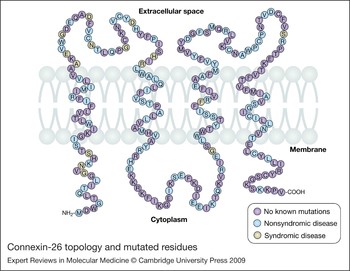
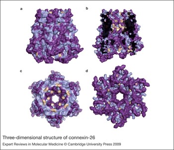
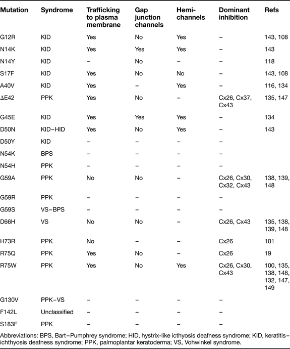
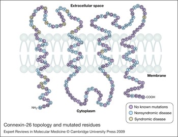
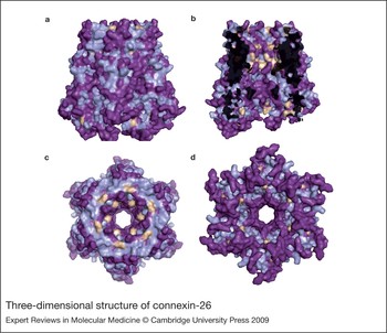
Cx26 mutations are present with a high frequency in the human population, and genetic screening for Cx26 is routine in cases of paediatric hearing impairment (Refs Reference Smith34, Reference Apps35, Reference Tranebaerg36). Mutations that cause nonsyndromic deafness are distributed throughout the Cx26 coding region, whereas syndromic mutations that cause skin disease are predominantly found in the N-terminus and first extracellular domain (Fig. 2). These domains have roles in channel gating at the cytoplasmic and extracellular openings of the pore, and syndromic Cx26 mutations are highly concentrated in these channel orifices (Fig. 3). There is ample evidence suggesting that simple loss-of-function mutations in GJB2 cause nonsyndromic deafness, but not the syndromic forms exhibiting epidermal pathology (Refs Reference Zhao7, Reference White37, Reference Bruzzone38). Both autosomal recessive and autosomal dominant nonsyndromic deafness have been associated with mutations in GJB2 (Refs Reference Kelsell39, Reference Denoyelle40). The most frequent recessive Cx26 mutation is a single base deletion (35ΔG), which results in a frameshift at residue 12 and premature termination of the protein. Other common recessive Cx26 mutations include nonsense mutations and small deletions or insertions, most of which also lead to premature termination of protein translation. Therefore, many of the commonly observed recessive mutations result in severely truncated connexin proteins that fail to retain any channel activity, and recessive hearing loss might largely be a result of null mutations in GJB2. By contrast, the Cx26 mutations causing skin diseases and deafness are all single amino acid changes, and the mechanism(s) whereby they lead to skin pathology are unclear. The identification of mutations in Cx26 that cause either nonsyndromic hearing loss, or syndromic deafness associated with skin disease, presents an interesting paradox that arises from the observation that complete loss of Cx26 function correlates with deafness, but not skin disease. Thus, human skin does not need functional Cx26 to maintain homeostasis. Some reports have noted that recessive mutations in Cx26 that lead to nonsyndromic deafness, but not skin disease, can result in thicker epidermis in the carriers, although the precise consequences of this phenomenon are not clear. (Refs Reference Meyer41, Reference Man42, Reference d'Adamo43). However, certain mutations in this gene dramatically perturb epidermal homeostasis, possibly through dominant gain-of-function mechanisms.
Connexin-26 topology and mutated residues. This illustration displays the location of amino acid residues in Cx26 relative to the membrane. Purple residues are not mutated. Blue amino acids are mutated residues found in nonsyndromic deafness and can be seen throughout the protein. Brown residues represent amino acids mutated in syndromic deafness, which are generally found within the N-terminus and first extracellular loop.
Three-dimensional structure of connexin-26. Syndromic and nonsyndromic mutations mapped onto the reported crystal structure of Cx26 (Ref. Reference Maeda150). Brown residues are reported syndromic mutations, blue residues are known nonsyndromic mutations, and purple residues are not reported to have a mutation. (a) Side view of a Cx26 hemichannel with six protein subunits. The structure has the extracellular portion of the protein at the top of the diagram and the cytoplasmic segment at the bottom. (b) Cut away side view of a Cx26 hemichannel with three protein subunits removed allowing a view of residues lining the pore domain. (c) View from the extracellular surface of the complete hemichannel looking down onto the protein and into the cell through the pore domain. (d) View from the cytoplasmic surface of the complete hemichannel looking out of the cell through the pore domain.
Nonsyndromic deafness
Nonsyndromic deafness is characterised by hearing loss in the absence of any other clinical manifestations. Cx26 mutations causing nonsyndromic deafness may be recessive or dominant. Approximately 80% of nonsyndromic deafness results from recessive mutations on over 100 different loci (Ref. Reference Yan and Liu44), whereas the remaining 20% is caused by dominant mutations on more than 30 loci (Ref. Reference Petit45). Mutations causing deafness have been linked to a number of different connexin genes (Refs Reference Kelsell39, Reference Grifa46, Reference Xia47), but up to 50% of genetic nonsyndromic hearing loss results from autosomal recessive mutations in GJB2. (Refs Reference Kelsell39, Reference Petersen and Willems48, Reference Chang49, Reference Guilford50, Reference del Castillo51). Over 100 unique recessive mutations in Cx26 have been identified to date (Fig. 2), but some of the most commonly occurring changes lead to severely truncated proteins as a result of nonsense mutations or deletions producing frameshifts (Ref. Reference Dai52). This makes the study of these specific mutations interesting because of their lack of effective protein function. Several of these mutations reoccur with high frequencies. For example, the 35ΔG allele of GJB2 is the most common mutation that has been observed. In Italian and Spanish populations it has been found to be present in 88% and 55% (respectively) of test subjects that reported hearing impairment (Ref. Reference Rabionet53). In a study of Greek patients with nonsyndromic hearing loss, the 35ΔG mutation of Cx26 made up 54% of the GJB2 alleles (Ref. Reference Antoniadi54). The 35ΔG mutation produces a frame shift that leads to a stop codon after residue 13, causing early termination and a complete loss of protein function (Ref. Reference Petit45).
The W24X mutation also seems to be prevalent in GJB2-related recessive deafness in certain populations. One investigation reported W24X mutations in 79% of the recessive deafness alleles studied in the Spanish Romani gypsy population (Ref. Reference Alvarez55). Studies in India revealed that 5–33% of the sensorineural deafness patients observed had the W24X mutation, and linked the mutation to a founder effect (Refs Reference Joseph and Rasool56, Reference Khandelwal57). A comprehensive study of the European Romani populations revealed that 23–93% of mutant alleles encoded W24X, and support the notion that founder effects might have contributed to the high frequency of GJB2 mutations associated with recessive nonsyndromic deafness (Ref. Reference Bouwer58). This mutation is a nonsense mutation that creates a severely truncated protein, similarly to 35ΔG and other common alleles such as 167ΔT and 235ΔC.
The Ashkenazi Jewish community contains another widely expressed GJB2 mutation that encodes a truncated form of the Cx26 protein, 167ΔT. The carrier frequency for the allele in this group of individuals can range from 2% to 4% (Refs Reference Bors59, Reference Dong60). It was reported that the 167ΔT mutation observed in unrelated Ashkenazi Jewish families was on the same haplotypic background and was also the result of a founder effect (Ref. Reference Lerer61). Although this was not the only Cx26 mutation found in the Ashkenazi population, it was estimated that up to 70% of Ashkenazi Jewish patients with nonsyndromic hearing loss possessed the 167ΔT mutation (Ref. Reference Lerer61). This is another GJB2 mutation that causes early truncation of the Cx26 protein, resulting in hearing loss.
Finally, the 235ΔC mutation has been found to have a high frequency of occurrence in Chinese patients with nonsyndromic hearing impairment. Up to 31% of cases possess the 235ΔC mutation in certain subregions of China, and the 235ΔC mutation is present in 5–15% of GJB2 alleles for all Chinese patients nationally (Refs Reference Dai52, Reference Dai62). Occurrence of 235ΔC is not limited to China, and the mutation has also been found in 73% of Japanese patients with nonsyndromic hearing loss compatible with recessive inheritance (Ref. Reference Abe63). This mutation is common in East Asian populations, probably as a result of founder effects (Ref. Reference Yan64). The 235ΔC mutation produces a truncated protein at the beginning of the second transmembrane domain (Fig. 2) and is similar to the other three common alleles described above. These nonsense mutations only result in nonsyndromic hearing loss, and suggest that complete loss of function causes nonsyndromic GJB2-associated deafness.
Nonsyndromic mutations produce a range of hearing impairment from profound deafness to loss of high frequencies. A genotype–phenotype correlation has been suggested, where 35ΔG homozygous mutations are more severe than heterozygous 35ΔG with another non-35ΔG mutation, or people with two non-35ΔG mutations (Ref. Reference Cryns65). This was supported by a comparison of the 235ΔC truncation and the V37I mutation, which suggested that truncated genotypes are more severe than missense mutations encoding full-length proteins (Ref. Reference Oguchi66). Later studies that analysed data from cohorts of individuals concluded that there was too much variability in the hearing loss of patients to determine statistically any genotype–phenotype relationship (Refs Reference Picciotti67, Reference Hilgert68, Reference Angeli69). Furthermore, one study conducted on patients with the 35ΔG and other deletion mutations from the same geographical area also found that the observed hearing loss was too variable to draw any meaningful correlations (Ref. Reference Hismi70).
The majority of observed mutations in Cx26 linked to nonsyndromic deafness are deletions that lead to frameshifts, and nonsense mutations that lead to early truncation of the protein. Thus, most mutations in GJB2 essentially result in gene knockout in homozygous patients, with the complete elimination of Cx26 function resulting in hearing loss (Ref. Reference Petit45). This is surprising, because other connexins are expressed in the inner ear with expression patterns that overlap with Cx26 (Refs Reference Kikuchi10, Reference Kikuchi13, Reference Lautermann71, Reference Forge72, Reference Xia73, Reference Ahmad74, Reference Kudo75, Reference Cohen-Salmon76, Reference Zhao77). Thus, the genetic ablation of Cx26 gap junctions must result in a very specific effect within the inner ear, possibly altering K+ ion transport in the cochlea, leading to a breakdown of the auditory process (Ref. Reference Zhao77). Excess K+ accumulation around the hair cells might cause cytotoxicity and cellular degeneration. However, truncation and loss of the Cx26 protein does not result in an associated skin disorder, and demonstrates that loss of Cx26 function alone cannot account for the skin disorders seen in syndromic deafness.
Syndromic deafness
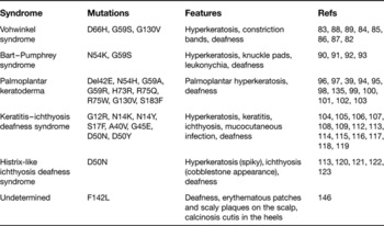
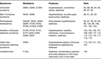
Syndromic hearing loss results in the disruption of normal hearing in addition to displaying other phenotype(s). Syndromic hearing loss caused by Cx26 mutations is associated with skin disorders and ectodermal dysfunction, in addition to deafness (Refs Reference Gerido and White78, Reference van Steensel79, Reference Richard80, Reference Lai-Cheong81). Syndromic hearing loss is autosomal dominant, and associated skin disorders can be mild to severe and include Vohwinkel syndrome (VS), Bart–Pumphrey syndrome (BPS), palmoplantar keratoderma (PPK), keratitis–ichthyosis deafness syndrome (KID) and hystrix-like ichthyosis deafness syndrome (HID). These conditions are all caused by dominant mutations in GJB2 (Fig. 2; Table 1). Since nonsyndromic deafness is predominantly a loss-of-function disorder, it follows that the syndromic mutants might show an alteration or gain-of-function to cause skin disease.
Dominant mutations in GJB2 that lead to syndromic deafness are infrequent, and therefore represent only a small portion of overall genetic hearing loss caused by connexin mutations. In contrast to the common recessive frameshift and truncation mutations described above, the dominant GJB2 mutations causing skin diseases and deafness are all single amino acid substitutions or deletions, and the mechanism(s) whereby they lead to skin pathology are poorly understood. Thus, although autosomal dominant inheritance of deafness linked to Cx26 is uncommon within the large spectrum of connexin mutations, it is the only mode of inheritance seen for cases of syndromic hearing loss.
Vohwinkel syndrome
Vohwinkel syndrome (OMIM 124500) is a skin disease associated with deafness that was first discovered in 1929 (Ref. Reference Vohwinkel82). It presents with several identifiable characteristics, such as sensorineural deafness, palmoplantar hyperkeratosis, constriction bands leading to autoamputation of the digits (pseudoainhum) and starfish-like keratoses (Refs Reference Korge83, Reference Maestrini84, Reference Maestrini85, Reference Peris86, Reference Drummond87). VS has been linked to the G59S, D66H and G130V mutations in GJB2 (Refs Reference Maestrini84, Reference Bondeson88, Reference Snoeckx89). The restriction of pathology to the palms and soles is shared by other syndromic deafness disorders caused by GJB2 mutations, although disorders such as PPK and BPS lack constriction bands and starfish-like keratoses (Refs Reference Korge83, Reference Drummond87). By contrast, the absence of ichthyosis and hair follicle defects, which are observed in other Cx26 disorders, such as HID or KID (Table 1), help to differentiate these diseases.
Connexin-26 syndromic deafness disorders
Bart–Pumphrey Syndrome
Bart–Pumphrey syndrome (OMIM 149200) is a syndromic deafness disease that is also linked to GJB2 mutations and shows a phenotype that displays overlap with VS. BPS patients also display a thickening of the palms and soles known as palmoplantar hyperkeratosis, but lack constriction bands and pseudoainhum seen with VS. These cases are further distinguished the presence of leukonychia (nail thickening and crumbling), as well as knuckle pads (Refs Reference Richard90, Reference Bart and Pumphrey91, Reference Ramer92). To date, there have been two observed mutated residues that are responsible for BPS. The N54K mutation was discovered in 2004 and is found in the first extracellular loop of Cx26, a domain that is frequently mutated in syndromic deafness (Ref. Reference Richard90). This mutation is thought to be at the very tip of the extracellular domain, where it influences docking, as well as pore formation. The G59S mutation was discovered in 2005, and lies within the same extracellular region as N54K (Ref. Reference Alexandrino93). The G59S mutation is not specific to BPS, and has also been described in cases of VS (Refs Reference Bondeson88, Reference Alexandrino93), and mutation of G59 to R or A residues has further been linked to PPK (Refs Reference Heathcote94, Reference Leonard95). This has led some to question the extent of differences observed between some forms of syndromic deafness associated with Cx26, because VS, BPS and PPK all share similar characteristics and mutation of the same residues.
Palmoplantar keratoderma with deafness
Palmoplantar keratoderma (OMIM 148350) is another syndromic complication of deafness linked to Cx26 dysfunction. This disease shows a relatively mild form of sensorineural hearing loss compared with the other Cx26 syndromic deafness disorders, and also presents with similar hyperkeratosis of the palms and feet as observed for VS and BPS (Refs Reference Sharland96, Reference Verbov97). As previously noted, there are shared mutations between all three diseases, and mutated residues linked to PPK, similarly to those in VS and BPS, are mainly found in the first extracellular loop. The following Cx26 mutations have been reported to cause PPK with deafness: N54H, G59A, G59R, H73R, R75Q, R75W, G130V and S183F (Refs Reference Heathcote94, Reference Leonard95, Reference Uyguner98, Reference Iossa99, Reference Richard100, Reference Zwart-Storm101, Reference Akiyama102, Reference Zwart-Storm103). The Cx26 mutations associated with PPK are similar in location and phenotype to those for VS and BPS, which might suggest that the classification of these disorders as three distinct syndromes in the premolecular era might need to be reconsidered. Patients with the same genetic mutation in GJB2 can be differently diagnosed as having PPK, VS or BPS (i.e. G59S or G130V), suggesting that these three syndromes may actually represent a single broad form of syndromic deafness with a heterogeneous phenotype.
Keratitis–ichthyosis deafness syndrome
Keratitis–ichthyosis deafness syndrome (OMIM 148210) expresses a distinctly different phenotype of syndromic deafness linked to Cx26 mutations than VS, BPS or PPK. KID syndrome presents with mild to severe sensorineural hearing loss associated with keratitis, loss of eyebrows and eyelashes, ichthyosis and an increased susceptibility to mucocutaneous infection (Refs Reference Grob104, Reference Nazzaro105, Reference Szymko-Bennett106). The keratitis can range from mild to bilaterally severe and can lead to complete blindness in some cases (Ref. Reference Janecke107). The skin lesions are not restricted to particular regions of the body (i.e. palms or soles) and show marked ichthyosis, which usually disrupts the barrier function and leads to increased susceptibility to mucocutaneous infection and carcinoma (Refs Reference Grob104, Reference Richard108, Reference van Steensel109). One KID mutation, G45E, frequently results in patient fatality within the first year, as a result of sepsis (Refs Reference Janecke107, Reference Gilliam and Williams110, Reference Griffith111, Reference Jonard112). This disorder is very different from the previous three syndromic conditions discussed, in terms of severity and location of phenotypes. There have been several Cx26 mutations associated with KID syndrome, and like the other syndromic mutations, some of them are found in the first extracellular loop. However, a few have also been located in the N-terminus. Eight mutations have been described so far: G12R, N14K, N14Y, S17F, A40V, G45E, D50N and D50Y (Refs Reference Janecke107, Reference Richard108, Reference Jonard112, Reference van Geel113, Reference van Steensel114, Reference Mazereeuw-Hautier115, Reference Montgomery116, Reference Stong117, Reference Arita118, Reference Yotsumoto119). Of these mutations, D50N is the most frequently observed and often leads to severe deafness with associated ichthyosis (Ref. Reference Mazereeuw-Hautier115). Although the other mutations were not as frequent, they all had a more severe phenotype than the mutations associated with the VS–BPS–PPK group of syndromic deafness disorders.
Hystrix-like icthyosis deafness syndrome
Hystrix-like icthyosis deafness syndrome (OMIM 602540) has also been described as a syndromic deafness mutation associated with Cx26. This condition is similar to KID syndrome and displays all of the common features of KID, except for keratitis. It was initially noted that HID displayed a distinct skin pattern of ichthyosis consisting of spiky hyperkeratotic masses with a cobblestone-like appearance that can appear throughout the body (Refs Reference van Geel113, Reference Schnyder and Gloor120, Reference Gulzow and Anton-Lamprecht121, Reference Traupe122, Reference Konig123). It was later determined that the only identified HID syndrome mutation in Cx26 was D50N, which is also commonly found with patients diagnosed as having KID syndrome (Ref. Reference van Geel113). The discovery that the only known HID mutation was a common KID mutation again supported the idea that these two syndromes might represent a single form of syndromic deafness with a heterogeneous phenotype. Combined with the similarities between VS, BPS and PPK, the emerging view is that there are two broad types of skin disorder associated with syndromic deafness: the VS–BPS–PPK group and the KID–HID group.
Clinical implications
For both syndromic and nonsyndromic deafness, the biggest challenge is how to treat the disorders. Patients suffering from nonsyndromic deafness typically receive cochlear implants. This approach may eventually bridge the gap for deafness patients, allowing full functionality and the possibility of extended audible ranges (Ref. Reference Clark124). One concern stems from the fact that connexin-related deafness, although prevalent in developed nations with access to implants, is also present in developing countries that have do not yet have access to the benefits of cochlear implant technology. The hope is that technical improvements will allow implants to be more cheaply available to deaf patients in areas not previously reached by such advancements (Ref. Reference Tarabichi125). In patients with syndromic deafness associated with Cx26, there is evidence that cochlear implants are also successful to treat the hearing loss associated with their disease (Refs Reference Barker and Briggs126, Reference Hampton127, Reference Cushing128).
The skin diseases associated with syndromic deafness have been shown to require a gain of function, because effective knockout mutations result in deafness, but not skin disorders (Ref. Reference Dai52). Therefore, treatments for one condition might not apply to the entire spectrum of skin disorders. New techniques could address current complications with this. To further complicate matters, the skin diseases seen in syndromic deafness patients are diverse and often complex in nature. Although patients with PPK might only observe hyperkeratosis of restricted areas of the skin, patients with KID syndrome may encounter keratitis or hair follicle defects. The diversity of symptoms has resulted in the lack of a cure for all Cx26 syndromic disorders and a narrower and more reactive treatment approach through application of retinoids, kertolyitics, corticosteroids and surgical techniques (Refs Reference Richard80, Reference Abdollahi129, Reference Braun-Falco130). Some techniques currently being developed could be useful to address the wide range of skin complications. If the mutant gene or its gained function can be inhibited, the epidermal pathology could improve. A study of the R75W mutation in Cx26 proteins both in vivo and in vitro displayed how siRNA might be able to knock down the expression of a mutated gene to restore hearing (Ref. Reference Maeda131).
Ultimately, a better understanding of the dysfunction of Cx26 associated with nonsyndromic and syndromic deafness is needed. Clinical observations may better identify and classify common trends in the observed symptoms. Since different associated skin disorders have several symptoms and mutations in common, there should be some clinical effort made to better understand the differences and similarities between these disorders.
Research in progress and outstanding research questions
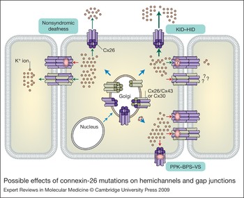
Current research has focused on defining the changes in channel properties induced by mutation that lead to the observed phenotypes for the different Cx26-related deafness disorders. To date, there have been many studies of Cx26 mutations, most of which have been carried out at a cellular level, using either the Xenopus oocyte expression assay or transfected mammalian cells (Refs Reference Richard108, Reference Montgomery116, Reference Mani132, Reference Mese133, Reference Gerido134, Reference Rouan135). In a study of dominant and recessive deafness causing mutations in GJB2, it was observed that the dominant mutation (W44C) failed to couple cells by itself and had a dominant-negative impact on coexpressed wild-type Cx26 (Ref. Reference Bruzzone136). Thus, it abolished the ability of the wild-type Cx26 channels to function properly, which is consistent with its dominant mode of inheritance (Ref. Reference Denoyelle40). By contrast, a recessive Cx26 mutation did not display the same dominant-negative inhibition of the wild-type protein, although it failed to make channels alone (Ref. Reference Bruzzone136). This study led to the idea that deafness could be caused by a disruption of normal Cx26-mediated gap junction communication, by disrupting the previously discussed flow of K+ ions within the inner ear through a loss-of-function effect (Fig. 4). As this loss of function does not cause skin disease in nonsyndromic patients, syndromic hearing loss would need to differ by having some sort of gain of function(s) in addition to the loss of gap junction coupling (Ref. Reference Dai52).
Possible effects of connexin-26 mutations on hemichannels and gap junctions. Purple connexin subunits are Cx26 proteins, and brown connexin subunits either Cx43 or Cx30. The brown dots represent K+ ions. Nonsyndromic mutations display a partial or complete loss of Cx26 gap junction activity. Most syndromic mutations found in Cx26 that result in KID–HID syndrome display large hemichannel currents and changes in gap junctional function. Most mutations in Cx26 that cause PPK–BPS–VS show a reduction of trafficking to the plasma membrane from the Golgi, cis-dominant inhibition of wild-type Cx26 and trans-dominant inhibition of other epidermal connexins.
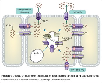
Syndromic mutations in GJB2 have been found to have additional functional characteristics thought to have a role in eliciting the different attributes of their associated disorder. There appears to be a common gain of function for the PPK–BPS–VS group of diseases. It has been shown that dominant mutations linked with these disorders have a cis-dominant negative effect on wild-type Cx26 proteins. In addition, the ΔE42, G59A, D66H and R75W mutations also have a trans-dominant negative effect on some of the other connexin proteins that are found in the epidermis, but might not have this effect on others (Cx43, Cx30 and Cx32) (Refs Reference Heathcote94, Reference Rouan135, Reference Richard137, Reference Marziano138, Reference Thomas139). For example, it was also observed that the D66H mutation resulted in an inhibition of wild-type Cx26 or Cx30 transport to the cellular membrane from the cytoplasm, which is consistent with a dominant mechanism of action (Ref. Reference Bakirtzis140). The mutations all result in a lack of functional Cx26 gap junction channels, which would explain their role in causing deafness (Table 2). These findings further suggest that mutations in GJB2 that result in PPK, BPS or VS gain the ability to trans-dominantly inhibit other connexin types found throughout the layers of the skin. Therefore, functional analyses have thus far identified distinct mechanisms between simple hearing loss mutations and the mutations linked to skin disorders found in patients with PPK, BPS or VS.
Known syndromic mutations in GJB2
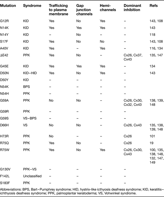
Abbreviations: BPS, Bart–Pumphrey syndrome; HID, hystrix-like icthyosis deafness syndrome; KID, keratitis–ichthyosis deafness syndrome; PPK, palmoplantar keratoderma; VS, Vohwinkel syndrome.
Since patients with KID–HID syndrome have a different skin phenotype than patients with PPK–BPS–VS, their Cx26 mutations might exhibit a different gain of function. A study of the Cx26 mutation A40V in Xenopus oocytes found that the expressed protein resulted in large hemichannel currents in single cells (Ref. Reference Montgomery116). This surprising feature was associated with an increase in cell death. An increase in cell death was also reported for the M163L mutation associated with nonsyndromic hearing loss, but it is unlikely to be due to hemichannel activity because the mutant did not traffic efficiently to the cell membrane (Ref. Reference Matos141). Although wild-type connexins can induce hemichannel currents that potentially have a role in normal cellular function (Refs Reference Saez4, Reference Bennett142), those recorded for A40V were much larger. The discovery of aberrant hemichannel currents caused by KID–HID-associated mutations, led to the idea that this additional role may be the gain of function that causes the disorders specific to KID–HID patients. Further analysis of G12R, N14K, G45E and D50N mutations revealed a common trend of aberrant hemichannel activity in cells expressing Cx26 mutations associated with these disorders (Refs Reference Stong117, Reference Gerido134, Reference Lee143). All GJB2 mutations related to KID–HID result in a disrupted gap junctional conductance (N14K and G45E have altered gating), which is a common feature among deafness mutations in Cx26 proteins (Table 2). In addition, Cx26 mutations G12R, N14K, G45E and D50N also result in an increase in cell death (Refs Reference Stong117, Reference Gerido134, Reference Matos141, Reference Lee143). This leads to the theory that KID–HID mutations in GJB2 result in aberrant hemichannel activity and an increase in cell death, which disrupts typical function in the epidermis and results in the phenotype specific to these patients.
To date, there has been only one in vivo study for syndromic mutations, which developed a mouse model expressing the VS-related D66H mutation in Cx26 (Ref. Reference Bakirtzis140). The animal displayed similar characteristics to those observed in patients with VS, such as constriction bands and hyperkeratosis. Further analysis showed that Cx26 and Cx30 failed to localise to the plasma membrane in epidermal cells, which is consistent with the theory of trans-dominant inhibition for VS–BPS–PPK mutations. Knockout mice have been created to model the loss of Cx26 protein expression within the inner ear; these mice displayed extensive cell death in supporting cells surrounding the inner hair cells, which triggered the death of hair cells and other cells within the inner ear (Refs Reference Cohen-Salmon144, Reference Wang145). The mice also showed hearing loss, as would be expected. Such studies are useful in making the ultimate link between in vitro theory and the observed clinical features of patients.
Acknowledgements and funding
Work in our laboratory is supported by National Institutes of Health grants EY13163 and DC06652. We thank the reviewers for their insight and thoughtful comments.