Introduction
Gastrointestinal helminths pose serious problems to ruminants because of the impact they have on meat and milk production and the high cost of management measures and control (Waller, Reference Waller1999; Araújo et al., Reference Araújo, Freitas, Vieira and Campos2006).
The cestode Moniezia sp. stands out among helminths as a very common infective agent in domestic ruminants worldwide; however, its pathological action on hosts has not been totally elucidated since its pathogenicity differs between young and adult animals (Beveridge, Reference Beveridge, Khalil, Jones and Bray1994). Approximately seven species of the genus Moniezia have been described to parasitize ruminants, but only Moniezia expansa and M. benedeni are considered important (Spasskii, Reference Spasskii1951).
New alternative control practices to minimize pasture contamination and reinfestation, such as biological control, can help to solve the current problems caused by this and other parasitic diseases (Gomes et al., Reference Gomes, Araújo and Ribeiro1999; Araújo et al., Reference Araújo, Freitas, Vieira and Campos2006). The use of nematophagous fungi appears therefore as a viable and promising alternative control for animals that are constantly being infected (Araújo et al., Reference Araújo, Guimarães, Campos, Sá and Assis2004). One of the several advantages of biological control using nematophagous fungi is the synergism with chemical control, which provides larger and wider action on the infective forms present in faeces, as well as on adult helminths parasitizing animals (Mota et al., Reference Mota, Campos and Araújo2003).
Gronvold et al. (Reference Gronvold, Wolstrup, Nansen, Henriksen, Larsen and Bresciani1993) reported that nematode-trapping fungi are universally common organisms with some negative effects on the transmission of a number of parasites and are considered as a viable alternative strategy for controlling animal-parasitic nematodes.
Nematophagous fungi comprise different types of fungi divided into predators, endoparasites and opportunists. These are cosmopolitan fungi, occurring in natural and agricultural soils and in all types of organic matter in decomposition (Araújo et al., Reference Araújo, Guimarães, Campos, Sá and Assis2004). Egg-parasitic fungi have been described as saprophytes and therefore do not depend on the presence of helminth eggs in the soil for their survival; they can be easily isolated and cultivated in laboratory. These fungi colonize the egg contents, even the developing larva within the egg (Lysek, Reference Lysek1978).
Fungal egg parasitism is an important biological phenomenon that can be used for biological control of helminths. The potential value of these microorganisms for biological control seems to be enormous (Lysek et al., Reference Lysek, Fassatiová, Pineda and Hernández1982). Their mechanism of action is based on three types of ovicidal activity: (1) physiological, with biochemical effect without morphological damage to eggshells; (2) lytic biochemical effect, with progressive morphological change in eggshells and embryos; and (3) lytic and morphological effects, with egg penetration and embryo mortality.
Among the promising species of this group, Paecilomyces lilacinus stands out as a potential biocontrol agent (Araújo et al., Reference Araújo, Santos and Ferraz1995; Ciarmela et al., Reference Ciarmela, Basualdo and Basualdo2000, Reference Ciarmela, Lori and Basulado2002). Paecilomyces lilacinus (Thom.) is a primary saprophytic filamentous fungus with cosmopolitan distribution, occurring more frequently in warmer regions. This fungus is nutritionally versatile and capable of penetrating and destroying helminth eggs (Fiedler & Sosnowska, Reference Fiedler and Sosnowska2007). Additionally, mechanical and enzymatic processes are involved, with the production of an alkaline serine protease during egg infection conferring on the fungus predatory capacity (Carneiro & Cayrol, Reference Carneiro and Cayrol1991; Park et al., Reference Park, Hargreaves, McConville, Stirling, Ghisalberti and Sivasithamparam2004). Although P. lilacinus has been reported in different types of cultivated or non-cultivated Brazilian soils, at depths of 0–40 cm or more (Carneiro & Cayrol, 1991) studies on its effectiveness against cestode eggs are still scarce.
The objective of this study was to evaluate the in vitro activity of the nematophagous fungus P. lilacinus against Moniezia sp. eggs.
Material and methods
Fungal cultures and cestode eggs
Paecilomyces lilacinus collected from a soil sample in Brazil was incubated in test tubes containing 2% corn-meal agar (2% CMA), in darkness at 4°C for 10 days.
Culture discs, 4 mm in diameter, were removed from grown fungal colonies, plated on to 9-cm diameter Petri dishes containing 20 ml of 2% potato-dextrose-agar (PDA 2%) and stored in the dark, at 25°C for 10 days. After colony growth, new culture discs, 4 mm in diameter, were transferred to 9-cm diameter Petri dishes containing 20 ml of 2% water-agar (2% WA) for 10 days.
Moniezia sp. eggs were analysed morphologically under an optical microscope using a 10 × objective lens. Eggs were recovered by dissection of an adult Moniezia sp. obtained from a goat that had died of natural causes.
Experimental assay with eggs of Moniezia
One hundred thousand eggs were analysed morphologically under an optical microscope using a 10 × objective lens and incubated on plates containing 2% WA medium with the isolates grown for 10 days and the control without fungus, with ten repetitions per group. Each plate in the treatments with the fungus contained 1000 eggs. After 5, 10 and 15 days post-incubation, approximately 100 eggs were collected from each plate with the fungus and from the control, as described by Araújo et al. (Reference Araújo, Santos and Ferraz1995), placed in glass slides with a drop of 1% Aman blue and evaluated under 10 × , 20 × and 40 × lenses according to Lysek et al. (Reference Lysek, Fassatiová, Pineda and Hernández1982): type 1, lytic effect without morphological damage to eggshells and hyphal adherence; type 2, lytic effect with morphological change in embryos and eggshells, without hyphal penetration of eggshells, and type 3, lytic effect with morphological change in embryos and eggshells, as well as hyphal penetration and internal colonization of eggs.
Data on each studied interval were examined by non-parametric Friedman 1% probability (Ayres et al., Reference Ayres, Ayres, Ayres and Santos2003).
Results and discussion
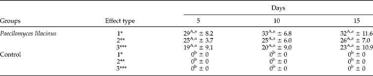
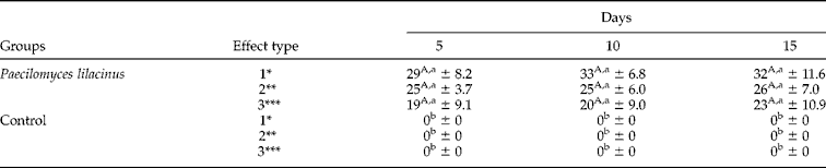
Percentages for type 1, 2 and 3 lytic effects of P. lilacinus ovicidal activity after 5, 10 and 15 days of interaction on Moniezia sp. eggs are shown in table 1.
Percentages and standard deviations of type 1, 2 and 3 effects of the fungus Paecilomyces lilacinus and the control without fungus on Moniezia sp. eggs after 5, 10 and 15 days post-incubation.
Percentages followed by same capital letters within the row are not significantly different by the Friedman test (P>0.01).
Percentages followed by same small letters within the column are not significantly different by the Friedman test (P>0.01).
* Lytic effect without morphological damage and hyphal adherence to eggshells.
** Lytic effect with morphological change in embryos and eggshells, without hyphal penetration of eggshells.
*** Lytic effect with morphological change in embryos and eggshells, with hyphal penetration and internal colonization of eggs.
Significant differences (P < 0.01) were found between the ovicidal activity of P. lilacinus against Moniezia sp. eggs over the intervals and the control group, characterizing the ovicidal activity of the fungus. The comparison of effects on eggs after 5, 10 and 15 days post-incubation showed no significant differences (P < 0.01), indicating that the activity of P. lilacinus occurred on the first day of observation (5 days post-incubation). However, the comparison between fungus-treated eggs and control eggs showed significant differences (P < 0.01) for all the intervals.
Although type 1 and 2 effects were found in the interaction between fungus and eggs, according to Lysek (Reference Lysek1976) a fungus is described as having ovicidal activity if it shows type 3 effects during the process of infection. After 5, 10 and 15 days post-incubation, type 3 effects were recorded as 19, 20 and 23%, respectively, which characterizes the fungus P. lilacinus as a potential biological control agent for this cestode.
According to Basualdo et al. (Reference Basualdo, Ciarmela, Sarmiento and Minvielle2000), the ovicidal activity of P. lilacinus against soil helminths points out its importance as a biocontrol agent. Studies have confirmed the P. lilacinus ovicidal activity against several helminth species (Olivares-Bernabeu & López-Llorca, Reference Olivares-Bernabeu and López-Llorca2002; Santiago et al., Reference Santiago, Homechim, Silva, Ribeiro, Gomes and Santoro2006). Araújo et al. (Reference Araújo, Santos and Ferraz1995) and Basualdo et al. (Reference Basualdo, Ciarmela, Sarmiento and Minvielle2000) reported high in vitro activity of P. lilacinus on eggs of Toxocara canis. Mizobutsi et al. (Reference Mizobutsi, Ferraz and Ribeiro2000), working with 64 isolates of 25 fungal species, among them P. lilacinus, found 77% of parasitism by this species on eggs of Meloidogyne javanica, showing better results than the other species tested. Ciarmela et al. (Reference Ciarmela, Thevenet, Alvarez, Minvielle and Basualdo2005) reported a negative biological activity of P. lilacinus on the in vitro viability of Taenia hydatigena oncospheres when compared with the control group.
This research is the first report of the action of P. lilacinus against eggs of Moniezia sp., identifying this fungus as a potential candidate for biological control of this helminth.