Introduction
The Ellipsocephaloidea Matthew, Reference Matthew1887 (sensu Adrain, Reference Adrain2011) comprises morphologically diverse trilobites that were major faunal components of the Cambrian Epoch 2 and 3 ecosystems (e.g., Suvorova, Reference Suvorova1956; Ahlberg and Bergström, Reference Ahlberg and Bergström1978; Zhang et al., Reference Zhang, Lu, Zhu, Qian, Lin, Zhou, Zhang and Yuan1980; Geyer, Reference Geyer1990; Jell, Reference Jell1990; Westrop and Landing, Reference Westrop and Landing2000; Żylińska and Masiak, Reference Żylińska and Masiak2007; Elicki and Geyer, Reference Elicki and Geyer2013; Høyberget et al., Reference Høyberget, Ebbestad, Funke and Nakrem2015). The classification and phylogeny of this group is still not fully understood, and attempts to find its unique synapomorphies have not yet been successful (Geyer, Reference Geyer1990). This is partially caused by insufficient knowledge of post-embryonic development in many members of this superfamily.
Developmental data are important for resolving phylogenetic relationships at various taxonomic levels (Edgecombe et al., Reference Edgecombe, Speyer and Chatterton1988; Fortey and Chatterton, Reference Fortey and Chatterton1988; Chatterton et al., Reference Chatterton, Siveter, Edgecombe and Hunt1990; Campbell and Chatterton, Reference Campbell and Chatterton2009) and for identifying homologous structures (Chatterton, Reference Chatterton1971; Mabee, Reference Mabee2000). Detailed studies of post-embryonic development have the potential to be fundamental for a phylogenetic analysis of ellipsocephaloid trilobites, but our current knowledge of development in this group is based on a few immature specimens (e.g., Suvorova, Reference Suvorova1956; Yi, Reference Yi1988; Geyer, Reference Geyer1990; Geyer and Landing, Reference Geyer and Landing2004) or comparatively late developmental stages only (Cederström et al., Reference Cederström, Ahlberg, Nilsson, Ahlgren and Eriksson2011; Laibl et al., Reference Laibl, Fatka, Budil, Ahlberg, Szabad, Vokáč and Kozák2015). More comprehensive data are available only for Ellipsostrenua gripi (Kautsky, Reference Kautsky1945), Ichangia ichangensis Chang, Reference Chang1957, Strenuaeva? spinosa Ahlberg and Bergström, Reference Ahlberg and Bergström1978, and Estaingia sinensis (Chang, Reference Chang1953) (see Kautsky, Reference Kautsky1945; Zhang and Pratt, Reference Zhang and Pratt1999; Cederström et al., Reference Cederström, Ahlberg, Babcock, Ahlgren, Høyberget and Nilsson2012; Dai and Zhang, Reference Dai and Zhang2012; respectively).
In this contribution we provide the first detailed description of the early post-embryonic development (protaspid and early meraspid stages) in Ellipsostrenua granulosa (Ahlberg, Reference Ahlberg1984). Consequently, we compare the development of E. granulosa with some other members of Ellipsocephalidae Matthew, Reference Matthew1887 and with members of the closely related Estaingiidae Öpik, Reference Öpik1975 in order to elucidate the developmental patterns in Ellipsocephaloidea.
Geological setting
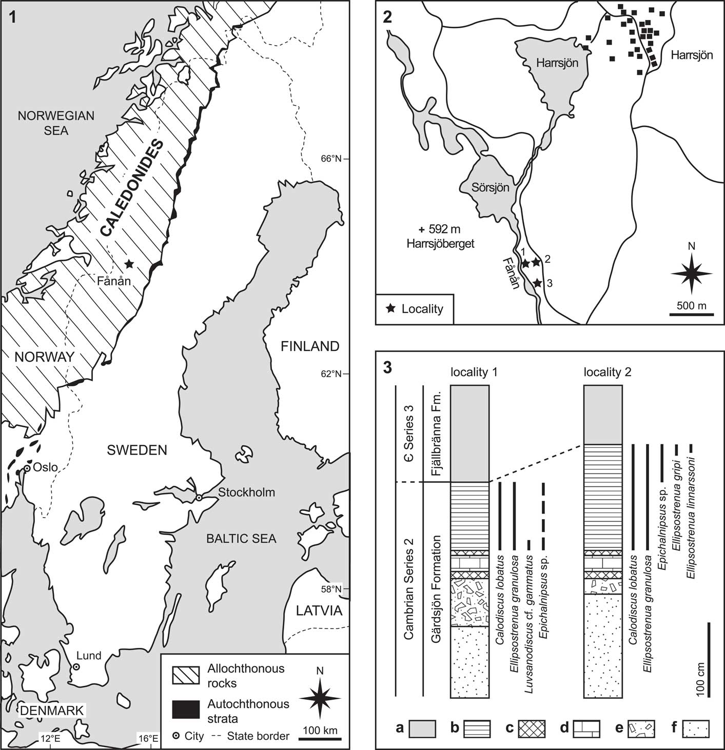
The material was collected by Peter Cederström from an ~1 m thick, gray calcareous mudstone at the top of the Gärdsjön Formation (Cambrian Series 2, Stage 4) in the Fånån area, south of Lake Sörsjön, northern Jämtland, Sweden (Fig. 1.1; see also Cederström et al., Reference Cederström, Ahlberg, Clarkson, Nilsson and Axheimer2009 for details). Hundreds of sclerites were collected at two localities (localities 1 and 2 in Fig. 1.2). The majority of sclerites are from the eastern bank of the Fånån rivulet, ~300 m south of Sörsjön (locality 1). This locality is situated ~300 m north of the trilobite-bearing locality (locality 3) described by Asklund and Thorslund (Reference Asklund and Thorslund1935) and Ahlberg (Reference Ahlberg1984). The second locality is a gravel pit situated 130 m east of locality 1. All trilobite sclerites from the Fånån area are generally slightly distorted tectonically. Associated trilobites include Calodiscus lobatus (Hall, Reference Hall1847); Luvsanodiscus cf. gammatus Korobov, Reference Korobov1980; Ellipsostrenua gripi (Kautsky, Reference Kautsky1945); E. linnarssoni (Kiær, Reference Kiær1917); and Epichalnipsus sp. (Fig. 1.3; see also Cederström et al., Reference Cederström, Ahlberg, Clarkson, Nilsson and Axheimer2009). The Fånån trilobites are from the Strenuaeva? spinosa Zone (Cambrian Series 2 zonation of Ahlberg et al., Reference Ahlberg, Cederström and Babcock2016). At both localities, the mudstone succession is vertically dipping and overlain by black shales of the Fjällbränna Formation (Fig. 1.3). This black shale succession has yielded fossils indicative of the Acadoparadoxides oelandicus Superzone (Stage 5).
Maps and stratigraphic section: (1) map of Scandinavia showing the location of the Fånån area (Jämtland, Sweden), and the distribution of allochthonous and autochthonous rocks in the Scandinavian Caledonides, based on Cederström et al. (Reference Cederström, Ahlberg, Clarkson, Nilsson and Axheimer2009); (2) detailed map of the Fånån area showing localities referred to in the text (locality 1, outcrop along the eastern bank of the Fånån rivulet, described by Cederström et al. (Reference Cederström, Ahlberg, Clarkson, Nilsson and Axheimer2009), locality 2, gravel pit described by Cederström et al. (Reference Cederström, Ahlberg, Clarkson, Nilsson and Axheimer2009), locality 3, type locality of E. granulosa, described by Asklund and Thorslund,Reference Asklund and Thorslund1935 and Ahlberg, Reference Ahlberg1984); (3) lithological succession and the stratigraphical distribution of trilobites in localities 1 and 2, based on Cederström et al. (Reference Cederström, Ahlberg, Clarkson, Nilsson and Axheimer2009) (a, black shale; b, calcareous mudstone; c, siltstone; d, limestone; e, conglomerate; f, sandstone).

Materials and methods
Material
The studied specimens are preserved as diagenetically slightly altered calcareous sclerites in a gray calcareous mudstone. All studied specimens were covered by ammonium chloride sublimate and photographed using an optical microscope (Keyence VHX-2000). Line drawings were produced directly from the photographs in Adobe Illustrator CS2.
Methods
The length and width dimensions of protaspid protocranidia (N=15) and meraspid cranidia (N=6) were measured from photographs by the optical image analyzer TpsDig2 (Rohlf, Reference Rohlf2006). In several protaspid and meraspid specimens (N=15) preserved with the intergenal spines (or at least their bases), the transverse distance between these spines was measured (intergenal spine separation sensu Webster, Reference Webster2007). The measured parameters were plotted on a bivariate diagram using the PAST package (Hammer et al., Reference Hammer, Harper and Ryan2001).
Additional data for the analysis of protaspid and early meraspid cranidial dimensions were collected from publications by Kautsky (Reference Kautsky1945), Zhang and Pratt (Reference Zhang and Pratt1999), and Dai and Zhang (Reference Dai and Zhang2012). The protocranidial and cranidial width was used as a proxy of protaspid and meraspid sizes. The width or width ranges were taken directly from the text descriptions of the mentioned publications.
Repository and institutional abbreviations
All figured specimens are numbered with the prefix Lund Original (LO) or Lund Reference (LR), and are housed at the Department of Geology, Lund University, Sweden.
Systematic paleontology
Morphological terminology used in the description generally follows Whittington and Kelly (Reference Whittington and Kelly1997), Palmer (Reference Palmer1998, for the terminology of macropleural segments), and Hughes et al. (Reference Hughes, Minelli and Fusco2006, for the terminology of the postcephalic part of the exoskeleton). Spines projecting from the postero-lateral corner of protaspid protocranidia (= posterior fixigenal spine of Palmer, Reference Palmer1962) or early meraspid cranidia (= intergenal spine) are here considered as homologous (cf., Størmer, Reference Størmer1942), and therefore in both cases are designated as intergenal spines. We follow the trilobite classification of Adrain (Reference Adrain2011).
Superfamily Ellipsocephaloidea Matthew, Reference Matthew1887
Family Ellipsocephalidae Matthew, Reference Matthew1887
Remarks
The concept of the family as presented by Jell and Adrain (Reference Jell and Adrain2003) is followed here. The whole group, however, needs a comprehensive revision supplemented by parsimony analysis.
Genus Ellipsostrenua (Kautsky, Reference Kautsky1945)
Type species
Strenuella (Ellipsostrenua) gripi Kautsky, Reference Kautsky1945 from the Grammajukku Formation (Cambrian Stage 4), Laisvall area, Lapland, northern Sweden.
Remarks
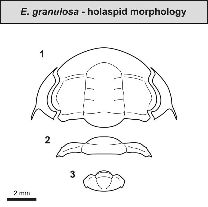
Kautsky (Reference Kautsky1945) erected Ellipsostrenua as a subgenus of Strenuella Matthew, Reference Matthew1887 with Strenuella (Ellipsostrenua) gripi Kautsky, Reference Kautsky1945 as type species. Henningsmoen (Reference Henningsmoen1959) raised Ellipsostrenua to generic level, and this approach was later followed by other authors (e.g., Sdzuy, Reference Sdzuy1961; Rushton, Reference Rushton1966). Ahlberg and Bergström (Reference Ahlberg and Bergström1978) considered Ellipsostrenua to be a junior subjective synonym of Ellipsocephalus Zenker, Reference Zenker1833. The type species of Ellipsostrenua differs, however, in several important respects from the type species of Ellipsocephalus, and Ellipsostrenua is here treated as a separate genus. The characters distinguishing Ellipsostrenua (Fig. 2) from Ellipsocephalus (cf., Šnajdr, Reference Šnajdr1958) include: (1) a tapering glabella without distinct anterolateral corners; (2) distinct genal spines; (3) pleurae terminating in sharp spines; and (4) a pygidium with narrow pleural regions, a wide, convex rhachis, and a pair of small spines.
Reconstruction of the adult morphology of Ellipsostrenua granulosa (Ahlberg, Reference Ahlberg1984): (1) cranidium and librigenae; (2) thoracic tergite; (3) pygidium. Based on Ahlberg (Reference Ahlberg1984). Surface sculpture omitted.

Ellipsostrenua granulosa (Ahlberg, Reference Ahlberg1984)
1984 Ellipsocephalus? granulosus Reference AhlbergAhlberg, p. 356, fig. 6.
2009 Ellipsocephalus? granulosus; Reference Cederström, Ahlberg, Clarkson, Nilsson and AxheimerCederström et al., p. 495, text-fig. 5.
non 2009 Ellipsocephalus? granulosus; Reference Cederström, Ahlberg, Clarkson, Nilsson and AxheimerCederström et al., p. 506, pl. 4, figs. 8–10.
2012 Ellipsocephalus granulosus; Reference Cederström, Ahlberg, Babcock, Ahlgren, Høyberget and NilssonCederström et al., p. 167, fig. 15AC–AF.
2015 Ornamentaspis? granulatus; Reference Høyberget, Ebbestad, Funke and NakremHøyberget et al., p. 39.
Holotype
An incomplete cranidium deposited in the type collections of the Geological Survey of Sweden, Uppsala, as SGU Type 3935 (Ahlberg, Reference Ahlberg1984, fig. 6A).
Diagnosis
See Ahlberg (Reference Ahlberg1984, p. 357).
Remarks
Ellipsostrenua granulosa is similar to closely related species E. gripi and E. linnnarssoni (cf., Kautsky, Reference Kautsky1945; Høyberget et al., Reference Høyberget, Ebbestad, Funke and Nakrem2015), but differs from them by: (1) strong granulation on the glabella, fixigenae, and thoracic tergites (in E. gripi and E. linnarssoni these parts of exoskeleton are generally pitted); (2) longer genal spines; and (3) more distinct eye ridges (see Ahlberg Reference Ahlberg1984).
Protaspides that were tentatively assigned to E. granulosa by Cederström et al. (Reference Cederström, Ahlberg, Clarkson, Nilsson and Axheimer2009, pl. 4, figs. 8–10) most likely belong to Calodiscus lobatus because they closely resemble protaspides of the latter species.
Post-embryonic development of Ellipsostrenua granulosa
Post-embryonic development in trilobites is traditionally divided into protaspid, meraspid, and holaspid periods based on the trunk articulation (for a comprehensive review see Whittington, Reference Whittington1957; Chatterton and Speyer, Reference Chatterton and Speyer1997; Hughes et al., Reference Hughes, Minelli and Fusco2006). We follow this division herein, although we are aware that these periods probably do not represent corresponding life phases among all trilobite taxa (for a detailed discussion, see Chatterton and Speyer, Reference Chatterton and Speyer1997; Park and Choi, Reference Park and Choi2011).
Protaspid period
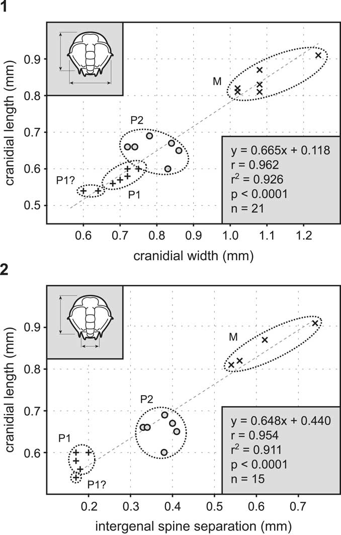
Among protaspides of E. granulosa it is possible to recognize early and late protaspid stages that are distinguished mainly by the presence of a distinct trunk portion in the late protaspid stage. Based on cranidial length and width measurements (Fig. 3.1), we cannot exclude the possibility that the two smallest specimens (marked as P1? in Fig. 3.1) may belong to an even earlier protaspid stage. However, the comparison of the cranidial length and intergenal spine separation (Fig. 3.2) shows slightly more distinct clusters. Because the intergenal spine separation undoubtedly reflects the width of the trunk more precisely than the cranidial width, we believe it is a more reliable indicator of the natural instars (provided that the development of the trunk is the most dynamic pattern during early development in all trilobites; see also Webster, Reference Webster2007, for comparable results in Nephrolenellus from the uppermost Dyeran Stage of the Great Basin U.S.A.). Therefore, a two stage division of the protaspid period in E. granulosa is employed herein.
Plots of Ellipsostrenua granulosa (Ahlberg, Reference Ahlberg1984): (1) scatter plot of cranidial width versus cranidial length of protocranidia and cranidia of Ellipsostrenua granulosa (Ahlberg, Reference Ahlberg1984); (2) scatter plot of intergenal spine separation versus cranidial length of protocranidia and cranidia of Ellipsostrenua granulosa (Ahlberg, Reference Ahlberg1984). Abbreviations: M, meraspid stages; P1, early protaspid stage; P2, late protaspid stage.
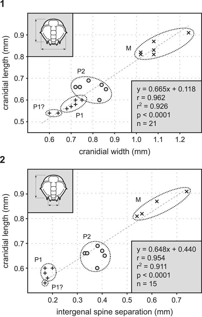
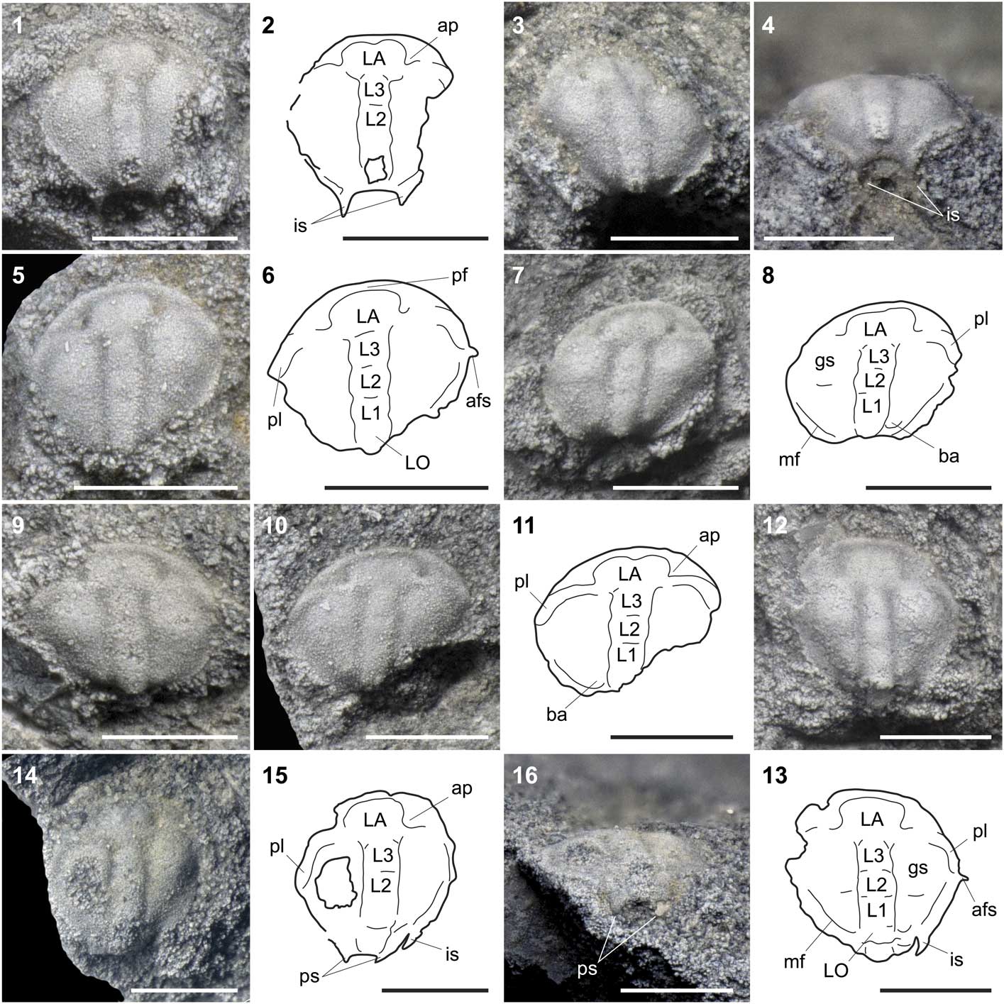
The early protaspid stage (Figs. 4.1–4.11, 6.1, 6.2) is sub-circular to sub-elliptical in outline and gently vaulted in lateral view. The protocranidium is 0.54–0.60 mm long and 0.60–0.75 mm wide. The axial furrows are sub-parallel and diverge anteriorly around a distinct frontal axial lobe (LA). Well-developed anterior pits are present lateral to LA. The axis of the early protaspid stage has five morphologically expressed segments, designated as LA–LO. In some specimens there is a shallow medial depression in the anterior margin of LA. The anterior border protrudes forward to form a very short preglabellar field. The proximal parts of the eye ridges originate behind the anterior pits and are connected to the posterior part of LA. Distally, the eye ridges are terminated by well-developed palpebral lobes that reach approximately to the exoskeletal mid-length. Minute anterior fixigenal spines are developed near the posterior tip of each palpebral lobe. The fixigenae are sub-oval with a slightly swollen anterior part. The bacullae are distinct and situated lateral to SO. The posterior border furrow extends from the bacullae, curves forward abaxially, and reaches the posterior tip of the palpebral lobe. A pair of postero-ventrally directed intergenal spines is developed on the posterior margin of the exoskeleton.
Photographs and interpretative drawings of protaspid stages of Ellipsostrenua granulosa (Ahlberg, Reference Ahlberg1984) from the Fånån area (Jämtland, Sweden): (1–11) early protaspid stage; (1, 2) LO 12403t, dorsal view; (3) LO 12404t, dorsal view; (4) LO 12404t, posterior view; (5, 6) LO 12405t, dorsal view; (7, 8) LO 12406t, dorsal view; (9) LO 12407t, dorsal view; (10, 11) LO 12408t, dorsal view; (12–16) late protaspid stage; (12, 13) LO 12409t, dorsal view; (14, 15) LO 12410t, dorsal view; (16) LO 12410t, posterior view. Abbreviations: afs, anterior fixigenal spine; ap, anterior pit; ba, baccula; gs, genal swelling; is, intergenal spine; LA–LO, glabellar lobes; mf, marginal furrow; pf, preglabellar field; pl, palpebral lobe; ps, pleural spine. All scale bars represent 0.5 mm.
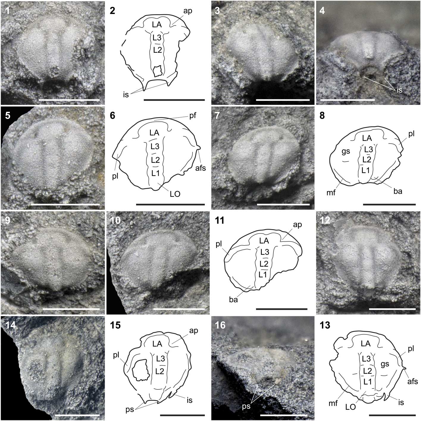
The late protaspid stage (Figs. 4.12–4.16, 6.3, 6.4) is similar to the early protaspid stage with the following exceptions: the protocranidium is 0.60–0.69 mm long and 0.72–0.86 mm wide; the palpebral lobes are slightly longer (exsag.); the glabella is clearly divided into LA–LO. The most striking difference is, however, the presence of a trunk, which occupies the space between the intergenal spines. The trunk bears one pair of short postero-ventrally directed pleural spines.
Meraspid period
The sclerites assigned to early meraspid stages of E. granulosa comprise early meraspid cranidia and pygidia.
Early meraspid cranidia (Figs. 5.1–5.5, 6.5) are sub-elliptical to sub-rectangular in outline, 0.81–0.91 mm long, and 1.02–1.24 mm wide. The glabella is narrow and divided into four glabellar lobes. LO to L3 are of similar width, LA being approximately twice as wide as LO–L3. A medial depression may be developed on the anterior margin of LA. The anterior cranidial border is comparatively wide (tr.) and long (sag.). Eye ridges are proximally connected to the posterior half of LA. The palpebral lobes reach the cranidial mid-length (exsag.). The anterior branches of the facial suture are short and sub-parallel. The posterior branch of the facial suture is almost imperceptible. The fixigenae are slightly vaulted, with distinct bacullae that are located next to the posterior margin of L1. The posterior border furrow is situated close to the intergenal spine, curved anteriorly, and reaches the posterior branch of the facial suture. The intergenal spines are generally broken off, but appear to have been short, directed horizontally, and situated at the postero-lateral edges of the cranidium.
Photographs and interpretative drawings of meraspid cranidia, meraspid pygidia and protaspid hypostomes of Ellipsostrenua granulosa (Ahlberg, Reference Ahlberg1984) from the Fånån area (Jämtland, Sweden): (1–5) early meraspid cranidia; (1, 2) LO 12411t, dorsal view; (3, 4) LO 12412t, dorsal view; (5) LO 12413t, dorsal view; (6–8) protaspid hypostomes; (6, 7) LO 12414t, ventral view; (8) LO 12415t, ventral view; (9–12) meraspid pygidia; (9, 10) LO 12416t, dorsal view; (11, 12) LO 12417t, dorsal view. Abbreviations: 2ms, second macrospine; ahr, articulating half-ring; ar, axial ring; aw, anterior wing; ba, baccula; is, intergenal spine; LA–LO, glabellar lobes; mb, middle hypostomal body; mf, marginal furrow; ms, marginal spines; pf, preglabellar field; pl, palpebral lobe; ps, pleural spine. All scale bars represent 0.5 mm.

Reconstruction of protaspid and early meraspid stages of Ellipsostrenua granulosa (Ahlberg, Reference Ahlberg1984) from the Fånån area (Jämtland, Sweden): (1) early protaspid stage, dorsal view; (2) early protaspid stage, posterior view; (3) late protaspid stage, dorsal view; (4) late protaspid stage, posterior view; (5) early meraspid cranidium, dorsal view; (6) early meraspid hypostome, ventral view; (7, 8) early meraspid pygidia, dorsal view.

The smallest known pygidia (Figs. 5.9, 5.10, 6.8) are 0.33–0.49 mm long and 0.47–0.70 mm wide. The axis is composed of three to five segments. The pleurae are separated by interpleural furrows and, with exception of the smallest pygidium, by distinct pleural furrows. The anteriormost pygidial segment is slightly longer (exsag.) than more posteriorly located segments and bears a pair of comparatively long macrospines. Minute pleural spines are developed in the segments posterior to the macrospinous segment. A comparison with other trilobites of the family Ellipsocephalidae (cf., Laibl et al., Reference Laibl, Fatka, Budil, Ahlberg, Szabad, Vokáč and Kozák2015) suggests that the macrospinous segment in these meraspid pygidia represents the second segment of the trunk, with the first segment already released into the thorax, making this an M1 meraspid pygidium. Hence, the width of these pygidia should roughly correspond to the width between the intergenal spines in early meraspid cranidia.
Later meraspid pygidia (Figs. 5.11, 5.12, 6.7) are semicircular in outline and ~0.5 mm long and 0.7 mm wide. The axis consists of six segments. The pleurae have distinct pleural and interpleural furrows and several pairs of pleural spines, except in some of the posterior-most segments.
Hypostomes
The small hypostomes assigned to E. granulosa are hourglass-shaped in outline, 0.35–0.50 mm long and 0.30–0.50 mm wide across the posterior lobe, and have an anterior margin suggestive of conterminant attachment (Figs. 5.6–5.8, 6.6). The middle hypostomal body is subtriangular and the posterior lobe is circular with at least four pairs of spines along its margin. The anterior wings project laterally. The size of these hypostomes suggests that they belong to early protaspides up to earliest meraspides.
Discussion
Comparative development of Ellipsocephalidae and Estaingiidae
In addition to Ellipsostrenua granulosa, the only other species of Ellipsocephalidae with sufficiently well-known early post-embryonic stages are E. gripi and S.? spinosa. Kautsky (Reference Kautsky1945) described protaspides, meraspid cranidia, and holaspid individuals of E. gripi from the top of the Grammajukku Formation (Cambrian Stage 4) in the Laisvall area, Lapland, northern Sweden. The early stages of E. gripi are morphologically nearly equivalent to the stages of E. granulosa described herein. The main difference is the comparatively longer intergenal spines in some meraspid cranidia of E. gripi (cf., Kautsky, Reference Kautsky1945, pl. 16, fig. 8, pl. 18, fig. 1). This difference can be attributed either to interspecific differences or, more likely, to the fragmentary nature of the early meraspid cranidia of E. granulosa resulting in frequent breakage of such spines. Indeed, the absence of intergenal spines in numerous other cranidia of E. gripi (cf., Kautsky, Reference Kautsky1945, pl. 16, figs. 4–7, 9, pl. 17, figs. 5–7) favors the latter possibility. Cederström et al. (Reference Cederström, Ahlberg, Babcock, Ahlgren, Høyberget and Nilsson2012) described disarticulated meraspid and holaspid sclerites of S.? spinosa from the Grammajukku Formation (Stage 4) in the Storuman and Laisvall areas, Lapland, Sweden. These meraspid cranidia are comparable to those of E. granulosa and E. gripi, but they have a slightly more pointed intergenal angle, a more rounded anterior border, and longer intergenal spines.
Sufficient developmental data, including numerous protaspid and early meraspid stages, are also known from some members of the Estaingiidae, which is believed to be phylogenetically closely related to Ellipsocephalidae (Paterson and Edgecombe, Reference Paterson and Edgecombe2006); both families are classified in the Ellipsocephaloidea Matthew, Reference Matthew1887 (cf., Adrain, Reference Adrain2011). Superbly preserved phosphatized sclerites of Ichangia ichangensis from the Shuigoukou Formation (Henan Province, South China plate) were described by Zhang and Pratt (Reference Zhang and Pratt1999) and Zhang and Clarkson (Reference Zhang and Clarkson2012). The protaspid period of I. ichangensis can be divided into two stages (stages 0 and 1 sensu Zhang and Pratt, Reference Zhang and Pratt1999). Dai and Zhang (Reference Dai and Zhang2012) described protaspides and meraspid cranidia of Estaingia sinensis from the Shuijingtuo Formation (Hubei Province, South China plate) and synonymized phosphatized material described by Zhang and Pratt (Reference Zhang and Pratt1999, p. 125) as “genus and species indeterminate 2” with E. sinensis. If the specimens described by both Zhang and Pratt (Reference Zhang and Pratt1999) and Dai and Zhang (Reference Dai and Zhang2012) are conspecific, the protaspid period of E. sinensis comprises an early stage (stage 0 sensu Zhang and Pratt, Reference Zhang and Pratt1999) and one (stage 1 sensu Zang and Pratt, Reference Zhang and Pratt1999) or two (anaprotaspid and metaprotaspid stages sensu Dai and Zhang, Reference Dai and Zhang2012) later stages.
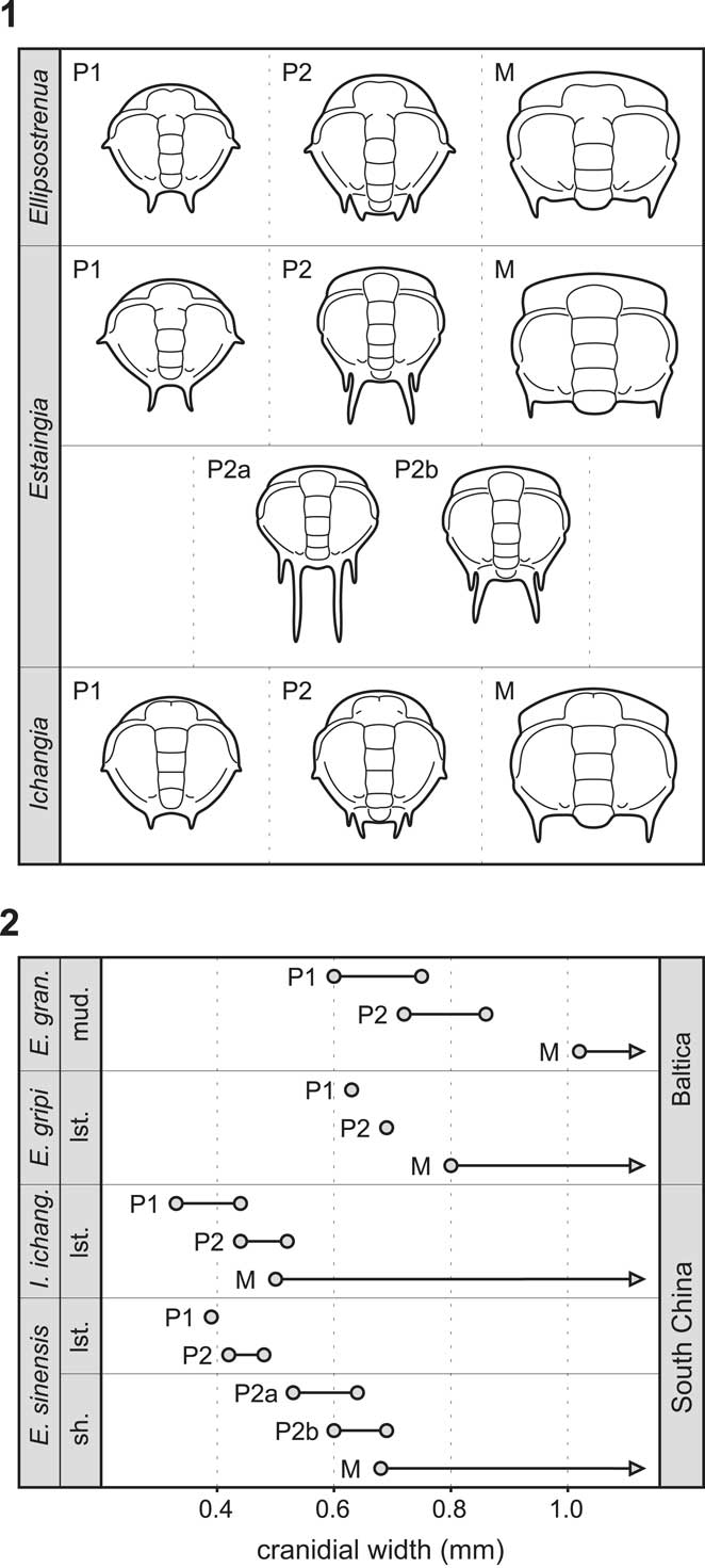
The earliest known protaspid stages of Ellipsostrenua, Ichangia, and Estaingia share a narrow axis with the same number of dorsally expressed segments (LA–LO). All of them have the same number of fixigenal spines in comparable positions. The trunk portion is not distinguishable. Hence, these earliest protaspides seem to represent homologous stages in all taxa discussed. Other morphological similarities (Fig. 7.1) are: (1) the early protaspides of these genera are all circular to elliptical and have distinct bacullae, (2) their palpebral lobes reach the exoskeletal mid-length, (3) the anterior parts of the fixigenae are gently swollen, (4) there is a short but distinct preglabellar field, and (5) there is an anteriorly curved posterior border furrow. The orientation of the fixigenal spines is identical: the anterior fixigenal spine is pointed laterally; the intergenal spine is directed postero-ventrally. The differences between early protaspides of these genera are minor and mainly include the shape of the LA and the length of the fixigenal spines (Fig. 7.1).
Comparative development of Ellipsocephalidae and Estaingiidae: (1) morphological comparison of protaspid and early meraspid stages of Ellipsostrenua, Estaingia, and Ichangia, based on Zhang and Pratt (Reference Zhang and Pratt1999), Dai and Zhang (Reference Dai and Zhang2012), Zhang and Clarkson (Reference Zhang and Clarkson2012), and data presented herein; (2) size comparison of protaspid and early meraspid stages of Ellipsostrenua granulosa, E. gripi, Ichangia ichangensis, and Estaingia sinensis, circles represent minimal and maximal cranidial width of the protaspid and early meraspid stages, based on Kautsky (Reference Kautsky1945), Zhang and Pratt (Reference Zhang and Pratt1999), Dai and Zhang (Reference Dai and Zhang2012), and data presented herein. Abbreviations: lst, limestone; M, meraspid stages; mud, mudstone; P1, early protaspid stage; P2, late protaspid stage; P2a, first late protaspid stage; P2b, second late protaspid stage; sh, shale.

Late protaspides of Ellipsostrenua, Ichangia, and Estangia (Fig. 7.1) are similar in their basic body plan. They have circular to sub-hexagonal protocranidia with a posterior border furrow curved forward abaxially, so that it reaches the posterior tip of the palpebral lobe. The protocranidia also share distinct bacullae, a prominent preglabellar field, and genal swellings. Protocranidia of Ellipsostrenua and Ichangia still bear two pairs of the fixigenal spines (the anterior fixigenal spines and the intergenal spines). The protocranidium in the late protaspid stage of Estaingia is, however, slightly different from those of Ellipsostrenua and Ichangia (e.g., it has a more parallel-sided glabella and lacks the anterior fixigenal spines). Because such a morphology is typical for later stages (meraspides) of Ellipsostrenua and Ichangia, the cranidial development of Estaingia was probably slightly accelerated in comparison with the two former genera. Estaingia may also possess two morphologically similar late protaspid stages, designated by Dai and Zhang (Reference Dai and Zhang2012) as the anaprotaspid and metaprotaspid stages, although only one late protaspid stage was reported by Zhang and Pratt (Reference Zhang and Pratt1999). The late protaspides of all three taxa also share a down-sloping trunk with one pair of pleural spines. These spines are relatively short in Ellipsostrenua and Ichangia, but extremely long in Estaingia, which may reflect an adaptation to a planktonic mode of life, as suggested by Zhang and Pratt (Reference Zhang and Pratt1999). In general, the morphological changes between early and late protaspid stages in all three of these genera are minor, including only slight allometric modifications and the development of a trunk portion.
Early meraspid cranidia (Fig. 7.1) of Ellipsostrenua, Estaingia, and Ichangia have forwardly diverging anterior branches of the facial suture (and therefore a comparatively wide anterior cranidial margin). They also have prominent palpebral lobes, intergenal spines, and bacculae. Near the intergenal spine, the posterior border furrow of the cranidium is still curved forward as in the late protaspid stage, forming a distinct intergenal angle. Consequently, the posterior branch of the facial suture is always very short in early meraspid cranidia and remains comparatively short up to the holaspid period (cf., Zhang et al., Reference Zhang, Lu, Zhu, Qian, Lin, Zhou, Zhang and Yuan1980, pl. 76, figs. 4–8; Cederström et al., Reference Cederström, Ahlberg, Babcock, Ahlgren, Høyberget and Nilsson2012, fig. 12; Dai and Zhang, Reference Dai and Zhang2012, fig. 3; Fig. 1.1 herein).
Recent studies suggest that conservativeness of early development is not always applicable (e.g., Poe, Reference Poe2006) and that even the earliest stages of closely related taxa or within a particular clade can be morphologically distinct (Scholtz, Reference Scholtz2005). Such early stage distinction has been reported also in some trilobites (e.g., Park and Kihm, Reference Park and Kihm2015; Laibl et al., Reference Laibl, Esteve and Fatka2017). Nevertheless, the complex and uniform morphology in early stages of several genera of the Ellipsocephaloidea suggests developmental conservativeness within this group. The only known exception seems to be a modification of late protaspides of Estangia to a planktonic mode of life, resulting in slight allometric re-pattering of its morphology.
Based on comparison with other contemporaneous Cambrian trilobites, the following characters of ellipsocephaloid trilobites are considered important for phylogenetic analyses: (1) conservative morphology of the early protaspid stage; (2) development of a trunk region with one pair of spines between the early and late protaspid stage; (3) only two pairs of fixigenal spines are present in the early protaspid stage (shared with some species of Paradoxididae; cf., Westergård, Reference Westergård1936; Laibl et al., Reference Laibl, Esteve and Fatka2017); (4) genal swellings are present (shared with members of Eodiscida; cf., Dai and Zhang, Reference Dai and Zhang2011; Zhang and Clarkson, Reference Zhang and Clarkson2012); and (5) prominent bacullae developed already in the early protaspid stage (shared with Eodiscida and Paradoxididae). An outgroup comparison is, however, necessary for resolving the polarity of these characters.
Variation in protaspid size of Ellipsocephalidae and Estaingiidae
Although the earliest known protaspid stages in Ellipsostrenua, Estaingia, and Ichangia likely represent homologous stages in the development of these genera (see above), they show variability in their size (Fig. 7.2). The width of early protaspides of E. granulosa and E. gripi from Sweden (Baltica) is comparable and varies from 0.60–0.75 mm. Similarly, the width of the smallest late protaspid specimens ranges from 0.69–0.86 mm. By contrast, the phosphatized protaspides from Henan (South China plate) are considerably smaller. The width of the early protaspid stage of both I. ichangensis and E. sinensis is always <0.45 mm (Zhang and Pratt, Reference Zhang and Pratt1999). Late protaspid stages of I. ichangensis and E. sinensis from Henan are 0.42–0.52 mm wide. The earliest known stage of E. sinensis from Hubei (South China plate), which is morphologically comparable to the late protaspid stage of the same species from Henan, is slightly larger, with a width varying between 0.53 and 0.64 mm (Dai and Zhang, Reference Dai and Zhang2012).
The size comparison shows that the protaspides in Ellipsocephalidae tend to be larger than their equivalents in Estaingiidae. Laibl et al. (Reference Laibl, Esteve and Fatka2017) showed that protaspid size may be related to taxonomy and, more significantly, to paleogeography. The variability in size between protaspides of Ellipsocephalidae and Estaingiidae can thus be either the result of taxonomic differences, or specific differences in paleogeographic settings. Since Baltica was located at higher latitudes than South China during Cambrian Epoch 2 (e.g., Torsvik and Cocks, Reference Torsvik and Cocks2013), it seems reasonable to assume the differences can be related to the different environmental settings of these continents. Laibl et al. (Reference Laibl, Esteve and Fatka2017), nonetheless, reported extremely small protaspides from Baltica. This may be a result of the small sample size from Baltica (only five trilobite species in Laibl et al., Reference Laibl, Esteve and Fatka2017) and/or taxonomic bias because the studied samples from Baltica contained protaspides of Eodiscida and Olenida only.
The size variability between late protaspides of E. sinensis from Henan and Hubei (assuming they are conspecific) may be a result of preservation and taphonomy (phosphatized in Henan versus internal molds in Hubei; see Zhang and Pratt, Reference Zhang and Pratt1999; Dai and Zhang, Reference Dai and Zhang2012) or environmental differences (micritic limestones in Henan versus black shales in Hubei). The variability in size of the earliest trilobite stages may also be a result of these factors. The number of species compared here is, however, insufficient to suggest some more general conclusions.
Acknowledgments
This manuscript benefited from constructive reviews by J.O.R. Ebbestad and P. Jell. We thank B. Hunda and N.C. Hughes for their editorial help with the manuscript. We are grateful to S. Pates for his English proofreading. L. Laibl was financially supported by Ministry of Culture of the Czech Republic (DKRVO 2017/06, National Museum, 00023272) and by Research Plan RVO 67985831 of the Institute of Geology of the CAS. Magn. Bergvalls Stiftelse (MBS) has supported research by P. Ahlberg.
Accessibility of supplementary material
Data available from the Dryad Digital Repository: https://doi.org/10.5061/dryad.36n6p34