Introduction
Atrial septal defect is a common type of CHD affecting adults. Transcatheter closure offers a minimally invasive approach for treating ostium secundum atrial septal defects, with outcomes comparable to surgical closure. However, challenges arise with larger defects that lack adequate margins and have a floppy septum for catheter closure. The floppy atrial septal defect margin can hinder achieving a stable, correctly aligned position for the device. To improve device alignment and successful occlusion of these larger defects, various modified deployment techniques have been developed. Assisted techniques are often employed to facilitate deployment. The balloon-assisted technique is one of the most popular techniques, although it has limitations. Reference Ananthakrishna Pillai, Upadhyay, Gousy and Handa1–Reference Wahab, Bairam and Cao6 The present study explored a novel assistance technique for atrial septal defect device closure inspired by the balloon assistance technique.
Material and methods
Aims and objective
To describe a novel assistance technique for atrial septal defect device closure in large atrial septal defects with challenging anatomy and its impact on procedural success and outcomes.
Study design and patient population
A retrospective, observational study was conducted at a tertiary care centre in eastern India between December 2023 and August 2024. The study analysed patients with large secundum atrial septal defects with challenging anatomy who underwent device closure using a novel device-assisted device closure technique.
Basal assessment and definitions
Patients underwent comprehensive clinical evaluation, including chest X-ray, electrocardiogram, and echocardiogram, to assess suitability for device closure. Detailed transthoracic and transesophageal echocardiograms were performed to measure the atrial septal defect size and surrounding margins for appropriate occluder selection. Large atrial septal defect was defined as atrial septal defect >25 mm. Reference Dalvi, Pinto and Gupta2 A thick margin measuring ≥5 mm was considered adequate. Margins were defined as deficient if they measured 3–5 mm, except for the retroaortic margin. Reference Sivakumar, Viswambaran and Bhattacharjya7 A “floppy rim” was a thin margin on transesophageal echocardiograms, which was felt to be insufficient to provide adequate support for device placement. Any patient with <3 mm margin (except anterosuperior rim) was not enrolled for device closure. Patients with large atrial septal defect and aneurysmal floppy margins or deficient posteroinferior margins were considered to have atrial septal defect with challenging anatomy for device closure. During the procedure, all patients underwent cardiac catheterisation and coronary angiography. Pulmonary artery pressure and left ventricular end-diastolic pressure were measured in all patients.
The assisting device
The device-assisted device closure technique aimed to support the left atrial disc in achieving a stable, parallel position close to the interatrial septum, facilitating smooth right atrial disc deployment. This approach prevented left atrial disc slippage into the right atrium and allowed for precise manoeuvrability during deployment. The assisting device was a 24-mm Cocoon atrial septal occluder (Vascular Innovations Co. Ltd., Thailand). The 24-mm device size was chosen because its left atrial disc (38 mm) could create a reasonable size wall for large atrial septal defect and also closely matched the size of the 33-mm equaliser balloon used in the popular balloon-assisted technique. The inner polypropylene fabric was removed using surgical scissors and forceps (Fig. 1A). This offered several advantages: ease of loading onto the long sheath along with the stiff wire, reduced risk of clot formation, and the ease of re-sterilizing the assisting device using the Ethylene Oxide sterilisation for reuse.
( a ) 24 mm Cocoon septal occluder with polypropylene fabric removed. ( b ) The occluder attached to a delivery cable with a super-stiff wire passed through the mesh close to the central hub. ( c ) A short loading sheath placed over the cable and wire combination. ( d , e ) The occluder is loaded into the short sheath underwater. ( f ) The occluder is introduced into the delivery sheath with the help of the cable while the stiff wire remains fixed.

The procedure
Double femoral venous access was obtained in all patients. The right femoral venous access was used to position the long sheath or delivery system in the left upper pulmonary vein for the target device. The left femoral venous access was used for the assisting device. A 0.035 Amplatz super-stiff wire (Boston Scientific Corporation, Marlborough, MA) was advanced through the left femoral venous access and parked in the left pulmonary vein. A 10 or 11 French delivery system or Cook Mullins sheath (Cook Medical LLC, Bloomington, IN) was passed over the wire and positioned in the left atrium near the pulmonary vein. The dilator was removed while the super-stiff wire remained in the pulmonary vein. The wire was maintained in the left upper pulmonary vein to anchor the delivery sheath and subsequently the device sheath combination in the correct alignment across the atrial septal defect.
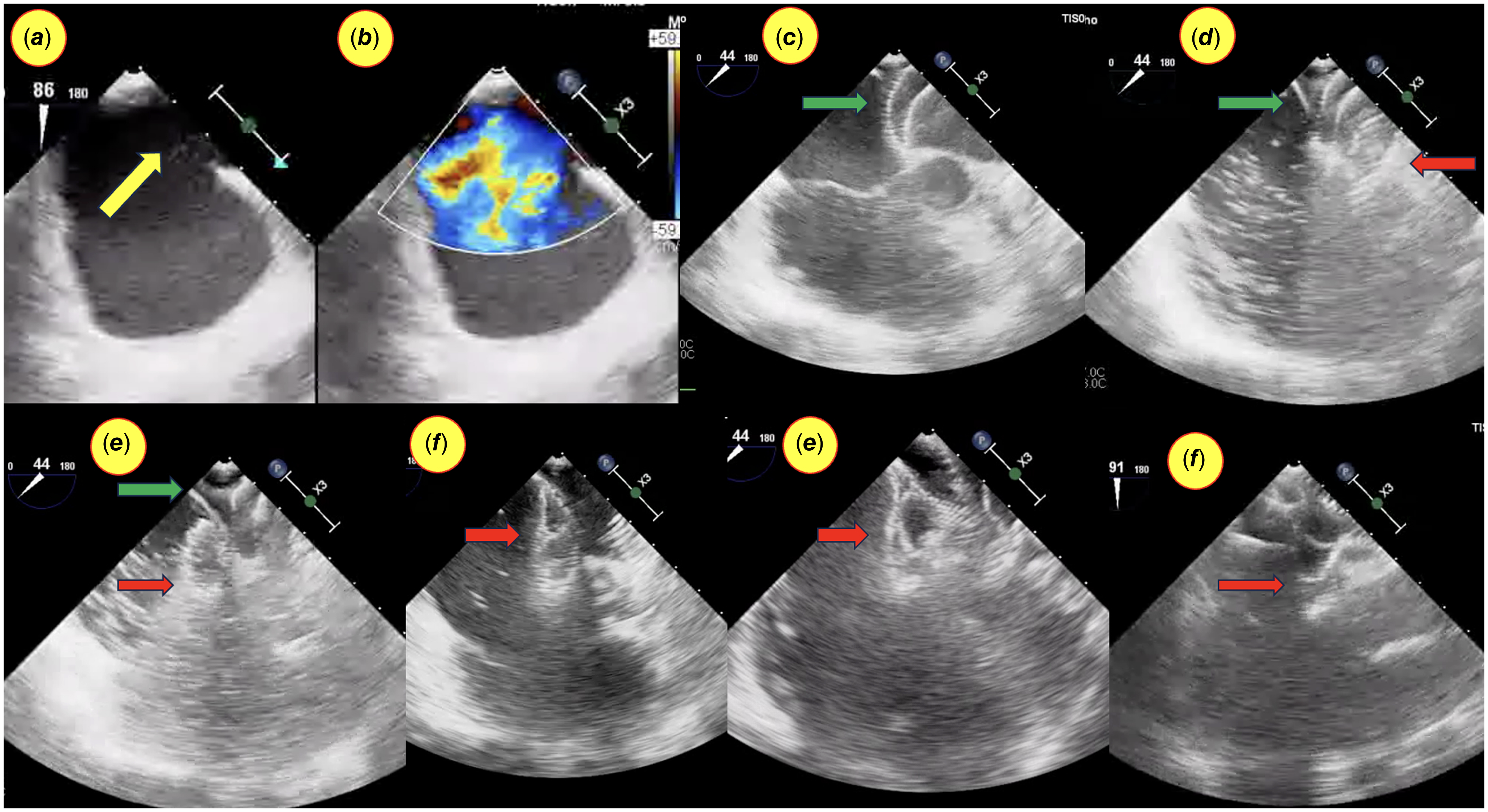
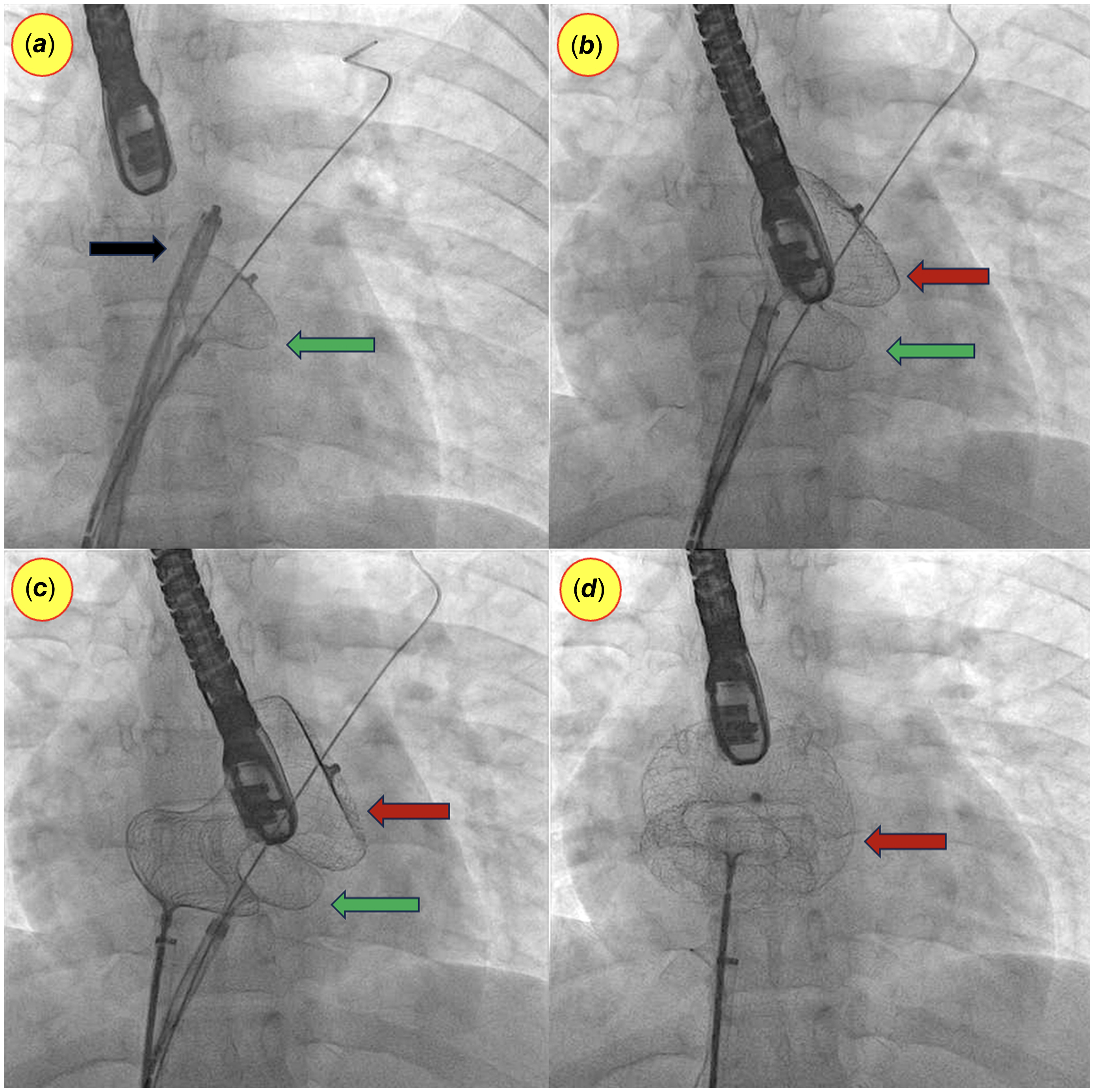
The assisting 24-mm device was loaded into the long sheath along with the super-stiff wire passing through the wire mesh under water (Fig. 1 B-F). The supporting 24-mm device was advanced across the left atrium, and the left atrial disc was deployed (Figs. 2A, 3A, 4A). The right atrial disc and waist were retained in the long sheath, and the cable was held in place at the cable-sheath junction outside. This partially deployed left atrial disc-sheath combination acted as a unit, maintained in the desired alignment and position by the stiff wire in the pulmonary vein. The assistant could manoeuvre this sheath-device combination unit during actual device deployment.
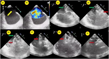
( a , b ) Transesophageal echocardiogram with colour Doppler showing a large atrial septal defect (ASD) (yellow arrow) with a thin, small posteroinferior margin. ( c ) The assisting septal occluder (green arrow) placed across the ASD. ( d ) The left atrial disc of the therapeutic septal occluder (red arrow) deployed over the assisting device. ( e ) The left atrial disc of the therapeutic device deployed. ( f , g , h ) The therapeutic septal occluder (red arrow) deployed slowly across the ASD.
Fluoroscopic image in anteroposterior view showing ( a ) the assisting septal occluder (green arrow) placed across the atrial septal defect under transesophageal guidance while the stiff wire is parked in the left upper pulmonary vein. The therapeutic device loaded in the long sheath is visible (black arrow). ( b ) The left atrial disc of the therapeutic septal occluder (maroon arrow) deployed over the assisting device (green arrow). ( c ) The left atrial disc of the therapeutic device (maroon arrow) deployed. ( d ) The fully deployed therapeutic septal occluder (maroon arrow).
Fluoroscopic image in left anterior oblique-cranial view showing ( a ) the assisting septal occluder (green arrow) placed across the atrial septal defect under transesophageal guidance while the stiff wire is parked in the left upper pulmonary vein. The therapeutic device loaded in the long sheath is visible (black arrow). ( b ) The left atrial disc of the therapeutic septal occluder (maroon arrow) deployed over the assisting device (green arrow). ( c ) The left atrial disc of the therapeutic device (maroon arrow) deployed. ( d ) The fully deployed therapeutic septal occluder (maroon arrow).
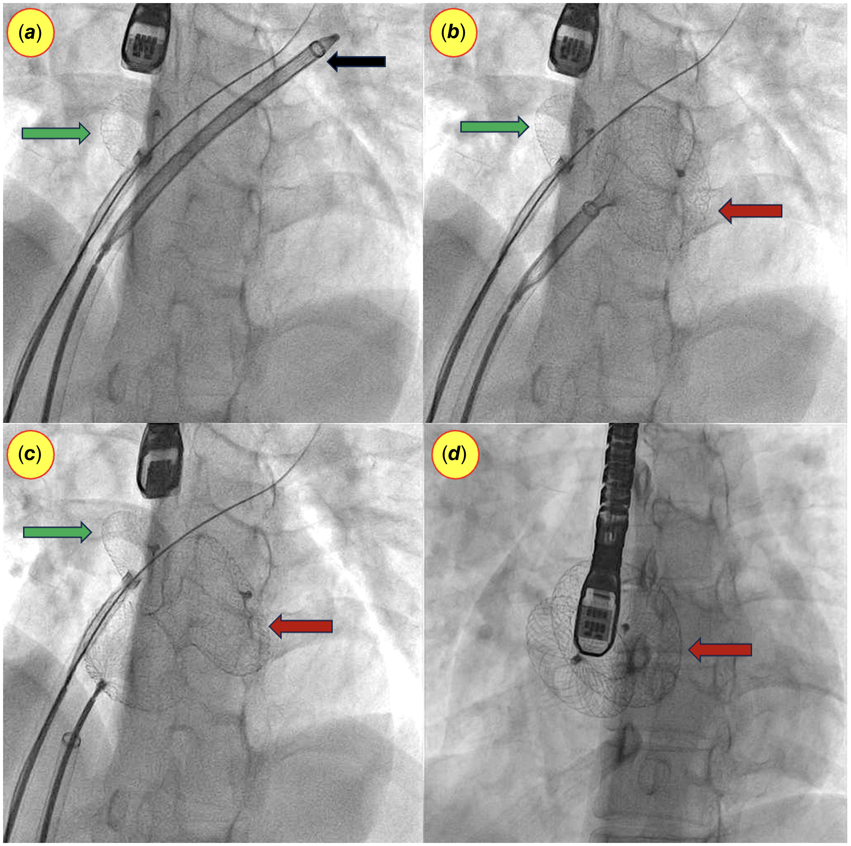
An appropriately sized device was advanced from the right femoral venous access, and the left atrial disc was deployed in the left atrium under transesophageal echocardiograms guidance (Fig. 2 A-D, 3B, 4B). As the left atrial disc of the target occluder approached the atrial septal defect, guarded by the artificial wall of the assisting device, the right atrial disc was deployed (Fig. 2E-G, 3C, 4C). Once both discs were positioned in the correct alignment, the left atrial disc of the supporting device was withdrawn into the long sheath along with the wire. The entire procedure was performed under fluoroscopic and transesophageal echocardiograms guidance. A stable, well-positioned device across the atrial septal defect with no significant residual flow was ensured before device release (Fig. 2H, 3D, 4D).
Ethical clearance
This retrospective study adhered to the approved protocol of the institutional Clinical Ethics and Research Committee (CERC/2021/Jun/iv). Patient confidentiality was ensured by anonymizing all data used in images and analyses. As the study utilised anonymized data, the ethics committee waived the requirement for individual patient consent.
Statistical method
Continuous variables were expressed as mean ± standard deviation and compared between the two groups using independent samples t-tests. Categorical variables were presented as frequencies and percentages, with comparisons made using either chi-square tests or Fisher’s exact tests, as appropriate. Statistical significance was set at a p-value less than 0.05 for all analyses.
Results
Patient baseline characteristics
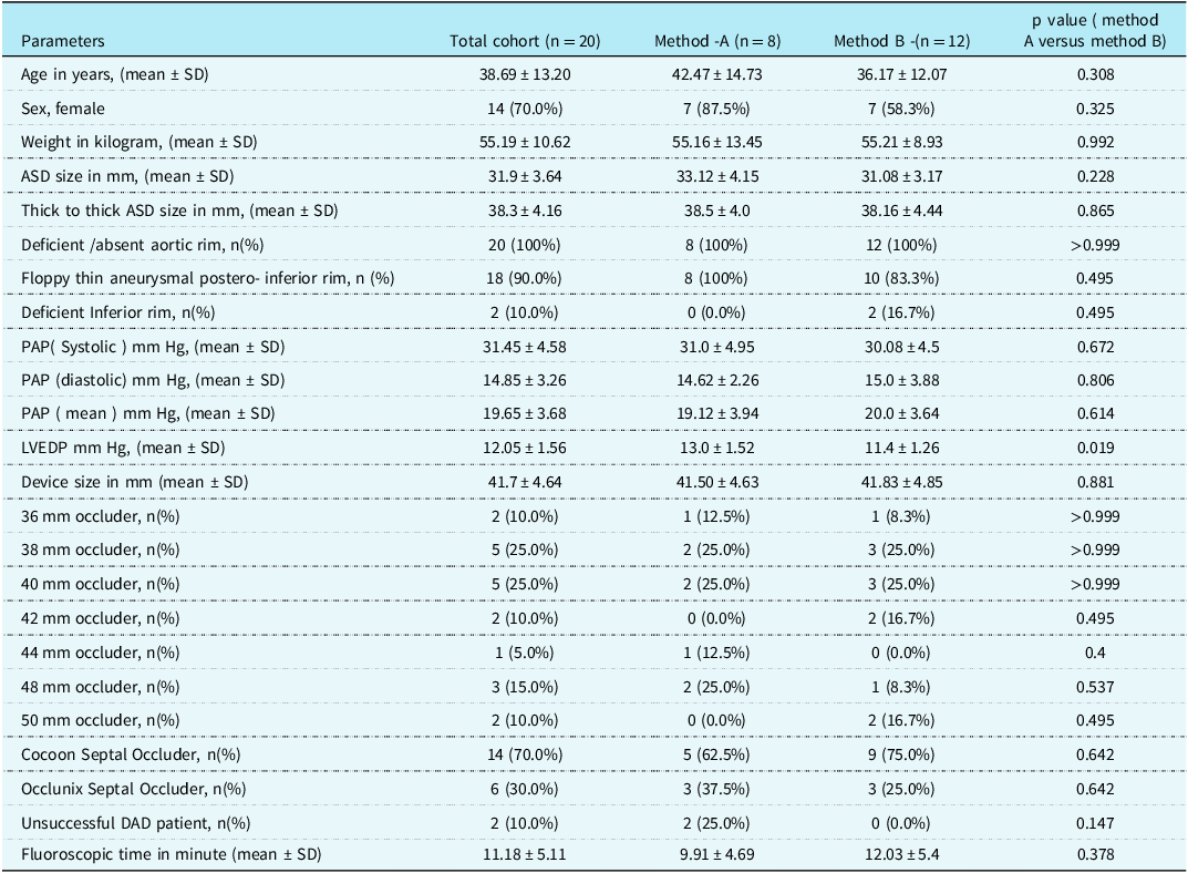
Between December 2023 and August 2024, 20 patients underwent device closure of secundum atrial defects, assisted by another atrial septal occluder. The average patient age was 38.69 years (range 18–66.5 years), and the average weight was 55.19 kg (range 31.7–75.4 kg). The atrial septal defect was, on average, 31.9 mm in diameter (range 26–38 mm) and 38.3 mm in thick-to-thick measurement (range 36–46 mm). Most patients (18) had thin, floppy margins in the posteroinferior septum. Two of them had deficient posteroinferior rims (Table 1). The size of the atrial septal occluder was determined based on the maximum thick-to-thick measurement in the transoesophageal echocardiogram. Pre-procedure haemodynamics revealed a mean pulmonary artery systolic pressure of 31.45 mm Hg and a mean pulmonary artery pressure of 19.65 mm Hg. The left ventricular end-diastolic pressure was 12.05 mm Hg. The coronary angiogram showed normal coronary arteries in 18 (90%) cases with no evidence of atherosclerotic disease. Two patients (10%) had mild atherosclerotic disease. One patient had mild right coronary artery stenosis, and another had mild left circumflex artery stenosis.
Characteristics of the patient and details of the haemodynamic and septal defect and occluder parameters

Group-A = device closure in anteroposterior fluoroscopic projection; Group- B = device closure in left anterior oblique-cranial fluoroscopic projection; ASD = atrial septal defect; PAP = pulmonary artery pressure; mean ± SD = mean ± standard deviation; LVEDP = left ventricular end-diastolic pressure; mmHg = millimetre of mercury; DAD = Device-assisted device closure.
Procedural characteristics
All patients underwent device closure of the atrial septal defect by the device-assisted device closure technique. The delivery sheath used for device deployment was 14 French, and the contralateral sheath used for device assistance was 10 or 11 French, depending on the availability of the long sheath or delivery system. The mean fluoroscopic time was 11.18 minutes (range 5.35–26.42). Device sizes ranged from 36 to 50 mm (median 40 mm). Fourteen devices were Cocoon septal occluders (Vascular Innovations Co. Ltd., Thailand) in the range between 36 and 42 mm, and the remaining six were 44 mm to 50 mm from Occlunix (Rivarp Medical, Bangalore, India). The device size was 0-6 mm larger (median and mode 4 mm) than the maximum thick-to-thick dimension of the atrial septal defect measured in the transoesophageal echocardiogram. Device-assisted device closure was successfully performed in 18 of the 20 patients (90%). The remaining two patients (10%) failed device closure with the device-assisted device closure technique, requiring other techniques (Table 1). In these two patients, the floppy posteroinferior margin was not allowing proper deployment and slipping inferior rim. In view of the new technique like device-assisted device closure for assisted device closure, repetitive attempts were not done, and the traditional balloon-assisted technique was used. In one of these two patients, additional support from the right upper pulmonary vein deployment approach was needed over the balloon support technique.
Of the 18 patients who underwent device-assisted device closure, two required gentle manipulation of the device for alignment of the device across the aortic margin. After the experience from the failure of the initial patients with the device-assisted device closure technique, it was felt that deploying the device in the anteroposterior fluoroscopic projection might not have allowed for correct alignment. Therefore, after the first eight patients, the left anterior oblique 30- cranial 30 view was used in conjunction with transesophageal echocardiograms guidance to better align the planes of the atrial defect and the atrial septal occluder.
Subgroup analysis
The patients were analysed in two groups and compared. In the first group (Group A) of eight patients having anteroposterior view deployment of the device, the device was successfully deployed in a single attempt in six patients. Two patients required balloon-assisted/additional techniques due to initial failure. The remaining 12 patients (Group B) had the device deployed in the left anterior oblique-cranial view. Eleven had single-attempt deployment, while one required two attempts. Two of the 12 patients required gentle manipulation due to disc malformation or aortic rim non-alignment. It is noteworthy that all patients had no residual flow after successful atrial septal defect closure. Statistically, the two groups of patients were comparable in most of the demographic, atrial septal defect anatomic, and haemodynamic parameters along with occluder type and size. Group A had two failures (25%) while Group B had no failures (Table 1), which was statistically not significant. Although the p-value was significant regarding left ventricular end-diastolic pressure, the clinical significance of the same is possibly not relevant.
Perioperative care and complication
There was no instance of device upsizing or downsizing. In two of the patients, the device after deployment appeared a little bulkier, and in hindsight, a one-size-lower device could have worked. In view of the significant floppy margin, downsizing was avoided. There was no embolisation episode among the cohort. Patients were discharged on dual antiplatelet therapy (Aspirin and Clopidogrel) in 17 cases, while one patient received only Aspirin. Two patients received clopidogrel and a non-vitamin K anticoagulant (Apixaban) in view of atrial arrhythmias observed post-device closure. No patients had any heart block during the follow-up period.
Two patients experienced complications in the one-month postoperative period. One patient developed fever, pneumonia, and pleural effusion one week after the procedure, unrelated to the procedure. The patient received conservative treatment from the respiratory team and recovered within two weeks. The septal occluder was stable in position with no residual flow and good left ventricular systolic function. Another patient had atrial arrhythmias with a fast ventricular rate, treated with intravenous amiodarone and anticoagulation. At the one-month follow-up, all patients were doing well with a good device profile, no residual flow, and good cardiac function.
Discussion
Transcatheter closure for secundum atrial septal defects has been well established in recent times. Numerous studies have demonstrated its safety and efficacy, with high success rates and minimal complications in cases of device closure of secundum atrial septal defect. Reference Chessa, Carminati and Butera8–Reference Singhi, Mahapatra, Kumar, Dey, Mishra and De10 However, transcatheter closure can be challenging in cases of large secundum atrial septal defects with deficient rims and floppy margins. These cases often require repeated attempts due to device misalignment, leading to prolonged procedures, elevated failure rates, and increased risks of complications, including device embolisation. Reference Dalvi, Pinto and Gupta2,Reference Quek, Wu, Chan, Ang, Ho and Yip9,Reference Narin, Baykan and Argun11,Reference Rossi, Cardoso Cde and Machado12
Large secundum atrial septal defects with deficient and floppy margins require specialised assisted techniques for device deployment. Over the past two decades, operators have explored various assisted techniques. These include the left upper pulmonary vein technique, right upper pulmonary vein technique, dilator-assisted technique (using the specially designed Hausdorf sheath), Tulip-Bud’s method, Greek manoeuvre, St Jude SL2 sheath technique, and the left atrial disc engagement-disengagement technique. These innovative approaches aim to improve device stability during deployment, leading to higher success rates and fewer complications. The need for multiple approaches highlights the challenges involved. Reference Ananthakrishna Pillai, Upadhyay, Gousy and Handa1,Reference Pinto, Jain and Dalvi5,Reference Quek, Wu, Chan, Ang, Ho and Yip9,Reference Narin, Baykan and Argun11,Reference Lee, Sutton and Lopez13–Reference Kammache, Mancini, Ovaert, Habib and Fraisse16
In the index study, the device-assisted device closure technique was successfully used in challenging large atrial septal defect cases with floppy and deficient margins. The procedural success rate (90%) with low rates of complications was felt encouraging for a novel procedure. We hypothesised that a novel support system, involving a temporary wall created using a smaller device’s left atrial disc, could assist in large occluder deployment. The device-assisted device closure technique can also aid in manipulating the target device using the combined support of the partially deployed device and sheath.
The device-assisted device closure technique was inspired by the popular balloon-assisted technique for transcatheter closure of large atrial septal defects (>25 mm). As described by Dalvi et al., the inflated balloon helps prevent the LA disc from adopting a horizontal orientation. However, in Dalvi et al.’s study, three patients experienced LA disc slippage into the right atrium after delivery outside the left upper pulmonary vein. They suggested that insufficient rims hinder the effective anchoring of the LA disc. Reference Dalvi, Pinto and Gupta2 The success rate of balloon-assisted technique was reported to be 87%–100% by different operators in various morphological variations. The balloon-assisted technique may be ineffective in small left atria and may warrant other techniques like the left atrial roof method and dilator-assisted support after the balloon technique fails. Reference Ananthakrishna Pillai, Upadhyay, Gousy and Handa1,Reference Dalvi, Pinto and Gupta2,Reference Kammache, Mancini, Ovaert, Habib and Fraisse16,Reference Pillai, Rangaswamy, Selvaraj, Saktheeswaran, Satheesh and Jayaraman17 Pillai et al. reported that in two failed balloon-assisted technique attempts, patients with defects larger than 40 mm experienced device instability after balloon deflation and withdrawal. These patients required elective surgical closure. Reference Pillai, Rangaswamy, Selvaraj, Saktheeswaran, Satheesh and Jayaraman17 The inflated balloon can reduce left atrial space and is not easily manoeuvrable, hindering device opening. Additionally, a smaller LA cavity may not accommodate larger devices. The curvature of the left atrium and the flexibility of the interatrial septum can also influence device anchorage. Reference Dalvi, Pinto and Gupta2,Reference Chan, Godman, Walsh, Wilson, Redington and Gibbs18 Other operators tried modifying the balloon-assisted technique using low-profile balloons like the Tyshak balloon. Kammache et al. described a modified sizing balloon technique (Meditech) to measure the defect and achieve atrial septal defect device closure with an 88% success rate. The disadvantage of the balloon-assisted technique is the risk of air embolism from improperly prepared balloons or ruptured low-profile balloons like the Tyshak balloon. Reference Kammache, Mancini, Ovaert, Habib and Fraisse16
Two patients in this series required additional manoeuvres along with device assistance. Some patients in published series required additional techniques, such as the pulmonary vein approach, in conjunction with balloon-assisted technique. Reference Dalvi, Pinto and Gupta2,Reference Narin, Baykan and Argun11 The success rate of the device-assisted device closure had a learning curve. Prior to the introduction of the device-assisted delivery closure technique, the balloon-assisted technique was the preferred assisted technique. Traditionally, balloon-assisted technique was performed in the anteroposterior projection, and consequently, device-assisted device closure was also initiated in this view. However, after encountering two failures in the initial eight device-assisted device closure cases, we recognised that the left anterior oblique-cranial view would provide a better visualisation of the atrial septal plane and device disc alignment. Subsequently, all cases were performed in this left anterior oblique-cranial view. The left anterior oblique-cranialview deployment had a relatively better success rate than the initial cohort of AP view deployment, although statistically it was not found significant.
The floppy and deficient margin defects are challenging and may warrant multiple attempts. In the index study, two failures and two patients requiring additional manoeuvres and one requiring two attempts reflect the challenge. Lopez et al. reported five unsuccessful attempts in the closure of large atrial septal defects with deficient rims, with two immediate device embolisations requiring surgical intervention. In two other cases, multiple alignment attempts failed to secure the device, leading to procedure termination and device retrieval. Reference Lopez, Dalvi and Balzer19 Combining modified techniques for percutaneous closure of large secundum atrial septal defects with deficient or absent rims is a way forward in the challenging cases. Szeliga et al. described a novel approach involving a combination of modified implantation techniques tailored for closing large secundum atrial septal defects characterised by deficient rims. Reference Szeliga, Dryżek, Rudziński and Góreczny20
The novel device-assisted device closure technique aims to improve success rates and provide more consistent application. The device-assisted delivery technique was inspired by the balloon-assisted technique, which offers static support. However, balloon-assisted technique, involving balloon inflation across the atrial septum, can encounter challenges like underinflation, tilting, or slipping of the balloon, hindering disc deployment and alignment. Moreover, the balloon can restrict space in the left atrium, complicating device manoeuvrability. In contrast, device-assisted device closure employs a long sheath and a partially deployed left atrial disc to form a stable unit against the atrial septal defect. This configuration enables gentle pushing and manoeuvring of the therapeutic device, facilitating the deployment and alignment of both atrial discs. This dynamic support, particularly visible in the left anterior oblique-cranial view, significantly contributed to the successful deployment of the device.
The index cohort of patients presented with large atrial septal defects and thin, floppy margins, which historically posed challenges for device closure. Previous attempts in such cases often resulted in device slippage or iatrogenic atrial septal damage. Consequently, these cases frequently required assistance, most commonly through balloon-assisted techniques. Based on the lead operator’s experience, patients with large atrial septal defects and floppy margins necessitating large devices were assigned to a direct assisted technique. Historically, this involved a direct balloon-assisted technique, but the current approach utilises a device-assisted device closure technique from the outset in these challenging atrial septal defect anatomies to facilitate successful device closure. The selection of patients requiring assistance techniques from the onset is subjective. Different operators decided on a direct assistance approach based on the anatomy of the defect and experience. The transesophageal echocardiograms could tell the different sizes of the defect but not the strength of the septum to hold the device. Sizing in the cases of floppy rims had significant operator bias based on individual experience. Reference Ananthakrishna Pillai, Upadhyay, Gousy and Handa1,Reference Haddad, Khraiche, Bonnet, Meot and Malekzadeh-Milani21
The 24 mm bare septal occluder without polypropylene fabric actually acted as a surgical metallic instrument that could be safely reused with ethylene oxide sterilisation. The same assistive device, introduced at the inception of the study, has been consistently employed in all enrolled patients. Throughout its utilisation, the device maintained its excellent functional condition and exhibited the capability to support further similar procedures. As a result, it provides a more economical and user-friendly alternative to traditional balloon-based techniques. Absence of fabric reduces the risk of blood clot formation. The reuse of ethylene oxide hardware in the catheterisation laboratory and reimplantation of medical devices after proper sterilisation techniques is a well-established practice in different parts of the resource-limited world. Reference Ribeiro, Graziano, Alfa and Goveia22–Reference Kapoor, Vora, Nataraj, Mishra, Kerkar and Manjunath24
The fluoroscopic time taken in the device-assisted device closure study included the time for cardiac haemodynamic assessment, coronary angiography, and the device closure. The fluoroscopic timings were comparable with timings from other operators. Reference Lopez, Dalvi and Balzer19 Embolisation while performing device closure of large atrial septal defects with challenging anatomy is not uncommon. Pillai et al. reported 15 episodes of device embolisation in a cohort of 346 patients that occurred within 12 hours post-implantation, with 12 cases having deficiencies in the posterior and inferior vena caval margins. Reference Ananthakrishna Pillai, Upadhyay, Gousy and Handa1 There was no embolisation of the device in the index series, which could be due to more aneurysmal septa than deficient septa in the series.
The anticoagulation practice for large atrial septal occluders varies among different series. We preferred dual antiplatelet agents for one year in the majority. Given the paucity of studies on large devices with extensive metal surfaces, some investigators chose to prescribe clopidogrel in combination with aspirin for 6 to 12 months. Reference Lopez, Dalvi and Balzer19
Limitations
This study has several limitations that should be acknowledged. Firstly, the retrospective study design represents a significant constraint. Secondly, the small sample size limits the generalizability of the findings. Additionally, there is potential for selection bias, as patients with larger defects may have been preferentially referred to the index unit for device closure, with decisions influenced by a single lead operator. The patients were taken directly for assisted device closure with the device-assisted device closure technique without trying the conventional method. There could be some selection bias in this regard. The selection of the assisting device as a 24 mm septal occluder was based on hypothetical extrapolation of the balloon-assisted technique. The device-assisted atrial septal defect closure technique is a newer approach inspired by balloon-assisted techniques, addressing their limitations. While a retrospective comparison between these two methods would be beneficial, frequent changes in our hospital’s digital database software restrict the availability of historical data, making such a comparison challenging. A prospective multicentre study with a larger sample size and multiple lead operators would likely provide more comprehensive insights into this topic.
Conclusions
The device-assisted device closure technique demonstrates promising results in the transcatheter closure of large secundum atrial septal defects. This retrospective study highlights the efficacy of device-assisted device closure technique in achieving successful closure even in challenging cases with large and complex atrial septal defects. While the left anterior oblique-cranial view appears to be a preferred approach, further studies are needed to establish definitive guidelines. Despite the encouraging outcomes, larger prospective trials are warranted to evaluate the efficacy and safety of device-assisted device closure in a wider patient population, particularly those with very large atrial septal defects with difficult margins.
Acknowledgements
We extend our sincere appreciation to the outpatient support staff of the Pediatric and Congenital Heart Disease Department, including Ms. Moumita Sil (Cardiology Technician), Ms. Pratima Hembram (Nursing Coordinator), and Ms. Mandira Mondal (OPD Coordinator), for their invaluable assistance with patient assessment and data collection. Mr. Vikas Tiwari and the team of the Department of Information Technology were very helpful in mining the digital data. Our heartfelt thanks go to Dr Sivakumar K of the Department of Pediatric Cardiology at Madras Medical Mission, Chennai, India, for his continuous academic and clinical guidance. We also extend our gratitude to Dr Somnath Dey, Senior Consultant Cardiac Anesthesiologist, for his support during the procedure. Finally, we express our deepest gratitude to the Medica Institute of Cardiac Sciences and the Hospital Authority for their unwavering support throughout the study. We are particularly grateful to the Dr Anil Singhi Foundation for their generous academic support of our scientific research endeavours.
Financial support
None.
Competing interests
The authors declare no conflict of interest.
Ethical statement
This scientific work adhered to the approved protocol of the institutional Clinical Ethics and Research Committee (CERC/2021/Jun/iv). Patient confidentiality was ensured by anonymising all data used in images and analyses. As the study utilised anonymised data, the ethics committee protocol waived the requirement for individual patient consent.