Introduction
Polycystic echinococcosis (PE) is a zoonosis endemic to the Neotropical region of the Americas, including the northern region of Brazil (Romig et al., Reference Romig, Deplazes, Jenkins, Giraudoux, Massolo, Craig, Wassermann, Takahashi and de la Rue2017). It is caused by infection with the metacestode stage of Echinococcus vogeli (Cestoda, Taeniidae), one of the species causing Neotropical echinococcosis. Echinococcus vogeli metacestodes typically grow as small cysts (0.6–6 cm in diameter) that can occur singly or as aggregates in viscera of suitable intermediate hosts (mainly Cuniculus paca), more often in the liver and lungs. Humans can be accidentally infected by ingesting eggs released by adult parasites through the feces of infected dogs (definitive hosts) in the environment (Thompson, Reference Thompson2017).
The number of reported human PE cases likely represents only a small fraction of current infections, since most countries where the disease occurs have no compulsory reporting. Moreover, the non-specificity of symptoms, the difficulty of laboratory diagnosis, the limited access to various endemic sites and misinformation of the affected populations all are involved in the difficulty of estimating the prevalence of human PE (Novo Alonso et al., Reference Novo Alonso, Ferreira, Fernández, Antequera, Blanco and Gajate1993; D'Alessandro & Rausch, Reference D'Alessandro and Rausch2008; Kern et al., Reference Kern, Silva, Akhan, Müllhaupt, Vizcaychipi, Budke and Vuitton2017). The E. vogeli metacestode usually grows in the liver, and the disease can have serious consequences for the patient (Genzini et al., Reference Genzini, Siqueira, Noujaim, Santos, Yamashita, Trevizol and Perosa2013). The growth of multiple parasite cysts can cause hepatic and vascular necrosis, infect the bile ducts and spread to the peritoneal cavity, disseminating to other abdominal and thoracic organs, and can even lead to death.
Due to its late and unspecific symptomatology, PE discovery usually occurs in advanced stages (Mihmanli et al., Reference Mihmanli, Idiz, Kaya, Demir, Bostanci, Omeroglu and Bozkurt2016). Clinical suspicion of PE occurs often during clinical investigations for other reasons. Initial diagnosis is based on clinical signs such as abdominal pain, palpable masses, jaundice and hepatomegaly, in addition to imaging tests such as X-ray, ultrasound and computed tomography in the presence of compatible epidemiology. These methods can be complemented by immunological assays based on the detection of circulating antibodies against the parasite. These assays are even more important in places with difficult access to more expensive radiological tests and with a lack of public health resources. The immunological assays are also important for the differential diagnosis of suspected liver and lung neoplasms, due to the similarity of the PE cysts with carcinomatous diseases in imaging tests (Almeida et al., Reference Almeida, Martins, Moraes, Viegas and Grilo1997; Pedrosa et al., Reference Pedrosa, Saíz, Arrazola, Ferreirós and Pedrosa2000). The confirmatory tests of PE can only be performed after the surgical removal of the cysts, and rely on the identification of the parasite material or on molecular diagnosis (Siles-Lucas et al., Reference Siles-Lucas, Casulli, Conraths and Müller2017).
There are currently several serological tests based on the detection of specific antibodies against Echinococcus antigens, but most of them are based on complex, variable and poorly characterized mixtures of native parasite antigens (D'Alessandro & Rausch, Reference D'Alessandro and Rausch2008; Thompson, Reference Thompson2017; Mubanga et al., Reference Mubanga, Mwape and Phiri2019). The most commonly used antigenic preparations for the serological tests for E. vogeli infections are protein extracts of hydatid fluid (HF) or native purified antigens from Echinococcus granulosus metacestodes (Li et al., Reference Li, Zhang and McManus2004; D'Alessandro & Rausch, Reference D'Alessandro and Rausch2008; Sarkari & Rezaei, Reference Sarkari and Rezaei2015). The diagnosis of cystic echinococcosis (CE), the most studied, has improved considerably during the past two decades, but is still problematic. The inherent heterogeneity of native crude or partially purified parasite antigens has led to inconsistencies in the immunodiagnosis of CE, due to the occurrence of both false positive and false negative results (Sarkari & Rezaei, Reference Sarkari and Rezaei2015). Similar disadvantages are observed when somatic or excretory secreting extracts of adult worms and protoscolices are used (Siles-Lucas et al., Reference Siles-Lucas, Casulli, Conraths and Müller2017). According to Sarkari & Rezaei (Reference Sarkari and Rezaei2015), tests with low sensitivity (up to 30% false negativity) and low specificity (up to 25% false positivity) generate results that are difficult to interpret. Nevertheless, serological tests still play an important confirmatory role in initial cyst image detection in the diagnosis of CE.
Very few PE antigens have been described in the literature. In this respect, only 86 E. vogeli protein entries are contained in GenBank (search date: 07/05/2021). Some of these proteins may have potential as antigens for the diagnosis of PE, but none of them have been tested so far, probably due to the limited access to parasite samples (Siles-Lucas et al., Reference Siles-Lucas, Casulli, Conraths and Müller2017).
The use of a more sensitive and specific antigen that can be more easily detected and also consistently produced on a large scale in different laboratories is essential to improve diagnosis by serological testing. In this sense, recombinant proteins have already been shown to be extremely valuable sources of antigens for serological diagnosis, reducing non-specific reactions and allowing standardization (Greene et al., Reference Greene, Hancock, Wilkins and Tsang2000; Obregón-Henao et al., Reference Obregón-Henao, Gil, Gómez, Sanzón, Teale and Restrepo2001; Silva et al., Reference Silva, Soares, Alves, Conde, Amaral and Teixeira2002; Virginio et al., Reference Virginio, Hernández, Rott, Monteiro, Zandonai, Nieto, Zaha and Ferreira2003; Savardashtaki et al., Reference Savardashtaki, Sarkari, Arianfar and Mostafavi2017). Besides dispensing with the need for parasite material for their production, they can be expressed and purified with relatively low cost, and have less variation compared to crude or purified native antigens.
The antigen B (AgB) is a secreted immunodominant component found in the HF that fills cysts or vesicles of Echinococcus spp. metacestodes (Lightowlers et al., Reference Lightowlers, Liu, Haralambous and Rickard1989). AgB is a multimeric thermostable lipoprotein composed of different 8-kDa subunits (Monteiro et al., Reference Monteiro, Cardoso, Follmer, da Silveira, Vargas, Kitajima, Zaha and Ferreira2012). In E. granulosus, there are five AgB subunits (EgAgB8/1–5) encoded by a multigenic family having five members (EgAgB8/1–5 genes) (Monteiro et al., Reference Monteiro, Zaha and Ferreira2008). Purified native EgAgB also has been used as antigen in the standardization of immunodiagnostic tests for CE (Folle et al., Reference Folle, Kitano and Lima2017). The recombinant EgAgB subunits (rEgAgB), produced by cloning and expression of the corresponding genes in Escherichia coli, have also been used for CE immunodiagnosis, either in enzyme-linked immunosorbent assays (ELISAs) or in immunoblot assays (Virginio et al., Reference Virginio, Hernández, Rott, Monteiro, Zandonai, Nieto, Zaha and Ferreira2003; Monteiro et al., Reference Monteiro, Zaha and Ferreira2008; Sarkari & Rezaei, Reference Sarkari and Rezaei2015). According to Siles-Lucas et al. (Reference Siles-Lucas, Casulli, Conraths and Müller2017), the achieved sensitivities and specificities ranged from 55% to 94.6% and from 80% to 91%, respectively, for rAgB8/1, and from 45% to 93% and from 86% to 98%, respectively, for rAgB8/2.
The amino acid sequences of AgB subunits from E. granulosus are fairly well conserved and immunologically cross-reactive with their orthologs from Echinococcus multilocularis (Jiang et al., Reference Jiang, Zhang, Liu and Feng2012). Therefore, E. granulosus recombinant AgB subunits are interesting candidates for assessment of their potential for PE diagnosis. Here, the recombinant AgB subunits rAgB8/1 and rAgB8/2, previously characterized as having value for the diagnosis of CE (Virgínio et al., Reference Virginio, Hernández, Rott, Monteiro, Zandonai, Nieto, Zaha and Ferreira2003), were tested for the diagnosis of PE. We also tested these recombinant antigens in total immunoglobulin G (IgG) ELISA with a panel of 43 sera from PE patients, and also performed specificity assays with 231 sera from clinically healthy individuals from endemic areas of the state of Acre, Brazil, and 11 positive sera from patients with other helminthiases. Our results showed that the tested recombinant antigens can be useful for the detection of specific antibodies in sera from suspected PE patients and may help to reduce problems that negatively influence PE serological diagnosis with native antigens.
Materials and methods
Serum samples
Positive control serum samples were obtained from 43 patients from endemic areas of Acre (Brazil) diagnosed with PE. The inclusion criteria of patients in the study were: (i) presence of cystic lesions with peripheral calcifications in imaging exams; (ii) positive serological diagnosis for echinococcosis by immunoblot tests using E. granulosus HF antigenic preparations; and (iii) epidemiological history, including current or previous residence in a rainforest area, engagement in subsistence hunting and presence of dogs at home. All criteria had to be met to include the patient in the study. All samples were kindly supplied by Dr Nilton Guiotti Siqueira from the Federal University of Acre, and followed the inclusion criteria established in previous studies (D'Alessandro, Reference D'Alessandro1997; Siqueira et al., Reference Siqueira, Siqueira, Rodrigues-Silva, Soares and Póvoa2013).
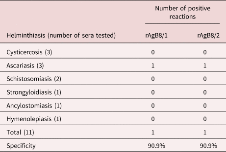
Sera from 231 healthy individuals also from endemic areas of Acre were included as negative controls. All donors had no symptoms suggestive of echinococcosis and had no complains about their health status at the time of serum collection. Although specificity tests for both antigens have already been performed (Virgínio et al., Reference Virginio, Hernández, Rott, Monteiro, Zandonai, Nieto, Zaha and Ferreira2003), we tested some samples present in our biobank to increase the evaluation of cross reactions (three sera from cysticercosis patients, three sera from patients with ascariasis, two sera from patients with schistosomiasis, two sera from strongyloidiasis patients, one serum from a hookworm patient and one serum from a hymenolepiasis patient).
Expression of the recombinant antigens
The rAgB8/1 and rAgB8/2, corresponding to the E. granulosus AgB subunits AgB8/1 and AgB8/2 (Fernández et al., Reference Fernández, Ferreira, Fernández, Zaha and Nieto1996), respectively, were expressed in E. coli as Glutathione S-transferase (GST)-fusion proteins from plasmid clones, as previously described by Rott et al. (Reference Rott, Fernández, Farias, Ceni, Ferreira, Haag and Zaha2000). The expression was carried out in E. coli BL21 Codon Plus Ril (Stratagene, La Jolla, California, USA) strains, and the recombinant proteins were purified by affinity chromatography followed by thrombin cleavage, as described by Smith & Johnson (Reference Smith and Johnson1998). Recovered protein concentrations were determined by spectrophotometry using a NanoDrop-ND1000 spectrophotometer (Johnstone & Thorpe, Reference Johnstone and Thorpe1982).
Potential immunodiagnosis of the recombinant antigens
ELISAs for detection of antibodies were performed as described by Virginio et al. (Reference Virginio, Hernández, Rott, Monteiro, Zandonai, Nieto, Zaha and Ferreira2003). Briefly, microtitration plates (Maxisorp, Thermo Scientific, Nunc, New York, NY, USA) were coated with 0.2 μg/well of rAgB8/1 or rAgB8/2, diluted in 0.1 M carbonate/bicarbonate buffer (pH 9.6). The sera samples were diluted 1:400 in blocking solution (phosphate-buffered saline containing 10% milk powder) and tested in triplicate. Positive sera from patients diagnosed with PE, negative sera from healthy individuals from endemic areas and conjugate controls were included in each plate. Anti-human IgG-peroxidase conjugate (Sigma-Aldrich, Burlington, MA, USA) diluted 1:15000 was used as a second antibody. O-phenylenediamine dihydrochloride (0.4 mg/ml in 0.1 M phosphate/citrate buffer, pH = 5) and hydrogen peroxide were used to visualize the antigen-antibody reaction. Optical density (OD) was registered at 492 nm in a Bio-Rad microplate reader (Model 680; Bio-Rad, Hercules, CA, USA) after the addition of stop solution (1 N sulphuric acid). The considered OD value represented the mean of three readings for each serum with less than 10% variation between them.
Determination of intra-assay and inter-assay variations
Coefficients of variation (CV) were calculated following the protocol of Flores et al. (Reference Flores, Carrillo, Tamez, Rangel, Rodríguez, Maldonado, Isibasi and Galán2016). For determination of the intra-assay coefficient of variation (IACV), ODs of ten randomly chosen samples were measured ten times each on the same plate. IACV was calculated by dividing the standard deviation (SD) of the ten measurements by their mean, and the obtained value was multiplied by 100. For determination of the inter-assay coefficient of variation (IECV), ODs from the same ten samples were measured on five different plates. IECV was calculated by dividing the SD of the ten measurements by their mean, and the value obtained was multiplied by 100. For the analysis of precision results, we used the acceptance criteria for the validation of the Food and Drug Administration (FDA) assays (USDHHS, 2001).
Statistical analysis
For the interpretation of seropositivity, cut-offs were calculated for each antigen based on receiver operating characteristic (ROC) curves (Hanley & McNeil, Reference Hanley and McNeil1982). Sensitivity levels were plotted against specificity levels at each cut-off point on a ROC curve. The threshold values used were those that gave the highest sum of sensitivity (%) and specificity (%), as described by Amagai et al. (Reference Amagai, Komai and Hashimoto1999). The area under the ROC curve (AUC) was the parameter used to define discriminatory antigen values between individuals with and without the disease.
Calculation of the critical index z for paired observations was used to test for differences between AUCs (Hanley & McNeil, Reference Hanley and McNeil1982), and the Kolmogorov–Smirnov adherence test was applied to assess specimen-specific distribution. By calculating the critical index z, it is possible to assess whether the difference in areas under two or more ROC curves derived from the same set of sera is random or real. A z-value below 1.96 is taken as evidence that two antigens are not significantly different (Hanley & McNeil, Reference Hanley and McNeil1982). Normal variables were described by mean ± SD, and in cases where the variable was not normally distributed, the median (interquartile range) was used. Comparisons between groups were performed using the Mann–Whitney test. A P-value < 0.05 was considered statistically significant.
All statistical calculations were performed using GraphPad Prism version 8.0.1 (GraphPad Software Inc., La Jolla, California, USA) and IBM SPSS Statistics for Windows, version 25.0 (IBM Corp., Armonk, New York, USA).
Results
Immunodiagnostic potential of the ELISA for rAgB8/1 and rAgB8/2
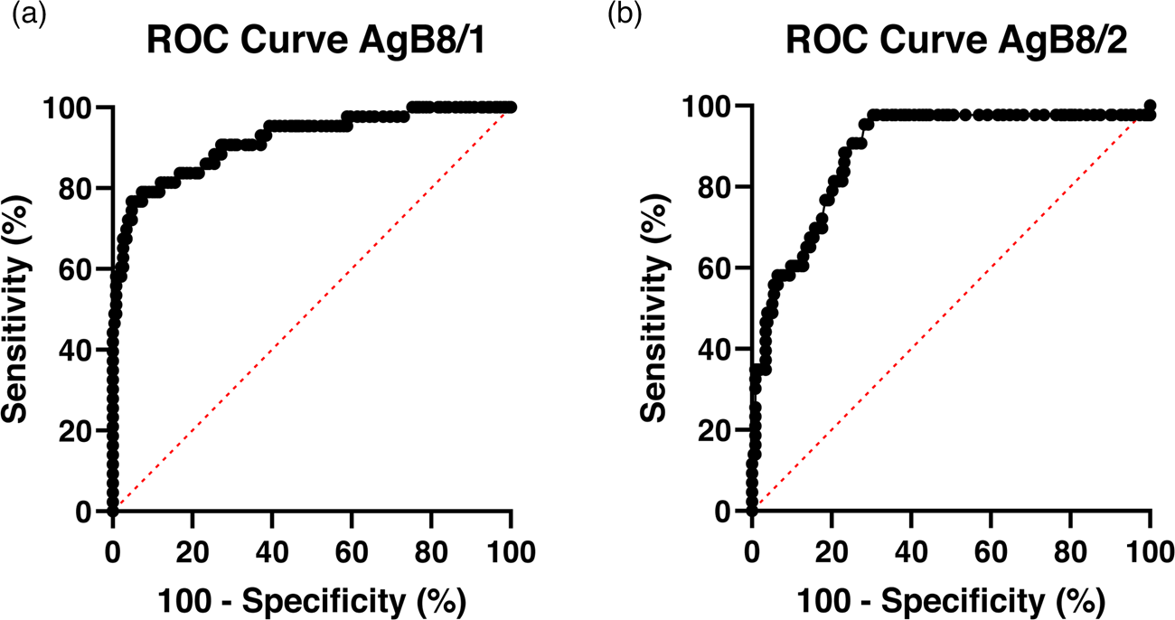
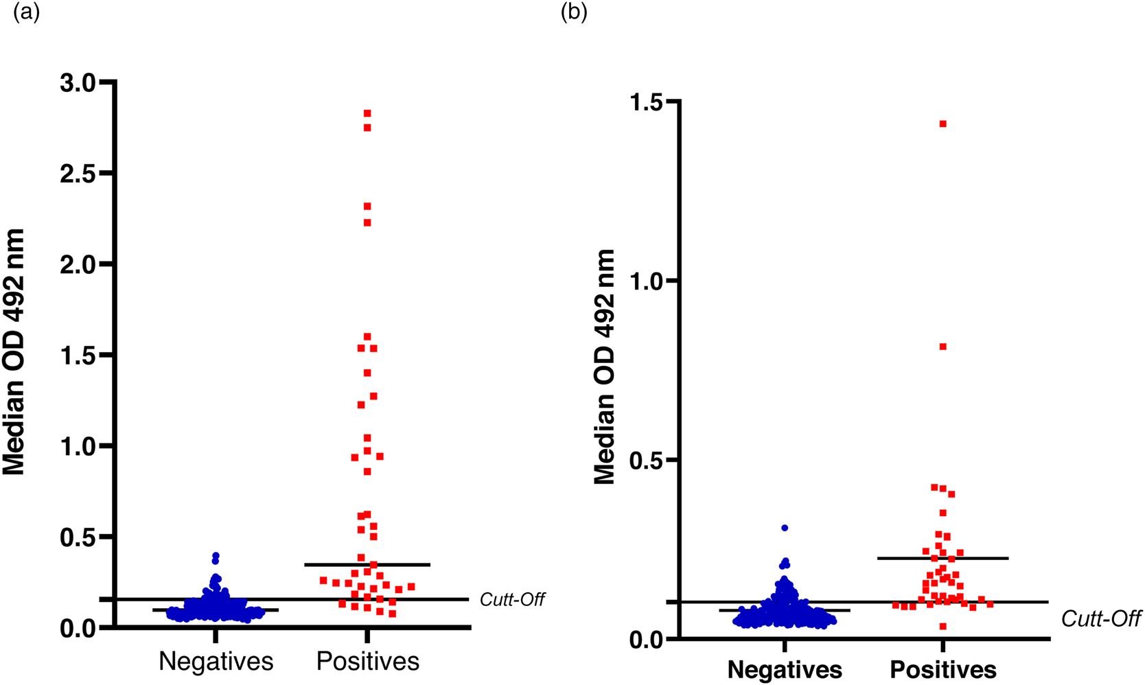
In the present study, 43 positive sera from patients with PE and 231 negative sera from clinically healthy patients residing in an endemic area were used to assess, by ELISA, the diagnostic value of the heterologous antigens EgAgB8/1 and EgAgB8/2 for the immunodiagnosis of PE. Yields of 1.83 and 2.50 mg/l of culture were obtained for rAgB8/1 and rAgB8/2, respectively. The specific IgG antibody responses against rEgAgB8/1 and rAgBB8/2 were investigated separately for each recombinant antigen. Based on the results expressed as OD measurements, the best cut-off values were calculated for both recombinant antigens using the ROC curve (fig. 1a, b). The cut-off values for rAgB8/1 and rAgb8/2 were 0.1565 and 0.1030, respectively, representing the maximization of the sum of sensitivity and specificity values. Higher OD measurements were present in the positive sera for both recombinant antigens. For comparison between the 231 sera from the negative and 43 positive samples for both heterologous antigens, the medians (interquartile) were calculated (fig. 2a, b). The absorbance value of rAgB8/1 was 0.097 (0.135–0.077) for negative and 0.346 (1.044–0.209) for positive samples (P < 0.001). In turn, for rAgB8/2 the absorbance was 0.067 (0.092–0.056) for negative and 0.158 (0.245–0.110) for positive samples (P < 0.001). The sensitivity of rAgB8/1 was 83.72% and the specificity was 83.12%, compared to 81.40% sensitivity and 80.09% specificity of rAgB8/2. The highest AUC value was for rAgB8/1 (0.9185), although it was not significantly different (z = 0.79) from the AUC of rAgB8/2 (0.8915). Sensitivity and specificity data and relevant parameters derived from the ROC curve are shown in table 1.
ROC curves used to determine cut-off values and areas under the curve for the two recombinant antigens rAgB8/1 (a) and rAgB8/2 (b) tested by IgG ELISA.

Representative plots of ELISAs with sera from healthy individuals (blue circles) and polycystic echinococcosis patients (red squares) against rAgB8/1 (a) and rAgB8/2 (b) antigens. Cut-offs: rAgB8/1 = 0.1565; rAgB8/2 = 0.1030. The horizontal bars in the sample distributions represent their median. OD, optical density.

Summary of parameters derived from the ROC curves of recombinant antigens tested by ELISA against sera from polycystic echinococcosis-positive patients and from healthy individuals.

CI, confidence interval.
Among the 43 sera from patients considered positive for PE used in this study, seven were negative for rAgB8/1 and eight were negative for rAgB8/2. However, only one showed negativity against both recombinant antigens. Therefore, basing the positivity of the test on a detectable reaction for both antigens (either rAgB8/1 or rAgB8/2) increased the sensitivity of the standardized total IgG ELISA to 97.8%. These results demonstrate that the combined use of the tests can be a useful tool to increase diagnostic sensitivity.
Intra-assay and inter-assay variations
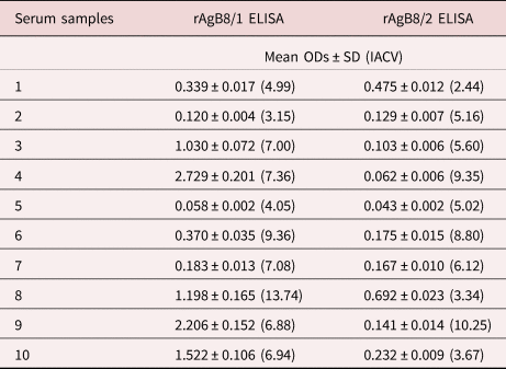
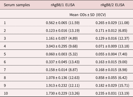
Intra-assay accuracy of rAgB8/1 assays had CV between 3.15% and 13.74%. In the rAgB8/2 assays, the CV was between 2.44% and 10.25%. The inter-assay precision of the rAgB8/1 assays had CV between 4.89% and 13.43%, whereas for rAgB8/2 assays, the CV was between 6.42% and 15.71%. Both IACV and IECV values (tables 2 and 3) met the acceptance criteria for FDA assay validation.
Intra-assay coefficients of variation.

Note: values are expressed as means ± SDs and IACV.
Inter-assay coefficients of variation.

Note: values are expressed as means ± SDs and IECV.
Cross-reactions
A specificity assay with 18 sera from toxocariasis and 47 sera from cysticercosis patients potentially having cross-reactive infections prevalent in CE endemic areas was previously performed by Virginio et al. (Reference Virginio, Hernández, Rott, Monteiro, Zandonai, Nieto, Zaha and Ferreira2003), and the specificity found was 97% for rAgb8/1 and 94% for rAgb8/2. We also tested some positive sera from patients with other parasitic infections, including other cestodes, present in our biobank, to assess the possibility of cross-reactions. Specificity was calculated from the ratio between the true negatives over the sum of the true negatives and false positives. The specificities of the tests performed are shown in table 4. Of these, only one ascariasis-positive serum also reacted to both recombinant antigens tested. Therefore, rAgB8/1 and rAgB8/2 antigens presented similar performance, suggesting that both antigens can be gender-specific and might be useful in diagnosing PE.
Specificity evaluation based on the reactivity of sera from patients with other helminthiases against rAgB8/1 and rAgB8/2 in total IgG ELISA.

Discussion
The complexity of echinococcosis and the heterogeneity of clinical cases require a thorough understanding of the parasite–host relationship and the parasite phenotype at each developmental stage, to choose the best available diagnostic tool acceptable for clinical practice. Early diagnosis of echinococcosis may decrease or even prevent the disease from worsening and minimize the risks to the patient (Siqueira et al., Reference Siqueira, Siqueira, Rodrigues-Silva, Soares and Póvoa2013). To date, several approaches have been described for laboratory diagnosis and follow-up of CE and alveolar echinococcosis (AE) infections, including detection of antibodies, antigens and cytokines (Siles-Lucas et al., Reference Siles-Lucas, Casulli, Conraths and Müller2017). However, there have been few studies on Neotropical echinococcoses, especially for laboratory diagnosis. This is due to factors like low endemicity of the diseases, and difficulties in accessing affected populations and obtaining parasite material for research.
To provide more dependable reagents for the diagnosis of human E. vogeli infections, here we validated two subunits of a major secretory antigen from E. granulosus (AgB), previously characterized as of diagnostic value for CE, for human PE diagnosis. These antigens (AgB8/1 and AgB8/2) produced as pure recombinant proteins (rAgB8/1 and rAgB8/2) in E. coli, allowed the detection of specific IgG antibodies in sera from PE patients in an ELISA. The use of recombinant antigens is a viable alternative for the development of more sensitive and specific methods for PE immunodiagnosis, with the advantage of intra-laboratory production of large amounts of pure antigen, dispensing with the use of animals and having less variation by lot in comparison with crude or purified antigens (Robles et al., Reference Robles, González, Govezensky, Mungia, Acero, Bobes, Gevorkian and Manoutcharian2005). Moreover, several studies have shown that AgB is highly gender-specific and is a good reagent for the immunodiagnosis of AE and CE (Brehm & Koziol, Reference Brehm and Koziol2017; Pagnozzi et al., Reference Pagnozzi, Tamarozzi and Roggio2018; Silva et al., Reference Silva, Cancela, Monteiro, Ferreira and Zaha2018).
Our study sampled 43 sera from patients diagnosed with PE, and we obtained sensitivity values of 83.72% and 81.40% against rAgB8/1 and rAgB8/2, respectively, with overall sensitivity reaching 97.8%. This sensitivity is higher than that achieved in a previous preliminary study using immunoblotting for AgB detection in crude E. granulosus HF from sheep liver cysts, which included six sera from PE patients, 44 from CE patients and 37 from AE patients (De la Rue et al., Reference De la Rue, Yamano, Almeida, Lesbich, Fernandes, Goto, Kouguchi and Takahashi2010). In that study, AgB corresponding bands with molecular weights of 8 kDa, 16 kDa and 24 kDa (monomer, dimer and trimer, respectively) were considered, and sensitivities ranged from 40% to 80% for each band, individually, with overall sensitivity of 83.3% in the patients with E. vogeli infection.
These results were quite satisfactory, even when compared with other serological methods tested in patients with CE, infected with the species from which the recombinant antigens were cloned. The reported sensitivities of various tests and antigens in the literature range from 60% to 90% in patients with CE, and the use of recombinant AgB and Ag5 antigens from E. granulosus allowed better diagnostic results (Wen et al., Reference Wen, Vuitton, Tuxun, Li, Vuitton, Zhang and McManus2019).
Mohammadzadeh et al. (Reference Mohammadzadeh, Sako, Sadjjadi, Sarkari and Ito2012) reported a high diagnostic odds ratio using rAgB8/1, in comparison with other antigens for diagnosis of CE in different endemic areas. They obtained 94.6% sensitivity and 93.9% specificity. Jiang et al. (Reference Jiang, Zhang, Liu and Feng2012) and Savardashtaki et al. (Reference Savardashtaki, Sarkari, Arianfar and Mostafavi2017) indicated that the rAgB8/1 antigen has high diagnostic value, and can thus be recommended for serodiagnosis of CE. Savardashtaki et al. (Reference Savardashtaki, Mostafavi-Pour, Arianfar and Sarkari2019) obtained 90% sensitivity and 93.5% specificity with rAgB8/2 in patients with CE, and stated that the use of rAgB8/2 in conjunction with other subunits of AgB can enhance the performance of serodiagnosis of human CE.
Compared with the results obtained by Virgínio et al. (Reference Virginio, Hernández, Rott, Monteiro, Zandonai, Nieto, Zaha and Ferreira2003) using rAgB8/1 and rAgb8/2 in ELISA for serodiagnosis of human CE samples, we obtained lower values of sensitivity and specificity for both antigens. In that study, sensitivity and specificity values of 84.5% and 91.2% were found for rAgB8/1, and 93.1% and 99.5% for rAgB8/2. This result makes sense, because in our study the antigens used were cloned from E. granulosus instead of from E. vogeli.
We found specificity of 90.9% for both recombinant antigens regarding positive sera for cysticercosis, ascariasis, schistosomiasis, strongyloidiasis, hookworm and hymenolepiasis. Of these, only one positive serum for ascariasis showed positivity for both antigens tested. It should be noted that the recombinant antigens used were the same ones used by Virgínio et al. (Reference Virginio, Hernández, Rott, Monteiro, Zandonai, Nieto, Zaha and Ferreira2003), who found 97% specificity for rAgB8/1 and 94% for rAgB8/2 when tested against a panel of cysticercosis and toxocariasis sera. Furthermore, we demonstrated high specificity values (83.12% and 80.09%, respectively) in relation to sera from healthy patients residing in an endemic area. Intra-gender cross-reactions were not tested, since we used E. granulosus recombinant antigens and the purpose of the tests was to evaluate the possibility of using these antigens against E. vogeli. A problem commonly found in echinococcosis serological testing is the high percentage of false positive results. Cross-reactions of sera from patients with other helminthiases with 8 kDa AgB subunits have been previously described, and AgB8/2 has been classified as more specific than the other subunits from AgB for E. granulosus (Lorenzo et al., Reference Lorenzo, Ferreira and Monteiro2005; Siles-Lucas et al., Reference Siles-Lucas, Casulli, Conraths and Müller2017).
The ELISA accuracy for both antigens was considered satisfactory, since the assay showed IACV between 3.15% and 13.74%, and IECV between 4.89% and 13.43 for rAgB8/1, and IACV between 2.44% and 10.25%, and IECV between 6.42% and 15.71% for rAgB8/2. Our results demonstrated sufficiently high reproducibility and accuracy to be considered valid according to the acceptance criteria for FDA assay validation (USDHHS, 2001), which state that at least 67% of control samples must be within 15% of their nominal values.
Note that a limitation of the study is the considerable difficulty to obtain large numbers of serum samples from patients with definitive diagnostic confirmation of PE. In the areas where the disease occurs, where the meat of wild animals, including that of pacas, is usually consumed as a source of protein, many people live in isolated and hard-to-access areas. This fact, combined with the lack of knowledge about the disease, often results in late or even incorrect diagnosis. The lack of systematic information about treatment also hinders the prognosis and evaluation of results in patients with peritoneal and hepatic injuries caused by E. vogeli (Siqueira et al., Reference Siqueira, Siqueira, Rodrigues-Silva, Soares and Póvoa2013). Therefore, the identification of alternative antigens with potential for the evaluation of serological tests should also consider other diagnostic evaluation parameters, such as imaging exams and patient history. In this study, all patients had history and imaging exams compatible with the disease, as well as positive reaction immunoblots using crude E. granulosus antigens.
The definitive diagnostic confirmation of PE in human hosts is achieved only by histological or molecular identification of parasite material from surgically removed cystic lesions (Craig et al., Reference Craig, Hegglin, Lightowlers, Torgerson and Wang2017). Human cyst material does not show viability for the isolation of antigens, and since the intermediate host is a wild animal present in areas with difficult access, the parasitic material to produce antigen preparations is very limited. The disease has complex clinical management, in which immunodiagnosis remains an important complementary method to imaging tests (Soares et al., Reference Soares, Moreira-Silva and Alves2004; Siles-Lucas et al., Reference Siles-Lucas, Casulli, Conraths and Müller2017).
This shortage of E. vogeli antigens is also due to the fact that the E. vogeli nuclear genome has not yet been sequenced, so there is a relatively small number of E. vogeli gene/protein entries in public databases (86 in the NCBI as of 17 March 2021; https://www.ncbi.nlm.nih.gov/protein/?term=echinococcus+vogeli). Of these sequences, only four (cytosolic malate dehydrogenase (MDH), AgB1, AgB2 and AgB4) have been recognized so far for their potential to diagnose PE based only on their similarity to ortholog proteins characterized as potential diagnostic antigens for E. granulosus and/or E. multilocularis (Siles-Lucas et al., Reference Siles-Lucas, Casulli, Conraths and Müller2017).
In this context, the use of heterologous antigens may be a good choice as alternative antigens for use in PE immunodiagnosis. Recombinant E. granulosus antigens, however, have not yet been tested for PE immunodiagnosis, as assessed here for rEgAgBs. AgB subunits were chosen because these antigens are highly immunogenic and relatively well conserved among different Echinococcus species (Esfedan et al., Reference Esfedan, Sarkari and Mikaeili2018; Savardashtaki et al., Reference Savardashtaki, Mostafavi-Pour, Arianfar and Sarkari2019). For instance, ortholog E. granulosus and E. multilocularis AgB1 and AgB2 subunits showed no significant differences in amino acid identity, having the same or similar domains and epitopes (Jiang et al., Reference Jiang, Zhang, Liu and Feng2012). Thus, it can be assumed that the degree of conservation would be similar with the so-far unsequenced E. vogeli AgB subunit orthologs. In line with that, sera samples from PE patients strongly reacted with E. granulosus AgB subunit oligomers in western blots (De la Rue et al., Reference De la Rue, Yamano, Almeida, Lesbich, Fernandes, Goto, Kouguchi and Takahashi2010; Debourgogne et al., Reference Debourgogne, Blanchet, Fior, Umhang, Simon and Aznar2017).
The immunoblot test is currently the most widely used for the genus-specific diagnosis of all echinococcosis cases and can achieve 90% sensitivity and 100% specificity for CE (Maddison et al., Reference Maddison, Slemenda, Schantz, Fried, Wilson and Tsang1989; Siles-Lucas et al., Reference Siles-Lucas, Casulli, Conraths and Müller2017). ELISA has also been shown to be very efficient because of its high sensitivity as well as the need for small amounts of antigen. Several authors have reported success using this technique with crude antigen preparations (Lightowlers et al., Reference Lightowlers, Liu, Haralambous and Rickard1989; Wen et al., Reference Wen, Vuitton, Tuxun, Li, Vuitton, Zhang and McManus2019), purified antigens (De la Rue et al., Reference De la Rue, Yamano, Almeida, Lesbich, Fernandes, Goto, Kouguchi and Takahashi2010; Manzano-Roman et al., Reference Manzano-Roman, Sanchez-Ovejero, Hernandez-Gonzalez, Casulli and Siles-Lucas2015; Iraqi, Reference Iraqi2016) and recombinant antigens (Faramarzi et al., Reference Faramarzi, Mobasheri, Yoosefy and Valadkhani2019; Wang et al., Reference Wang, Zhu and Zhan2019). Petrone et al. (Reference Petrone, Tamarozzi, Vola, Morales, Ludovisi, Fard, Mariconti, Brunetti and Goletti2021) evaluated the accuracy of a whole-blood stimulation test based on interleukin-4 and other immune/growth factors detection in response to AgB of E. granulosus sensu lato, to discriminate cyst viability and detect cyst reactivation in patients with hepatic CE. They concluded that some of these immune/growth factors significantly decreased in active CE during follow-up. These analyses could also be performed for PE; however, it would be important to consider the difference in the staging classification of the cysts, as this is currently performed based on the classification used for E. multilocularis (Siqueira et al., Reference Siqueira, Siqueira, Rodrigues-Silva, Soares and Póvoa2013).
Finally, our results demonstrated that rAgB8/1 performed slightly better than rAgB8/2 in anti-IgG ELISA. However, by calculating the critical index z, we found no significant differences between the AUC values. This demonstrates the possibility that both antigens have similar diagnostic values for PE, unlike what was seen in other studies evaluating the diagnostic value of these antigens for CE, which obtained better results with rAgB8/2 (González-Sapienza et al., Reference González-Sapienza, Lorenzo and Nieto2000; Chemale et al., Reference Chemale, Haag, Ferreira and Zaha2001, Virgínio et al., Reference Virginio, Hernández, Rott, Monteiro, Zandonai, Nieto, Zaha and Ferreira2003).
By using the tests with both antigens separately in parallel, we greatly increased the overall sensitivity. This finding is of great importance for possible use in routine laboratory tests, and the use of a combination (pool) of antigens may achieve a significant increase in the sensitivity of the test. Standardization of other recombinant antigens also has the potential to further increase diagnostic efficiency and could represent a tool (Lorenzo et al., Reference Lorenzo, Ferreira and Monteiro2005) to confirm weakly positive or negative serum samples classified as doubtful after initial screenings, with ODs close to the cut-off value – a common situation in disease serology.
In conclusion, as far as we know, this is the first study that has assessed the use of recombinant antigens for the diagnosis of E. vogeli infections in human patients. Our results showed that, despite their heterologous nature, rAgB8/1 and rAgB8/2 antigens performed reasonably well in indirect ELISA for the detection of specific antibodies (total IgGs) in sera from PE patients, within the established parameters required for FDA assay validation. Therefore, these antigens are potential substitutes for the currently used crude antigenic preparations derived from HF or purified native AgB, either for clinical diagnosis or in epidemiological surveys. Combining the use of both antigens improves sensitivity and will be of particular interest to confirm clinical PE diagnosis.
Acknowledgements
The authors would like to thank Paulo Eduardo Ferlini Teixeira and the Federal Institute of Acre for helping with sample collection logistics.
Financial support
This study was funded by the Oswaldo Cruz Foundation, Brazil.
Conflicts of interest
None.
Ethical standards
All stages of this project were approved by the Research Ethics Committee of Oswaldo Cruz Institute, and all participants signed a free and informed consent form.
Author contributions
D.D.G., H.B.F. and R.R.S. conceived the study and designed the experiments; D.D.G. and V.G.V. carried out the production of the recombinant antigens; N.G.S. provided the positive sera samples; D.D.G., F.B.O. and R.R.S. collected sera samples from healthy people; D.D.G. and V.G.V. carried out ELISA standardization and validation; D.D.G., H.B.F. and R.R.S. drafted the manuscript. All authors critically revised it for intellectual content and approved the final version of the manuscript.