Introduction
Inflammation, derived from the Latin word ‘inflammare’ (to set on fire), is part of the body's response to harmful stimuli, often an infectious agent. Maternal viral or bacterial infections are common during the 9-month human gestation period.Reference Lazzarotto and Lanari 1 The association between maternal infection and increased risk of intrauterine infection (chorioamnionitis), preterm birth and low birth weight has been well established.Reference Gajendra and Kumar 2 – Reference Leitich, Bodner-Adler and Brunbauer 8 Recent epidemiological studies conducted in human populations reveal an association between maternal infection in pregnancy and the development of psychiatric and neurologic disorders including schizophrenia, autism and cerebral palsy in the offspring.Reference Mednick, Machon, Huttunen and Bonett 9 – Reference Yoon, Jun and Romero 17
Several mechanisms have been proposed to explain the mechanism by which maternal infection is associated with adverse fetal outcomes, including transplacental passage of bacteria or cell wall lipopolysaccharide (LPS) with initiation of a fetal inflammatory response.Reference Beloosesky, Weiner and Khativ 18 It also has been proposed that the placenta might act as a site of cytokine production, following activation by LPS or maternally derived cytokines. Alternatively, transplacental passage of maternal proinflammatory cytokines or reactive oxidative species may evoke further inflammatory responses in the fetus.
The immune system is classically divided into two arms: the innate and adaptive immune systems. During pregnancy, the innate system is the first to develop in the fetus, thus becoming the main defense mechanism against infections during fetal and early neonatal life.Reference Morein, Blomqvist and Hu 19 , Reference Philbin and Levy 20 The innate immune system provides immediate, though nonspecific, defense against infection via recruitment of inflammatory cells through the production of cytokines [e.g. interleukin 1β (IL-1β), IL-6, IL-8 and tumor necrosis factor α (TNF-α)], activation of the complement cascade through the binding of C-reactive protein (CRP), removal of foreign substances by activated phagocytes using reactive oxygen species and, ultimately, activation of the adaptive immune system. 21 The adaptive immunity is not completely functional until after birth.Reference Morein, Blomqvist and Hu 19 , Reference Philbin and Levy 20
Our previous animal studies indicate that simulated maternal infection with LPS evokes fetal systemic, amniotic and placental inflammatory responses, evident by increase in the proinflammatory cytokine mRNA levels and protein levels in the fetal tissues.Reference Beloosesky, Weiner and Khativ 18
As the fetal immune system is in a state of development, early activation and stressors may affect subsequent development. Among the stressors that may impact the developing fetus, fetal glucocorticoid exposure or in utero hypoxemia may program offspring physiology and disease expression.Reference Drake, Walker and Seckl 22 In a rat model of maternal protein restriction throughout pregnancy and lactation, expression of a large number of genes associated with immune system and inflammation were reduced in the offspring.Reference Gajendra and Kumar 2 In view of these findings, we speculate that inflammatory and oxidative maternal stress during pregnancy exposes the fetal immune system to early stressors, which in turn might alter the development of the immune system in the offspring.
In our current study, we used a previously reported model of LPS-induced acute maternal inflammation to determine whether the offspring's oxidative state and CRP levels are programmed by gestational inflammation.
Methods
Animals and treatments
Sprague-Dawley pregnant rats (Harlan Sprague Dawley, Inc.) were obtained at gestational day 11 (term = 21) and allowed to acclimate for 7 days before beginning of experiments. Animals were maintained in temperature (37°C) and light (6 am lights on; 6 pm lights off) controlled facilities with access to food (LabDiet 5001 Rodent Diet, PMI Nutrition International, LLC) and water ad libitum throughout the study.
At gestational day 18, the pregnant rats received intraperitoneal (i.p.) injections of physiological saline (Sal) or LPS. LPS (Escherichia coli, serotype 0111:B5, Calbiochem) was reconstituted in physiological saline and administered at 500 μg/kg’ (maternal LPS dose found to induce impaired immune response in the offspring).Reference Beloosesky, Maravi and Weiner 23 There were four pregnant rats in each group. Male and female pups were delivered spontaneously (E21) and allowed to mature until postnatal day 24. At day 24, half of the offspring in each group received LPS 100 μg/kg’ (LPS dose found to induce inflammation in the offspring in the neonatal periodReference Beloosesky, Maravi and Weiner 23 ), whereas the other half were studied under basal conditions. There was only a single blood draw (at sacrifice) from each of the offspring. Two groups were used to compare basal values, offspring whose mothers received saline and offspring whose mothers received LPS. Similarly, two groups were used to compare offspring responses to LPS.
At 4 h following LPS, pups were anesthetized with pentobarbital and the heart and peritoneal cavities were exposed via midline incision. Blood was collected via cardiac puncture and centrifuged at 4°C to isolate serum. Subsequently, all samples were stored at −20°C for further processing and analyses.
Sample collection
Serum lipid peroxides formation (PD) and CRP levels were determined in the offspring before and at 4 h following LPS injection. This time frame was selected based upon prior studies in which we demonstrated the highest serum cytokine response 3–4 h following LPS injection. 21 , Reference Beloosesky, Gayle and Ross 24 , Reference Awad, Khatib and Ginsberg 25 For the CRP measurement, at least 12 pups were measured in each group. For the oxidative stress (OS) measurement, at least six were measured in each group (for PD measurement to be accurate, we used only the clearest serum samples, with no sign of hemolysis or red blood cells). Approximate equal number of males and females were used in both groups.
The protocols and procedures for this study were approved by the Institutional Animal Care and Utilization Committee at Rappaport Research and Education Institute.
Serum lipid peroxidation
Serum was diluted 1:4 (v:v) with phosphate buffered saline and then incubated for 2 h at 37°C. Serum lipid peroxidation was determined by measuring the generated amount of PD using spectrophotometric methods.Reference Aviram and Vaya 26 The PD test analyzes lipid peroxide formation by their capacity to convert iodide to iodine measured at 365 nm.Reference el-Saadani, Esterbauer and el-Sayed 27
CRP Determinations
Helica rat CRP assay (Helica Biosystems) was used to determine levels of plasma CRP according to the manufacturer's protocol. Briefly, aliquots of plasma were incubated in wells precoated with rat CRP antibodies followed by rinse and subsequent incubation with rabbit anti-rat CRP conjugate. After rinsing to remove unbounded conjugate, substrate was added to yield a colored product quantified by absorbance readings at 450 nm.
Data analyses
Blood levels of inflammatory biomarkers, CRP and OS, were compared between groups at baseline and after LPS injection. As responses were normally distributed, the results were analyzed by t-test. All results are expressed as means ± s.e.m. Differences were considered to be significant only for P < 0.05.
Results
There was no difference in the litter size or gestational age at birth between the two groups of dams. Although no preterm births were noted in the LPS dams, the birth weight was significantly lower in the LPS group as compared with the Sal group (6.1 ± 0.5 v. 7.0 ± 0.3 g; P < 0.05).
OS
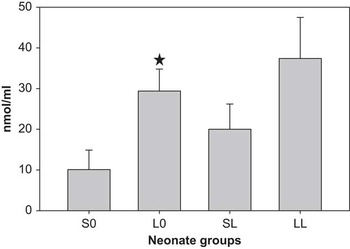
At baseline, neonates whose dams have been exposed to LPS had significantly higher levels of OS than control (Sal) neonates (PD 29.4 ± 5.4 v. 10.1 ± 4.8 nmol/ml; P < 0.05). In response to neonate LPS, the OS level among neonates whose mothers received LPS was not significantly higher than that of the neonates whose mothers received saline (37.4 ± 10.1 v. 20.0 ± 6.2 nmol/ml). There were no significant differences noticed between male and female pups (Fig. 1).
Lipid peroxides formation (PD). Neonatal PD at baseline or 4 h following lipopolysaccharide (LPS) injection. Two groups were used to compare basal values, offspring whose mothers received saline (S0) and offspring whose mothers received LPS (L0). Similarly, two groups were used to compare offspring responses to LPS, SL and LL for offspring whose mothers received saline and offspring whose mothers received LPS, respectively. *P < 0.05, significantly different between S0 and L0 groups.

CRP
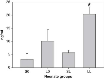
The basal CRP levels in the neonates whose dams have been exposed to LPS trended higher than control (Sal) neonates (10.1 ± 4.1 v. 3.2 ± 2.2 ng/ml; P = 0.09) but the difference did not reach statistical significance. In response to LPS, neonates whose dams have been exposed to LPS had three-fold greater CRP levels than the neonates whose mothers received saline (20.4 ± 2.8 v. 5.7 ± 1.0 ng/ml; P < 0.01). There were no significant differences noticed between male and female pups (Fig. 2).
C-reactive protein (CRP) levels. Neonatal CRP levels at baseline or 4 h following lipopolysaccharide (LPS) injection. Two groups were used to compare basal values, offspring whose mothers received saline (S0) and offspring whose mothers received LPS (L0). Similarly, two groups were used to compare offspring responses to LPS, SL and LL for offspring whose mothers received saline and offspring whose mothers received LPS, respectively. *P < 0.05, significantly different between SL and LL groups.

Discussion
The results of this study demonstrate that maternal inflammation during pregnancy (LPS administration on day 18) significantly increases offspring basal oxidative state, with a trend toward higher basal serum CRP levels. In response to re-exposure of the neonates to LPS (LPS administration at 24 days), CRP levels increased three-fold in the offspring of dams exposed to LPS as compared with the offspring of control dams. There was no difference in the OS response following LPS injection between the two groups, they had a similar increase in the PD following LPS.
OS is attributed to a discrepancy between the production of damaging reactive oxygen species and the capacity to mitigate their damaging effects. Systemic LPS administration leads to activation of the innate immune system. Binding of LPS to toll-like receptor 4 (TLR4) on monocytes/macrophages stimulates the generation of reactive oxygen species and OS. PD are products resulting from peroxidation (oxidation) of lipids. In serum, low-density lipoprotein is the major carrier of cholesteryl esters, thus being a substrate for lipid peroxidation.
Previous studies have demonstrated a correlation between maternal inflammation and increased fetal OS levels, though not in the offspring neonatal period. For example, markers indicating placental OS have been found after maternal inflammation,Reference Xu, Wang and Zhao 28 – Reference Ejima, Koji and Tsuruta 30 and OS elevation has been observed in rat fetuses brains several hours and days after deliveryReference Ejima, Koji and Tsuruta 30 – Reference Lante, Meunier and Guiramand 34 following maternal LPS injection.
It is well accepted that in addition to genetic inheritance, the intrauterine environment has a crucial role in programming fetal development.Reference Fowden, Giussani and Forhead 35 The elevation in basal offspring OS levels following maternal i.p. LPS injection implies an increased inflammatory basal state among the neonate born to mothers who suffer from inflammation during the pregnancy period. The implications of increased OS in utero may contribute to intrauterine fetal death, fetal growth restriction and preterm labor. The elevation of OS in the fetal brain may explain the increased incidence of offspring psychiatric and neurologic disorders after maternal inflammation during pregnancy. Notably, it is well established that high OS is associated with arteriosclerosis and cardiovascular diseases.Reference Aviram 36 In recent years, with the recognition that arteriosclerosis is an inflammatory process,Reference Ross 37 a plethora of studies have demonstrated that inflammatory plasma marker elevation (such as CRP and IL-6) can be a significant predictor for future risk of developing cardiovascular events,Reference Ridker, Cushman, Stampfer, Tracy and Hennekens 38 – Reference Liuzzo, Biasucci and Gallimore 42 colon cancer and metabolic syndrome. Thus, the elevation in the basal level of OS may have adverse consequences for physiological function later in life.
CRP is an acute-phase protein that increases during systemic inflammation. CRP is synthesized by the liver and facilitates complement activation, opsonization and apoptotic clearance.Reference Dempsey, Vaidya and Cheng 43 CRP is moderately elevated in obesity, metabolic syndrome, diabetes and fatty liver.Reference Sun, Koike and Ichikawa 44 Clinical studies have shown that elevated CRP levels in healthy populations predict vascular events such as myocardial infarction and stroke as well as the development of diabetes. Recent evidence has emerged implicating CRP directly in atherogenesis. CRP has been found in human atherosclerotic plaque and CRP has been shown to cause endothelial cell dysfunction, oxidant stress and intimal hypertrophy in experimental models. Induction of CRP in hepatocytes is principally regulated at the transcriptional level by IL-6, an effect that can be enhanced by IL-1β.Reference Kushner, Jiang, Zhang, Lozanski and Samols 45 Few studies have demonstrated that chronic inflammatory states are associated with only elevated CRP with no difference in the baseline cytokines,Reference Escobar-Morreale, Luque-Ramirez and Gonzalez 46 similar to our findings. The mechanism for the elevated basal CRP is unknown but may occur as a result of permanently altered hepatic function. Thus, both elevation in OS and a trend to elevated CRP may have adverse consequences for physiological function later in life.Reference Wilson, Ryan and Boyle 47
In response to offspring LPS, CRP levels nearly doubled in both groups of neonates whose mother received LPS and neonates whose mother received saline, and the OS elevation was similar in both groups. Overproduction of OS might be a deleterious process that can be an important mediator of damage to cell structures. Alternatively, beneficial effects of OS occur at low/moderate concentrations and involve physiological roles in cellular responses to noxia, as for example, in defense against infectious agentsReference Valko, Leibfritz and Moncol 48 that might be lacking in the offspring of dams exposed to LPS.
In prior studies, 21 , Reference Beloosesky, Maravi and Weiner 23 we demonstrated that maternal LPS exposure induces maternal systemic inflammation evident by increase of the inflammatory cytokines and results in offspring with a suppressed cytokine response to inflammatory stimuli. Specifically, pups of LPS-injected dams had significantly reduced IL-6, IL-1, TNF-α and IL-10 responses to a subsequent LPS injection when compared with pups of control (saline-injected) dams. Our previous findings of lower cytokine response to LPS and the current findings of higher baseline OS and a trend to higher CRP levels can be explained by recent studies that suggests that mild elevation in CRP levels imply a mild degree of tissue stress or injury, suggesting the hypothesis that the presence of distressed cells, rather than a resulting inflammatory response, is commonly the stimulus for CRP production.Reference Kushner, Rzewnicki and Samols 49 Studies on human cell lines have demonstrated that chronic inflammatory states are associated with only elevated CRP with no difference in the basal cytokines.Reference Kushner, Jiang, Zhang, Lozanski and Samols 45 We speculate that in utero exposure to IL-6 may program the liver and change the offspring OS and CRP response. Thus, activation of the fetal macrophage TLR4 system can induce LPS tolerance later in life.Reference Kirsten, de Oliveira and de Oliveira 50 Similar attenuation of the inflammatory response to LPS despite increased basal CRP levels was demonstrated in offspring of nutrient-restricted dams.Reference Desai, Gayle, Casillas, Boles and Ross 51
The limitation of our study is that it focuses specifically on maternal inflammation-induced fetal injury, not direct fetal infection. If the primary infection was fetal, we would expect that there would be direct injurious effects of the infectious agent, as well as the potential for a similar programming effect of the inflammatory mediators, similar to that demonstrated in our study. Further studies are warranted to compare the programming effects of maternal, intraamniotic and fetal infection on the offspring immune function.
In summary, we demonstrated that maternal inflammation during pregnancy increases offspring basal OS state and increases offspring serum CRP in response to an inflammatory stimulus. Higher CRP and OS levels imply an increased risk for developing future illnesses, including cardiovascular, psychiatric and neurologic disorders.
Acknowledgments
This research was supported by The Legacy Heritage Clinical Research Initiative of the Israel Science Foundation (Grant no. 1814/07).