Introduction
The dog is one of the potential hosts for parasitic zoonoses, such as Toxocara canis, Ancylostoma caninum, Giardia sp., Cryptosporidium sp., Echinococcus sp., Dipylidium caninum and Uncinaria spp. Surveys for helminth zoonoses in dogs have been carried throughout the world, in countries such as Brazil (Oliveira-Sequeira et al., Reference Oliveira-Sequeira, Amarante, Ferrari and Nunes2002), Australia (Palmer et al., Reference Palmer, Thompson, Traub, Rees and Robertson2008), Argentina (Soriano et al., Reference Soriano, Pierangeli, Roccia, Bergagna, Lazzarini, Celescinco, Saiz, Kossman, Contreras, Arias and Basualdo2010), Kyrgyzstan (Ziadinov et al., Reference Ziadinov, Deplazes, Mathis, Mutunova, Abdykerimov, Nurgaziev and Torgerson2010), Lithuania (Bružinskaite et al., Reference Bružinskaite, Sarkunas, Torgerson, Mathis and Deplazes2009), Hungary (Fok et al., Reference Fok, Szatmári, Busák and Rozgonyi2001), the Czech Republic (Dubná et al., Reference Dubná, Langrová, Nápravník, Jankovská, Vadlejch, Pekár and Fechtner2007), Greece (Papazahariadou et al., Reference Papazahariadou, Founta, Papadopoulos, Chliounakis, Antoniadou-Sotiriadou and Theodorides2007), Spain (Martínez-Moreno et al., Reference Martínez-Moreno, Hernández, López-Cobos, Becerra, Acosta and Martínez-Moreno2007), Belgium (Claerebout et al., Reference Claerebout, Casaert, Dalemans, De Wilde, Levecke, Vercruysse and Geurden2009), Italy (Rinaldi et al., Reference Rinaldi, Pennacchio, Musella, Maurelli and Cringoli2011), Germany (Barutzki & Schaper, Reference Barutzki and Schaper2011) and several European countries (Dyachenko et al., Reference Dyachenko, Pantchev, Gawlowska, Vrhovec and Bauer2008). The most prevalent worms reported in dogs are Ancylostomidae and Trichuris sp., followed by Toxocara canis (Oliveira-Sequeira et al., Reference Oliveira-Sequeira, Amarante, Ferrari and Nunes2002; Martins et al., Reference Martins, Sousa and Portugal2005a; Crespo et al., Reference Crespo, Rosa, Morgado, Ferreirinha, Cerejo and Madeira2006, Reference Crespo, Rosa and Almeida2010; Bajer et al., Reference Bajer, Bednarska and Rodo2011; Ferreira et al., Reference Ferreira, Pereira-Baltasar, Parreira, Padre, Vilhena, Tavira, Atouguia and Centeno-Lima2011; Rinaldi et al., Reference Rinaldi, Pennacchio, Musella, Maurelli and Cringoli2011).
Human toxocariasis is an important zoonosis that has been reported in several countries, such as Argentina (Radman et al., Reference Radman, Archelli, Fonrouge, Guardis, del and Linzitto2000), the USA (Walsh, Reference Walsh2011) and European countries (Deplazes et al., Reference Deplazes, Knapen, Van Schweiger and Overgaauw2011), being a common parasitosis in children (Castelo et al., Reference Castelo, Dinis and Rocha2008; Deplazes et al., Reference Deplazes, Knapen, Van Schweiger and Overgaauw2011). Dog parasite infection and environmental contamination have been evaluated in several countries in rural areas or in cities, demonstrating their importance and stressing the related zoonotic risk factors (Oliveira-Sequeira et al., Reference Oliveira-Sequeira, Amarante, Ferrari and Nunes2002; Dubná et al., Reference Dubná, Langrová, Nápravník, Jankovská, Vadlejch, Pekár and Fechtner2007; Palmer et al., Reference Palmer, Thompson, Traub, Rees and Robertson2008). In southern Portugal, throughout a 10-year period (1995–2005), official laboratory serodiagnosis results revealed 21.9% positive sera for Toxocara sp. infection in 457 suspicious samples (Martins et al., Reference Martins, Vilares, Júlio, Correia, Ferreira, Reis, Gargaté, Gomes and Ângelo2005b). No human seroprevalence surveys have been carried out in Portugal (Castelo et al., Reference Castelo, Dinis and Rocha2008). The T. canis and T. cati ascarids infect humans through the ingestion of embryonated eggs, either by regular contact with dogs and cats, or by contaminated soil. Rural regions, lower socio-economic background, geophagy pica, young age and owning dogs are the main risk factors for seropositivity (Nelson et al., Reference Nelson, Greene and Ernhart1996; Eberhardt et al., Reference Eberhardt, Bialek, Nägele and Dichgans2005; Walsh, Reference Walsh2011; Manini et al., Reference Manini, Marchioro, Colli and Nishi2012). Toxocara canis can reach high prevalence values due to the large number of eggs excreted and egg resistance to environmental conditions. Embryonated eggs hatch in the intestine of dogs or cats to form larvae, which may develop into adult worms and release eggs that are then excreted with faeces. Humans, rodents and other mammals are paratenic hosts that can suffer tissue damage in various organs (visceral larva migrans and ocular toxocariasis) caused by the larval stage of T. canis. Children may exhibit severe infection and suffer from seizures, myocarditis, diminished lung function (Walsh, Reference Walsh2011) and eosinophilic meningomyelitis (Eberhardt et al., Reference Eberhardt, Bialek, Nägele and Dichgans2005) depending on the location of the larvae.
Cutaneous larva migrans or creeping eruption is a zoonotic infection caused by some human and animal hookworm species, namely A. caninum and Uncinaria stenocephala, two geohelminths transmitted by dogs and cats (definitive hosts). Released into the environment (soil, sand, grass, etc.) through the faeces, under favourable conditions of moisture and warmth, the eggs hatch and become third-stage infective larvae in 6–12 days. These larvae can survive for 3–4 weeks under favourable conditions. Penetration of infective larvae in human skin causes infection that can affect all ages. Rodents are paratenic hosts that can be an important source of infection for dogs and cats. According to Kwon et al. (Reference Kwon, Kim, Lee, Choi, Chai, Nakamura-Uchiyama, Nawa and Choi2003), larval penetration usually occurs in the feet, buttocks and genitalia. Larval migration originates in a characteristic serpiginous skin eruption, usually erythematous and sometimes vesicular, leading to intensive pruritus and secondary bacterial infections, causing a localized eosinophilic inflammatory reaction. In some cases, in humans, A. caninum migrates to the intestine causing eosinophilic enteritis or diffuse unilateral subacute neuroenteritis (Centers for Disease Control & Prevention, 2012).
Hydatidosis is an important zoonosis in the Mediterranean region (World Health Organization, 2002), the most affected region in Europe (Jenkins et al., Reference Jenkins, Romig and Thompson2005), and is one of the five most frequent zoonoses in this area (Dakkak, Reference Dakkak2010), despite being sub-notified in Portugal. Throughout a 25-year period (1979–2003), Morais (Reference Morais2007) presented the Évora region as yielding a mean incidence of 12.2 cases/100,000 inhabitants per year. Central Portugal is referred to by Morais (Reference Morais2010) as having the second highest number of cases in official statistics (9.2%) and the third highest in his personal clinical data (6.2%). Echinococcus granulosus data in dogs are not published and up-to-date abattoir data in the intermediate hosts are not available. The dog is the final host of the domestic cycle, and E. granulosus eggs are shed in their faeces and ingested by intermediate hosts (humans, ruminants and pigs), inside which the larvae develop cystic echinococcosis. The majority of the most recent data on this subject refers to environmental contamination (Crespo et al., Reference Crespo, Rosa, Morgado, Ferreirinha, Cerejo and Madeira2006; Cardoso et al., Reference Cardoso, Costa, Costa, Conceição and Castro2011). Carried out both in rural and city areas, these studies established that Ancylostomidae and Ascaridae were the most prevalent zoonotic helminths (72 and 16%, respectively).
The aim of the present study was to evaluate the occurrence and intensity of infection of gastrointestinal parasites in rural dogs, in the county of Cantanhede, northern Portugal.
Materials and methods
Study area
The county of Cantanhede was selected for this cross-sectional study because of the occurrence of cystic echinococcosis in home-slaughtered pigs (Castro et al., Reference Castro, Silva, Veloso, Freire, Rodrigues, Silva, Agante, Conceição and Correia da Costa2005; Conceição et al., Reference Conceição, Silva, Veloso, Castro, Silva, Cravo, Bastos and Correia da Costa2005) and the presence of adult worms of E. granulosus in stray dog necropsies (personal data, unpublished).
The county of Cantanhede belongs to the District of Coimbra, and is located in northern Portugal. The main town is Cantanhede (40°21′N and 8°36′W) and the region has 38,920 inhabitants dispersed over 390.89 km2. Roast piglet (leitão da Bairrada) and the Bairrada vineyards are the main source of income in this rural area, and the home slaughtering of pigs is a frequent practice. In 2008, there were 2573 breeding pigs registered as family units, and also two intensive piggeries (with 898 reproducers). In addition, the small-ruminant population was 2123, distributed in 382 flocks, and there were approximately 8000 bovines (official data from veterinary services).
Collection and examination of faecal samples of dogs
Small-ruminant farms were chosen for the gastrointestinal helminth evaluation of dogs, since sheep are the main risk for E. granulosus infection and so far only G7 (the pig strain) has been found in the region (Castro et al., Reference Castro, Silva, Veloso, Freire, Rodrigues, Silva, Agante, Conceição and Correia da Costa2005). Moreover, the size of the dog population in Cantanhede is unknown. The sample size was evaluated assuming 50% parasite prevalence (P= 50%), with a 95% confidence level and 4% error.
Between November 2008 and September 2009, 234 small-ruminant farms were visited with the purpose of collecting dog faecal samples. In total, 484 dogs from different breeds, crosses and ages were taken into account from 15 of the 19 subregions of Cantanhede. Faeces (n= 301) were collected from the ground in kennels, fenced pastures, or close to other non-specific sites where dogs were confined. Whenever farmers had more than one dog, it became impossible to associate the samples with a specific dog. Finally, in some cases farmers had no dogs, and in a few other cases no samples were collected. Therefore, samples were collected from 165 small-ruminant farms, and hence only this subset was taken into account in the final study.
Faecal samples were stored in plastic bags in refrigerated containers, sent to the laboratory and analysed using the McMaster (Soulsby, Reference Soulsby1982), Willis (Bajer et al., Reference Bajer, Bednarska and Rodo2011) and the Ritchie (formol–ether sedimentation) (Golvan & Ambroise-Thomas, Reference Golvan and Ambroise-Thomas1984) techniques, followed by microscopy. Each technique was performed with 2 g of faeces.
Percoll fractionation of faecal samples
Percoll (reference 17-0891-01; GE Healthcare, Amersham Biosciences Limited, Amersham, UK) step gradients were used to perform a partial clean-up and concentration of parasite eggs. Percoll step gradients were based on helminth egg density. An evaluation of Percoll fractionation was performed using step gradients composed of 2 ml of Percoll at a density of 1.3 g/ml, followed by 1 ml of Percoll with densities of 1.2, 1.18, 1.16 and 1.14 g/ml, in a conical glass tube (internal diameter 1.4 × length 15 cm). Stool samples from dogs (about 2 g) containing helminth eggs (Toxocara spp., n= 7; Trichuris spp., n= 9; Taenia spp., n= 4) were re-suspended in 4 ml of phosphate-buffered saline (PBS) with 1% Triton X-100 and filtered through gauze. A stool suspension of 2 ml was placed over the Percoll step gradient and sedimented for 2 h at room temperature. Up to 1 ml of each phase was collected into a 15 ml tube and mixed gently with 10 ml of PBS. Samples were centrifuged at 1000 rpm for 10 min. The pellet was re-suspended in 1 ml of PBS and analysed to evaluate cleaning and helminth structure concentration. Helminth eggs were recovered mainly at the density of 1.18 g/ml (recovery >75% of helminth eggs), with the remaining parasitic structures being found in the sediment and in the phase of density 1.16 g/ml.
The methodology was further simplified using a step gradient composed of 2 ml of Percoll at 1.3 g/ml followed by 1 ml at 1.18 g/ml and 1 ml of 1.16 g/ml. The phase corresponding to 1.18 g/ml density was collected (about 1 ml) and treated as described above. This simplified methodology was used for the stool samples positive for Taenidae.
Molecular detection of Echinococcus granulosus
Stool material isolated using Percoll gradients was used for further evaluation of the presence of E. granulosus. For extraction of total DNA, the QIAamp DNA Mini Kit (Qiagen, GmbH, Hilden, Germany) was used according to the protocol provided by the manufacturer for the purification of tissues. The presence of E. granulosus DNA in stool samples was evaluated using the polymerase chain reaction (PCR) technique. For each sample we performed PCR reactions for the amplification of a NADH dehydrogenase I fragment (Bowles et al., Reference Bowles, Blair and McManus1992), cytochrome c oxidase 1 fragment (Bowles & McManus, Reference Bowles and MacManus1993) and E. granulosus repeated sequence (Abbasi et al., Reference Abbasi, Branzenburg, Campos-Ponce, Abdel Hafez, Raoul, Craig and Hamburger2003). Results were analysed using electrophoresis, and DNA analysed from samples of E. granulosus cysts and also from faeces positive for E. granulosus was used as positive controls. These controls were detected using PCR followed by sequencing of amplified DNA fragments.
Questionnaire
The epidemiological survey was conducted by distributing a questionnaire to 234 small-ruminant farmers to evaluate risk behaviours, with reference to the feeding habits of dogs, including offal (raw organs or meat), the frequency, if any, of anthelmintic treatment and the number of dogs per farm. Slaughtering practices were also registered. The type, age and breed of dogs were not recorded since most rural dogs are guard dogs of mixed breed and, most of the time, when several dogs are present, faecal identification is not possible. The presence of farm animals other than small ruminants was registered. This enquiry was designed mainly to survey the presence of E. granulosus.
Data analysis
Data analysis was performed using the statistical analysis software IBM SPSS v. 19.0 (SPSS Inc., Chicago, Illinois, USA). Fisher's exact test was used to compare nominal binary variables in 2 × 2 tables, and the odds ratio was used to estimate the probability of occurrence of a given risk behaviour, at 95% confidence intervals and for 165 farmers who provided faecal samples of dogs.
Results
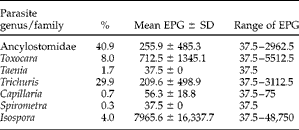
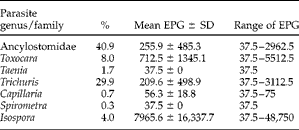
Seven species of intestinal parasites were detected with coprological techniques and the most prevalent was Ancylostomidae, with a prevalence of 40.9% (table 1). Of the faecal samples (n= 301), 58.8% (n= 177) were positive for at least one type of parasite and Taeniidae eggs were present in 1.7% (n= 5), although the PCR analysis proved that none of them was E. granulosus. Toxocara sp. had a mean elimination of 712.5 eggs per g (EPG) (range 37.5–5512.5 EPG) followed by Ancylostomidae with 255.9 EPG (range 37.5–2962.5 EPG) and Taeniidae eggs with 37.5 EPG. Besides the zoonotic parasites, Isospora sp. had a high oocyst elimination (mean 7965.6 EPG; range 37.5–48,750 EPG), occurring in 4.0% of the samples. A high prevalence was found for Trichuris sp. (29.9%), with a mean of 209.6 EPG (range 37.5–3112.5 EPG). Additionally, Spirometra sp. and Capillaria sp. were found to have low egg shedding (table 1).
The prevalence (%) and mean faecal counts of oocysts/eggs per g (EPG) of gastrointestinal parasites in 301 dogs from the Cantanhede region; SD = standard deviation.
Of the farmers who owned dogs (n= 195) from which samples were collected, 48.5% (n= 80) had at least one dog with one or two zoonotic parasites (Ancylostomidae or Toxocara sp.). Dogs with Taeniidae eggs belonged to different farms. The questionnaires showed that of the 234 farmers questioned, 83.3% (n= 195) owned at least one dog, with a mean of 2.5 dogs (SD = 1.9 dogs) per farmer and a maximum of 10 dogs. Most farmers (66.7%) stated that they treat their dogs with anthelmintic drugs, 23.6% performing the treatment at least twice per year, but only a few of them (5.5%) doing it three times per year. It was not possible to confirm which drugs were used.
The results of the questionnaire showed that 43.6% of the farmers fed their dogs with viscera and, of these, 23.9% with raw viscera and 75% fed them with small-ruminant viscera and/or pig viscera. Pig home slaughtering was found to be a common practice (57% of those questioned). Most pig slaughtering was carried out once a year (68.1%), but was performed twice or three times a year by the remainer of the farmers (31.9%). Moreover, farmers who do not perform home slaughtering feed their dogs with viscera from slaughtering performed by neighbours or family (10.7%). Some of the farmers questioned (23.5%) had already seen cysts in the liver of home-slaughtered pigs. The survey additionally included a question regarding the farmer's knowledge of possible human hydatidosis infections, to which 7% answered positively.
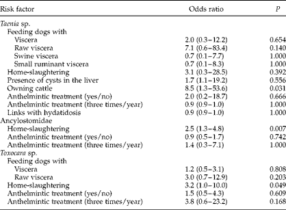
The presence of cattle in the farms was quite low (16.4%), and most of the farmers had only one animal for meat production and in-house consumption. In the risk analysis results (table 2) the ‘owning cattle’ factor yielded a statistically significant result for Taenia sp. (P= 0.031). ‘Home slaughtering’ was the only factor presenting a statistically significant result for Ancylostomidae (P= 0.007) and for Toxocara sp. (P= 0.049).
The association between host variables and infections with Taenia sp., Ancylostomidae and Toxocara sp. infections; 95% confidence intervals given in parentheses.

Discussion
The overall parasitic infection of dogs in the region of Cantanhede (58.8%) is slightly higher than in other rural areas of Portugal (Crespo et al., Reference Crespo, Rosa, Morgado, Ferreirinha, Cerejo and Madeira2006), with seven different species of dog parasites and a high level of co-infections. This fact may be related to rural dog provenance. In fact, a higher Ancylostomidae prevalence (40.9%) than Toxocara sp. (8%) was found in the present study, which is consistent with the results of Dubná et al. (Reference Dubná, Langrová, Nápravník, Jankovská, Vadlejch, Pekár and Fechtner2007), but differs from those of Sousa et al. (Reference Sousa, Sanches and Martins2005). This might be due to the origin of the samples, since faeces were collected but not soil and grass. This circumstance reflects different levels of egg resistance to environmental effects, since it is known that Toxocara canis and Trichuris sp. eggs have more environmental resistance than Ancyclostomidae eggs (Deplazes et al., Reference Deplazes, Knapen, Van Schweiger and Overgaauw2011), which may influence the egg parasite count. In the present study, dogs were confined to a restricted area, since they were not shepherd dogs, providing conditions for strong environmental contamination and a geohelmintic infection for the three nematode species. In fact, they were mostly guard dogs, and whenever more than five dogs were owned (12.4%), these were generally used for hunting.
In spite of its low prevalence (8%), Toxocara sp. shows a high intensity of infection in some dogs, with a maximum of 5512.5 EPG (table 1), which represents a health risk for the farmers. A similar case was observed for Trichuris sp., with a maximum egg shedding of 3112.5 EPG, which suggests health problems for the dogs. Isospora sp. was the only protozoan observed in this study in 4% of sampled faeces, but with a very high intensity of infection (maximum 48,750 EPG) and a mean elimination of 7965.6 EPG.
A total of five samples were positive for Taenia sp. with a very low count (37.5 EPG) and none for E. granulosus, as determined by the PCR technique. Considering that adult E. granulosus were observed in stray dogs in Cantanhede's kennel, this result indicates that further studies must be undertaken. The irregular trait of Taeniidae proglottid elimination, given that eggs are expelled while enclosed within the proglottids, renders an actual EPG count irrelevant for the evaluation of parasite burden. On the other hand, the McMaster technique has poor sensitivity (Bružinskaite et al., Reference Bružinskaite, Sarkunas, Torgerson, Mathis and Deplazes2009), although saturated saline solution is the technique advised for the analysis of dog faeces (Bajer et al., Reference Bajer, Bednarska and Rodo2011). The present study combines the McMaster and Willis techniques with the formol–ether technique of Ritchie, which provides egg concentration and sedimentation. The Willis, and especially the Ritchie, techniques allowed for an effective detection of Taeniidae. Taenia sp. is related to farm dogs that have access to offal and rodents. Up to 37.2% of farmers claimed to carry out home slaughtering in Cantanhede. This practice proved to have a statistical significance for Toxocara sp. and Ancylostomidae infection, a fact that has also been stressed in other studies (Dakkak, Reference Dakkak2010; Deplazes et al., Reference Deplazes, Knapen, Van Schweiger and Overgaauw2011), and acknowledged to be a key element for the perpetuation of the life cycle of E. granulosus (Bružinskaite et al., Reference Bružinskaite, Sarkunas, Torgerson, Mathis and Deplazes2009). Toxocara canis infection through the accidental consumption of poultry, small-ruminant and swine viscera may also occur (ESCCAP, 2010). This was shown by Taira et al. (Reference Taira, Saeed, Permin and Kapel2004) with pigs experimentally infected with Toxocara larvae, and by Lloyd (Reference Lloyd2006), who found a relatively high rate of T. canis antibody levels and, in a previous study, even parasitic granulomata, in sheep (Lloyd et al., Reference Lloyd, Martin, Walters and Soulsby1991), which may represent an additional risk of infection for dogs and, by inference, for humans, whenever raw or insufficiently cooked pig and poultry organs or meat are consumed. In field conditions, this risk was not evaluated, and neither was the prevalence of infections in farm animals with dog nematode larvae. It is assumed to be possible in the present rural conditions, but this would need further investigation. The present study revealed poor hygiene conditions and animal housing, with a great diversity of domestic animals. The presence of cattle in the farms was investigated, since they are also intermediate hosts of several Taeniidae, including E. granulosus. Moreover, in abattoirs in northern Portugal, hepatic cysts are frequently observed in cattle (personal data, unpublished). The statistical significance of the presence of cattle for Taeniidae egg shedding in dogs may therefore suggest the presence of other species (Christofi et al., Reference Christofi, Deplazes, Christofi, Tanner, Economides and Eckert2002), given the frequency of hunting with dogs.
During the survey, while talking informally with the farmers, we were told that canine parasite control is usually performed simultaneously with rabies vaccination, which suggests the possibility of confusion with anthelmintic treatment. Additionally, we found that when farmers treat their dogs by themselves, most of them (66.7%) claim to use anthelmintic drugs and ignore the indications of the drugs they administer and their respective spectra, a practice that leads to drug misuse. The combination of these factors may explain the statistical non-significance of the effect of ‘anthelmintic treatment’ on the infection by zoonotic parasites investigated in this study.
In the Mediterranean region, the key factors for the development of zoonotic helminths are associated with the diversity of livestock production, close proximity of man to animals, illegal home slaughtering and the lack of adequate health education (Dakkak, Reference Dakkak2010; Deplazes et al., Reference Deplazes, Knapen, Van Schweiger and Overgaauw2011). We found all these risk factors in the region of Cantanhede. Humans in rural areas are exposed to helminth eggs through different routes, the most important of which are water, food and soil, and also direct contact with dogs. In addition, in Portugal, the impact of wildlife in zoonotic helminth transmission is unknown, even though it has been referred to as an important route in other countries (Ziadinov et al., Reference Ziadinov, Deplazes, Mathis, Mutunova, Abdykerimov, Nurgaziev and Torgerson2010). Up-to-date information on the regional prevalence of intestinal parasites is vital for the development of strategies concerning parasite control. The results presented here suggest that, particularly in the region of Cantanhede but also in similar areas, it is important to promote an increased integration of medical and veterinary services to provide adequate educational standards and veterinary control.
Acknowledgements
The authors wish to acknowledge the collaboration provided by the Farmers Health Organization of Tocha and Cantanhede, in particular the veterinary support given by Victor Nunes in fieldwork, as well as to thank Conceição Cravo for her invaluable laboratory support.