Milk and dairy products are important constituents of a daily diet for many people in Western countries. Recently, focus has been directed to the fact that milk contains endogenous estrogens such as estrone, estriol, 17α-estradiol and 17β-estradiol (E2) and their conjugated metabolites (Malekinejad et al. Reference Malekinejad, Scherpenisse and Bergwerff2006; Farlow et al. Reference Farlow, Xu and Veenstra2009; Tso & Aga, Reference Tso and Aga2010). Milk also contains exogenous estrogens in the form of plant derived phytoestrogens (Hoikkala et al. Reference Hoikkala, Mustonen, Saastamoinen, Jokela, Taponen, Saloniemi and Wahala2007; Andersen et al. Reference Andersen, Weisbjerg, Hansen-Møller and Sejrsen2008; Steinshamn et al. Reference Steinshamn, Purup, Thuen and Hansen-Moller2008; Nielsen et al. Reference Nielsen, Norgaard, Purup, Frette and Bonefeld-Jorgensen2009), a diverse group of non-steroidal naturally occurring compounds with structural and functional similarities with mammalian estrogens. Concern has been raised about the possible adverse effect of milk-borne hormones especially estrogens in relation to breast cancer by some epidemiological found correlations (Boyd et al. Reference Boyd, Martin, Noffel, Lockwood and Trichler1993; Gaard et al. Reference Gaard, Tretli and Loken1995; Cho et al. Reference Cho, Spiegelman, Hunter, Chen, Stampfer, Colditz and Willett2003) supported by results from animal studies (Qin et al. Reference Qin, Xu, Wang, Ganmaa, Li, Wang, Kaneko, Hoshi, Shirai and Sato2004, Reference Qin, Xu, Tezuka, Li, Arita, Hoshi and Sato2007, Reference Qin, Xu, Tezuka, Wang and Hoshi2008; Ma et al. Reference Ma, Katoh, Zhou and Wang2007; Zhou et al. Reference Zhou, Qin, Tang, Ma, Wang and Wang2007). Estrogens are potent mitogens in both normal and malignant mammary cells and proliferation of malignant cells is suggested as an explanation of why estrogens can increase breast cancer risk. It is also possible that in normal cells, increased proliferation induced by estrogens may lead to accumulation of DNA adducts and eventually mutations and thereby cancer (Hilakivi-Clarke et al. Reference Hilakivi-Clarke, Cabanes, Olivo, Kerr, Bouker and Clarke2002).
The estrogen level in milk reflect the pregnancy status of the cow and during the second and third trimester of pregnancy the milk content of total estrogens, which include both free and conjugated forms, increase from 0 to 60 ng/l in non-pregnant and early pregnant cows to more than 1600 ng/l (Malekinejad et al. Reference Malekinejad, Scherpenisse and Bergwerff2006). We previously calculated that only 42% of the total delivered milk from modern Western dairy farms is produced by pregnant cows with only 21% being produced by cows in their second and third trimester of pregnancy (Nielsen et al. Reference Nielsen, Khan, Davis, Michels and Hilakivi-Clarke2011). Although the per cent-wise contribution to total delivered milk may be low, the very high level of estrogens in milk from second and third trimester pregnant cows is assumed to be the main contributor to the considerable levels of endogenous estrogens found in commercial milk (Farlow et al. Reference Farlow, Xu and Veenstra2009) since commercial milk is a mix of milk from cows at all different stages of lactation and pregnancy. Consequently, milk and dairy products have been estimated to be the main source of dietary estrogens accounting for 60–70% of the daily intake of estrogens (Hartmann et al. Reference Hartmann, Lacorn and Steinhart1998).
The potential estrogenic activity of milk has only been investigated in biological assays in a few studies. Commercial low-fat milk increased the uterine weight, which is used as a measure of estrogenic activity, with 18% in pre-pubertal rats and with 53% in ovariectomized rats compared with control animals fed artificial milk (Ganmaa et al. Reference Ganmaa, Tezuka, Enkhmaa, Hoshi and Sato2006). Another study found no uterotrophic effect of milk differing in phytoestrogen content compared with water exposed control animals (Nielsen et al. Reference Nielsen, Norgaard, Purup, Frette and Bonefeld-Jorgensen2009). In vitro, extracts of milk with a high content of the phytoestrogen equol showed significant indirect estrogenic activity by activating an estrogen-receptor (ER) reporter gene in human breast cancer cells (MVLN) (Nielsen et al. Reference Nielsen, Norgaard, Purup, Frette and Bonefeld-Jorgensen2009), whereas extracts of commercial milk only resulted in minor induction of the ER reporter gene in T47D.luc cells (Dip et al. Reference Dip, Lenz, Antignac, Le Bizec, Gmuender and Naegeli2008).
The in vitro assay with human breast cancer cells (MCF-7) has been identified as a sensitive method to screen for estrogenic activity of for example single potential estrogenic compounds (Gutendorf & Westendorf, Reference Gutendorf and Westendorf2001) and extracts of human serum where endogenous steroids was separated from xenestrogens (Rasmussen et al. Reference Rasmussen, Nielsen, Andersen, Nielsen, Weihe and Grandjean2003). MCF-7 cells proliferate in response to estrogens or estrogen-like stimulation and provide an easy measurable endpoint of both estrogenic but also potential anti-estrogenic activity. Since milk (whey) contains other hormones and growth factors, such as insulin-like growth factor 1 (IGF-1) and progestogenes (Dupont & Le, Reference Dupont and Le2001; Chen et al. Reference Chen, Chien, Chen and Ng2011) that also influences the proliferation of MCF-7 cells, it is not possible to test the isolated estrogen-induced effect of whey in these cells, but the overall proliferative effect of milk can be assessed. The aim of our study was to use MCF-7 cells as a model system to determine the proliferative and anti-proliferative effect of whey from milk obtained during the first or second half of pregnancy where milk estrogen levels are expected to differ significantly, in order to add knowledge to the discussion on the potential role of milk intake on human breast cancer development.
Materials and Methods
Milk samples
Milk samples were collected from 11 Danish Holstein and 11 Red Danish multiparous cows, 12 samples represent cows in their first half of pregnancy and 10 samples represent cows in their second half of pregnancy. All cows were fed a total mixed ration where the roughage part consisted of clover grass silage and maize silage (1:1) and the main concentrates were soybean meal and rolled barley. Milk from individual cows was obtained during the morning and evening milking and pooled to one milk sample.
Milk was centrifuged at 3500 g at 4°C for 20 min and skim milk aspirated and frozen at −20°C for later use. Milk contains large amounts of fat and casein that cause unfavourable culture conditions for cells in vitro and therefore the estrogenic activity of milk was assessed using whey where fat and casein have been removed. Skim milk was titrated with 5 m acetic acid to precipitation at pH 4·2, then centrifuged at 9000 g for 30 min at 4°C. The acidic whey was then neutralized with 3 m and 1 m NaOH to pH 7·4 and centrifuged again at 9000 g for 30 min at 4°C. The neutralised acidified whey was frozen at −20°C for use in the in vitro assay.
MCF-7 cell proliferation assay
The estrogenic activity of whey samples was screened in MCF-7 BUS cells (MCF-7) which were a kind gift from Professor Eva C. Bonefeld-Jørgensen, Aarhus University. Cells were cultivated in Dulbecco Modified Eagle Medium (DMEM) containing 1000 mg D-glucose/l and 110 mg sodium pyruvate/l and supplemented with 15 mm HEPES, 4 mm glutamax (Invitrogen A/S, Tåstrup, Denmark), 6·4 mg gentamicin/100 ml (Sigma-Aldrich, Brøndby, Denmark) and 10% heat-inactivated foetal calf serum (FCS; Invitrogen). Cells were grown in 75 cm3 flasks in a cell incubator (37°C) with humidified air and a CO2 concentration of 5% until approximately 90% confluence.
For the experiment, cells were seeded in 96-well plates at a density of 1500 cells/well in medium containing 10% dextran-coated charcoal treated FCS (DCC-FCS) (VWR-Bie & Berntsen A/S, Herlev, Denmark) (0·1 ml medium/well). After 24 h, 0·1 ml of medium containing two times the desired whey concentration (due to the 1:1 dilution of whey) was added to the wells.
Whey from individual cows was tested in concentrations of 0·1, 0·25, 0·5, 1·25, 5 and 10% of the cell culture media in triplicate wells. A medium with 20% whey was sterile filtrated (0·2 μm mesh) and used for preparation of the other whey concentrations. The effect of whey was screened in the absence and presence of E2 (Sigma-Aldrich) in the medium in order to detect both potential estrogenic and anti-estrogenic properties. A growth curve with E2 ranging from 0·1 pm to 50 μm was performed to determine the concentration of E2 resulting in a growth stimulatory effect approximately half of the maximum response to allow for detection of both additive and inhibitory effects of whey. E2 was dissolved in ethanol at a concentration of 10 mm before being supplemented with experimental medium. The final concentration of ethanol in the cell culture medium did not exceed 0·01% and did not affect cell growth.
On day seven, cell viability was determined by a resazurin metabolism assay (AlamarBlue, BioSource, AH diagnostic, Aarhus, Denmark). AlamarBlue reagents were added to wells according to the manufacturer's instructions and after an incubation time of 6 h measured with a fluorometer. The amount of fluorescence measured is proportional to the number of living cells and thereby indirectly to cell proliferation.
Statistical analysis
Data on the fluorescence of cells exposed to whey relative to the control treatment were analyzed by one-way ANOVA followed by Fisher's LSD test. This was done separately within each of the whey concentrations tested. Results are expressed as means±sem, and significance was judged at P<0·05.
Results
In order to establish the maximum concentration of whey tolerated by MCF-7 cells, a range of whey concentrations (0–10%) in the medium were tested (data not shown). When 1·25% or more whey was added to the medium, cell growth dropped to approximately half the response induced by the control medium with no whey and therefore we preceded our investigations with a maximum of 0·5% whey added to the medium.
Figure 1 shows the effect of increasing concentrations of E2 on the cells. At 100 pm (27·2 pg E2/ml) cell growth was four times higher than of control cells, whereas 10 pm (2·72 pg E2/ml) induced a factor two response compared with the control treatment. Based on this result, 10 pm E2 was selected as the background for testing the potential anti-estrogenic effect of whey samples.
Proliferative response of MCF-7 cells to increasing concentrations of 17β estradiol (E2). The control treatment is without E2. 10 pm E2 induced a response corresponding to approximately 50% of the maximum response and therefore this concentration was chosen as the background when testing the potential anti-proliferative effect of whey. Mean values±sem are shown.

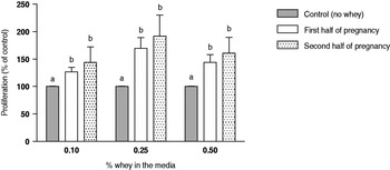
Whey from milk obtained during the first half of pregnancy resulted in a 27–70% increase in cell proliferation relative to the control treatment (P<0·05) dependent on the concentration of whey included in the medium (Fig. 2). The increase in cell proliferation was 44–92% for milk obtained during the second half of pregnancy compared with the control treatment (P<0·05) dependent on the whey concentration in the medium (Fig. 2). There was no significant difference in the proliferative effect of whey from the two stages of pregnancy (P=0·24, P=0·31, P=0·31 for 0·1, 0·25 and 0·5% whey, respectively).
Effect of different concentrations of whey from milk obtained during the first or second half of pregnancy on the proliferation of MCF-7 cells relative to control cells not exposed to whey. Statistical significant differences (P<0·05) within each whey concentration tested are indicated by different letters (A, B). Mean values± sem are shown.

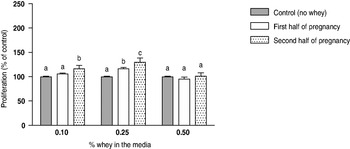
Figure 3 show the proliferative response to whey from milk at the two stages of pregnancy with 10 pm E2 included in the growth medium expressed relative to control cells also exposed to E2. The growth stimulating effect of whey when E2 was present in the medium was much less pronounced than without E2, but still confirming the estrogenic effect of whey mentioned earlier. Milk from the first half of pregnancy resulted in a 16% increase in proliferation (P=0·001) at a whey concentration of 0·25 in the medium, whereas there were no significant differences to the control at any other whey concentration tested. Milk from the second half of pregnancy stimulated cell growth by 16 and 29% (P<0·05) when 0·1 and 0·25% whey were tested, but there were no significant differences at 0·5% whey included in the medium. Overall, there was no anti-proliferative effect of milk regardless the stage of pregnancy, since cell growth with whey plus E2 was not lower than the control treatment plus E2.
Effect of different concentrations of whey from milk obtained in the first or second half of pregnancy on the proliferation of MCF-7 cells relative to control cells not exposed to whey, when a background of 10 pm estradiol (E2) was included in the culture medium at all treatments. Note that inclusion of 10 pm E2 in the medium increased proliferation of MCF-7 cells by 100% compared with no E2 according to Fig. 1, but here in Fig. 3 the control treatment is still set to 100% to show the relative proliferation. Statistical significant differences (P<0·05) within each whey concentration tested are indicated by different letters (A, B). Mean values± sem are shown.

Discussion
Concerns about the negative health effects of exogenous estrogens in foods such as milk have been raised (Andersson & Skakkebaek, Reference Andersson and Skakkebaek1999; Fritsche & Steinhart, Reference Fritsche and Steinhart1999), especially in relation to estrogen sensitive cancers like breast cancer (Ganmaa & Sato, Reference Ganmaa and Sato2005). Indeed it is well established that milk contains various endogenous estrogens (Malekinejad et al. Reference Malekinejad, Scherpenisse and Bergwerff2006; Courant et al. Reference Courant, Antignac, Maume, Monteau, Andre and Le2007; Farlow et al. Reference Farlow, Xu and Veenstra2009) and feed derived phytoestrogens (Andersen et al. Reference Andersen, Nielsen, Purup, Kristensen, Eriksen, Søegaard, Sørensen and Fretté2009; Nielsen et al. Reference Nielsen, Norgaard, Purup, Frette and Bonefeld-Jorgensen2009) that potentially contribute to an estrogenic effect of milk upon ingestion. In the present study we found that whey induced a significant proliferative response in MCF-7 cells, which may be caused, at least in part, by the estrogens present in milk. To our knowledge no studies have previously investigated the proliferative effect of whey in estrogen-sensitive breast cancer cell lines, but it has been reported that extracts of milk with a high content of phytoestrogens caused significant estrogen receptor transactivation in MVLN cells, which is a direct measure of estrogenic activity (Nielsen et al. Reference Nielsen, Norgaard, Purup, Frette and Bonefeld-Jorgensen2009).
The level of endogenous estrogens in milk increase as pregnancy of the cow proceeds (Malekinejad et al. Reference Malekinejad, Scherpenisse and Bergwerff2006) and therefore we hypothesized that there would be an increase in the proliferative effect of whey from the first to the second half of pregnancy. But there was no significant difference in the proliferative effect of whey regardless of stage of pregnancy. The growth of estrogen-sensitive MCF-7 cells is influenced by other hormones and growth factors such as insulin-like growth factor 1 (IGF-1) and progestogenes (Dupont & Le, Reference Dupont and Le2001; Chen et al. Reference Chen, Chien, Chen and Ng2011). Especially for IGF-1 and estrogens, their signalling pathways are intertwined and they both act as stimulators of MCF-7 cell proliferation resulting in a synergistic effect (Dupont & Le, Reference Dupont and Le2001). Chen et al. (Reference Chen, Chien, Chen and Ng2011) showed that progestogen-induced apoptosis in MCF-7 cells may be partly inhibited in the presence of certain concentrations of estrogen indicating a complex interaction between those two hormones in regulating cell growth. Commercial milk contains both IGF-1 and progesterone (Vicini et al. Reference Vicini, Etherton, Kris-Etherton, Ballam, Denham, Staub, Goldstein, Cady, McGrath and Lucy2008) although it was not measured in this experiment, and therefore we cannot rule out the possibility that interactions between milk estrogens and other hormones and growth factors, may have masked the isolated proliferative effect of estrogens.
We did not observe any anti-proliferative effect when whey was tested in the presence of E2 in the culture medium. Some of the phytoestrogens (genistein, enterolactone and biochanin A) that are found in milk in low concentrations (Antignac et al. Reference Antignac, Cariou, Bizec and Andre2004; Andersen et al. Reference Andersen, Nielsen, Purup, Kristensen, Eriksen, Søegaard, Sørensen and Fretté2009) have been reported to exert growth inhibitory effects on MCF-7 cells when tested in concentrations from 20 to 90 μm in the presence of E2 (Wang & Kurzer, Reference Wang and Kurzer1997; Wang & Kurzer, Reference Wang and Kurzer1998) indicating anti-estrogenic properties of these phytoestrogens. However, we observed a minor stimulatory effect of certain combinations of pregnancy stage and whey concentration when tested in the presence of E2 (Fig. 3). Therefore, our study does not support the findings of phytoestrogens having anti-estrogenic effects at least not when found naturally incorporated in milk.
Overall, we used whey in the MCF-7 cell assay because cells cultured in vitro do not tolerate whole milk added to the culture medium. When the concentration of whey in the medium was 1·25% or more, the fluorescence response dropped to only 50% of control cells exposed to no whey, indicating very unfavourable culture conditions with higher concentrations of whey. Previous unpublished observations from our laboratory show that MCF-7 cells tolerated up to 10% whey in the medium – we do not know what may cause this difference. Nevertheless it is important to emphasize that when biological samples like milk are screened for bioactivity in vitro, the bioactives in milk are diluted substantially in the cell culture medium and thereby the potential response is also diluted.
According to Abeyawardene et al. (Reference Abeyawardene, Hathorn and Glencross1984) the level of free estradiol is approximately half in skim milk and approximately one fourth in whey compared with whole milk. Thus, when we test the effect of whey, any effect of free estradiol is likely to be up to four times lower than it would have been with whole milk containing milk fat. More recent studies confirm that free estrogens are positively correlated with milk fat percentage (Hartmann et al. Reference Hartmann, Lacorn and Steinhart1998; Fritsche & Steinhart, Reference Fritsche and Steinhart1999; Malekinejad et al. Reference Malekinejad, Scherpenisse and Bergwerff2006; Pape-Zambito et al. Reference Pape-Zambito, Magliaro and Kensinger2007). But as much as 98% of the total estrogens in milk has been reported to be in conjugated non-free forms (Farlow et al. Reference Farlow, Xu and Veenstra2009) more likely to be associated with the aqueous milk fraction. Therefore, one can assume that the difference in total estrogen bioavailability between whole milk containing fat and whey is less pronounced than suggested by Abeyawardene et al. (Reference Abeyawardene, Hathorn and Glencross1984) and that the proliferative effect of whey that we observe reflects relatively well the proliferative effect of whole milk containing fat.
A variety of in vivo models have been used to assess the potential estrogenicity of milk. In the rodent uterotrophic assay, an increase in the uterus weight in pre-pubertal or ovariectomized mice or rats is used as a measure of estrogenic exposure (Diel et al. Reference Diel, Schmidt and Vollmer2002). By applying this assay Ganmaa et al. (Reference Ganmaa, Tezuka, Enkhmaa, Hoshi and Sato2006) and Zhou et al. (Reference Zhou, Qin, Ma, Wang and Wang2010) showed that intake of commercial milk lead to an increase in the uterus weight in ovariectomized rats, thereby concluding that milk possessed estrogenic activity. In a previous study with pre-pubertal mice we were not able to detect any difference in the uterotrophic activity between milk having a high or low content of the phytoestrogen equol and water exposed controls (Nielsen et al. Reference Nielsen, Norgaard, Purup, Frette and Bonefeld-Jorgensen2009). Animal studies of carcinogen-induced mammary tumorigenesis suggest that milk possess estrogenic effects by promoting carcinogen-induced breast cancer in adult rats (Qin et al. Reference Qin, Xu, Wang, Ganmaa, Li, Wang, Kaneko, Hoshi, Shirai and Sato2004, Reference Qin, Xu, Tezuka, Li, Arita, Hoshi and Sato2007, Reference Qin, Xu, Tezuka, Wang and Hoshi2008; Ma et al. Reference Ma, Katoh, Zhou and Wang2007; Zhou et al. Reference Zhou, Qin, Tang, Ma, Wang and Wang2007). However, milk intake before and during puberty showed breast cancer protective effects later in life in another rat study (Nielsen et al. Reference Nielsen, Khan, Davis, Michels and Hilakivi-Clarke2011), and together these results correspond well with the overall theory, that a dietary estrogenic exposure can both increase and reduce mammary tumorigenesis depending on when during a lifespan animals are exposed to it (Hilakivi-Clarke et al. Reference Hilakivi-Clarke, Cabanes, Olivo, Kerr, Bouker and Clarke2002; Cabanes et al. Reference Cabanes, Wang, Olivo, De Assis, Gustafsson, Khan and Hilakivi-Clarke2004).
Cell-based models such as the one we apply in the present study have the limitation that they do not incorporate the effects of biological processes such as bioavailability, pharmacokinetics, metabolism, distribution and interaction with binding and transport proteins as it occurs in the intact organism, which may all influence the in vivo concentration of ingested milk estrogens and their potential biological effects. Therefore we find it highly speculative to estimate the in vivo concentration of milk estrogens assuming for example complete absorption and their potential effect in for example mammary tissue. Instead the implications of our results should be studied directly in animals or humans to elaborate the possible biological effects of milk estrogens. Maruyama et al. (Reference Maruyama, Oshima and Ohyama2010) recently found that men and children drinking 600 ml milk/m2 body surface showed significantly increased levels of estrone, estradiol, estriol and pregnanediol in urine and in men serum estrone also increased significantly after milk intake indicating that milk estrogens were absorbed. More studies in this area are clearly warranted.
In summary we found that whey, irrespective of the pregnancy stage from which the milk was obtained induced a significant proliferative response in MCF-7 cells and no anti-proliferative effect, which may be caused, at least in part, by estrogens present in milk. The implications of our findings in relation to, for example, breast cancer will have to be studied further in other model systems preferentially in vivo.
The authors wish to thank Annette K. Nielsen for her excellent technical assistance in the MCF-7 cell proliferation assay. The project was funded by The Danish Agency for Science, Technology and Innovation.