Introduction
Malignant laryngeal tumours are usually of squamous cell origin. Sarcomas of the larynx are extremely unusual neoplasms, occurring in less than 1 per cent of cases.Reference Harris1 Rhabdomyosarcoma, a malignant mesenchymal neoplasm, is the most common soft tissue sarcoma in children and adolescents. These tumours often resemble the muscle cells found in 7- to 10-day-old embryos.Reference Ruske, Glassford, Costello and Stewart2 Most rhabdomyosarcomas of the head and neck region occur in the orbit, nasopharynx and nose.Reference Masson and Soule3 However, occurrence of this tumour type in the larynx, and especially in the subglottis, is rare.
The four principal histological variants of rhabdomyosarcoma are embryonal, alveolar, pleomorphic and botryoid. In general, the embryonal subtype occurs in the younger population and the pleomorphic subtype in the older population.Reference Ruske, Glassford, Costello and Stewart2
Similar to rhabdomyosarcomas affecting other anatomical regions, laryngeal rhabdomyosarcoma has traditionally been treated by radical surgery followed by radiotherapy and/or chemotherapy. Recent advances in combined therapy raise the possibility that this tumour may now be successfully treated with a combination of high-dose radiotherapy and pulse chemotherapy, thus avoiding the need for organ-damaging surgery.Reference DeGroot, Frazier and Wood4
We report a rare case of subglottic embryonal rhabdomyosarcoma in a 13-year-old girl treated with chemoradiation alone, and review the literature reflecting a move towards organ preservation. As far as we know, this is the first documented case in the English language of subglottic embryonal rhabdomyosarcoma in an adolescent female treated with chemoradiation alone.
Case report
A previously healthy 13-year-old girl presented to our tertiary care referral centre complaining of coughing and hoarseness of voice, which was initially treated with antibiotics. Owing to persistent complaints and difficulty in breathing, fibre-optic laryngoscopy was performed one month later. This revealed a soft tissue mass confined to the subglottis, with reduced vocal fold mobility. Tracheostomy was performed to relieve stridor. Computed tomography imaging of the neck revealed well-circumscribed soft tissue thickening of the subglottis causing near-complete luminal obliteration (Figure 1). There was no clinically appreciable cervical lymphadenopathy and other aspects of the physical examination were normal. Direct laryngoscopic examination was performed under anaesthesia and a biopsy taken of the lesion. The biopsy showed an undifferentiated malignant round cell tumour. Immunohistochemical analysis showed that tumour cells were positive for desmin and myogenin and negative for cluster of differentiation 45 glycoprotein (leukocyte common antigen), suggesting embryonal rhabdomyosarcoma.
Flexible endoscopic (a) and axial computed tomography (b) images of tumour at diagnosis. P = posterior

Owing to the patient's age, the subglottic extent of the tumour, and the impressive results of Intergroup Rhabdomyosarcoma Study IV (‘IRS-IV’), total laryngectomy was not performed; the patient was treated with chemoradiation alone. Two cycles of induction chemotherapy comprising ‘pulse’ vincristine, cyclophosphamide and doxorubicin (‘VAC’) were administered over a two-month period. Three-dimensional conformal radiotherapy was administered over a six-week period to provide a total dose of 60 Gy in 30 fractions. The tumour mass almost completely disappeared, although there was diminished abduction of the left vocal fold (Figure 2). The patient was decannulated. Maintenance chemotherapy, comprising monthly doses of vincristine, ifosfamide and etoposide, was started. After six months of chemotherapy, the left vocal fold regained motion and the voice improved. After 24 months, the patient is now tumour free (Figure 3).
Flexible endoscopic (a) and axial computed tomography (b) images of larynx after six months of chemoradiotherapy. P = posterior
Flexible endoscopic image of larynx at two-year follow up.
Discussion
Sarcomas account for approximately 1 per cent of all laryngeal neoplasms, of which half are diagnosed as fibrosarcomas.Reference Harris1 Rhabdomyosarcomas are the rarest sarcoma type to be diagnosed in the larynx. Documentation of this rare disease is important for our understanding and future management of such diseases. Historically, many rhabdomyosarcomas of the larynx have not been well documented by either micrographs of histological features or complete descriptions of clinical and/or radiography findings. We decided to include only well-documented cases of embryonal rhabdomyosarcoma in this review.
Rhabdomyosarcoma, the most common soft tissue sarcoma in childhood, is a malignant neoplasm arising from embryonal mesenchyme that normally gives rise to striated muscle.Reference Hicks and Flaitz5 Histologically, it resembles developing muscle in the fetus at 7–10 weeks. A third of all rhabdomyosarcomas occur in the head and neck region, of which 60 per cent occur in the orbit, nasopharynx and nose.Reference Sivanandan, Kong and Kaplan6 Rhabdomyosarcoma of the larynx is extremely rare. Laryngeal rhabdomyosarcomas have predominantly been reported in the supraglottis and glottis. Exclusive involvement of the subglottis, as in our case, is extremely uncommon.
The four principal histological variants of rhabdomyosarcoma, depending on their degree of cellular differentiation and maturity, are embryonal, alveolar, pleomorphic and botryoid. The embryonal variant is predominant in paediatric and adolescent patients, whereas the pleomorphic type is mainly found in adults.Reference Ruske, Glassford, Costello and Stewart2 Laryngeal rhabdomyosarcoma has occurred in most age groups, with the youngest being under 1 year and the oldest 72 years of age. However, most cases have been reported in the first two decades of life, with a second peak in the sixth decade, and most have been reported in males. Embryonal rhabdomyosarcoma shows a bimodal distribution, with one peak occurring in the first decade of life and a second, smaller peak in adolescence, noted for males only.
Rhabdomyosarcoma presentation in the larynx often occurs with more advanced symptoms compared with the more common squamous cell carcinoma of the larynx. This may be a consequence of the fast-growing nature of rhabdomyosarcoma or of its position in the larynx. Despite this, laryngeal rhabdomyosarcoma tends to be less aggressive than rhabdomyosarcoma located elsewhere in the head and neck. Symptoms of airway obstruction such as shortness of breath, stridor or respiratory distress are usually the presenting symptoms. Other common symptoms are hoarseness, dysphagia, weight loss and pain.Reference Ruske, Glassford, Costello and Stewart2
• Rhabdomyosarcoma is a rare laryngeal malignancy
• A case of subglottic embryonal rhabdomyosarcoma in an adolescent female treated with chemoradiation alone is described
• Surgical control of rhabdomyosarcoma has been successful in some cases
• Combination therapy also seems effective, while preserving laryngeal function
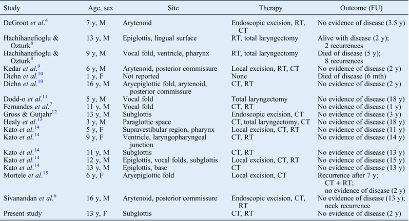
Therapy for laryngeal rhabdomyosarcoma can be variable. Although surgery continues to form a major part of head and neck rhabdomyosarcoma treatment, its potential morbidity and loss of voice do not justify its use for treating the paediatric larynx, especially since more viable modalities are now available.Reference Fernandes, Tandon, Tickoo and Rath7 Radiotherapy has been used as both a primary modality and an adjuvant to surgery for managing laryngeal rhabdomyosarcomas. Over the years, chemotherapy has gained a significant adjunctive role. According to Intergroup Rhabdomyosarcoma Studies IV, rhabdomyosarcoma treatment has changed from surgery to an emphasis on organ preservation with the use of multimodality treatment protocols. The literature on embryonal rhabdomyosarcoma of the larynx over the past few decades reflects this shift towards organ preservation. Embryonal rhabdomyosarcoma of the larynx appears to be highly responsive to chemoradiation, with a good prognosis. Seventeen case reports of laryngeal embryonal rhabdomyosarcoma were identified, and a summary of the pertinent aspects of treatment and outcome are summarised in Table I. Here, we present the first well-documented case of subglottic embryonal rhabdomyosarcoma in an adolescent female to be treated by chemoradiation alone.
Case reports of embryonal rhabdomyosarcoma of the larynx
FU = follow up; y = years; M = male; RT = radiotherapy; CT = chemotherapy; F = female; mth = months
Following consideration of the Intergroup Rhabdomyosarcoma Study results, the generally favourable response of the embryonal cell type to chemotherapy and the young age of the patient in the present case, laryngectomy was not performed; the patient was instead treated with chemoradiation. There was no local or regional recurrence after 24 months of follow up. While we cannot yet claim a complete cure, our study demonstrates the effectiveness of induction chemotherapy, radiotherapy and maintenance chemotherapy in controlling the tumour while preserving laryngeal function.