Introduction
Lung adenocarcinoma with enteric differentiation was described for the first time in 1991 by Tsao and Fraser who reported a ‘primary well-differentiated pulmonary adenocarcinoma with enteric differentiation’ after the histological examination of a lung nodule excised from the right upper lobe in a 40-year-old man. Microscope examination revealed typical features of differentiated small intestine epithelium including goblet, Paneth and enterochromaffin cells with ultrastructural features of ‘numerous long and straight surface microvilli’ (Ref. Reference Tsao and Fraser1). After appropriate diagnostic procedures to exclude the intestinal origin of the neoplasm, the authors formulated the diagnosis of ‘primary pulmonary adenocarcinoma with enteric differentiation’ (PAED) (Ref. Reference Tsao and Fraser1).
Since then, several case reports have been published, highlighting the role of immunohistochemistry (IHC) to formulate a differential diagnosis between PAED and colorectal cancer metastases (Refs Reference Weidner2, Reference Inamura3, Reference Yousem4, Reference Satoh5, Reference Maeda6, Reference Li7, Reference Canney8, Reference Wei9, Reference Tominaga10).
Based on these reports, PAED and ‘pulmonary enteric adenocarcinoma’ (PEAC) definitions, which are interchangeably used (Refs Reference Bian11, Reference Nottegar12), officially entered within the non-small-cell lung cancer (NSCLC) classification in 2011 according to the American Thoracic Society (ATS), the International Association for the Study of Lung Cancer (IASLC) and the European Respiratory Society (ERS) (Ref. Reference Travis13). An international panel of thoracic surgeons, oncologists, pneumologists, pathologists and radiologists representing the three scientific societies established that this pathological entity could be defined by an entero-like morphology in more than 50% of tumour cells, together with the positive IHC staining for at least one of the major markers of intestinal differentiation, namely cytokeratin 20 (CK20), Caudal Type Homeobox (CDX2) and/or Mucin 2 (MUC2) (Refs Reference Travis14, Reference Truini15). The positive staining for Cytokeratin 7 (CK7) and Thyroid Transcription Factor-1 (TTF-1) in approximately half cases also favours the differential diagnosis (Refs Reference Travis13, Reference Travis14, Reference Truini15, Reference Wang16, Reference El Hammoumi, El Ochi and and Kabiri17).
In 2015, this NSCLC variant was included in the World Health Organization (WHO) classification of lung malignancies as a less frequent subtype of adenocarcinoma, separated from the more common lepidic, acinar, papillary, micropapillary or solid variants (Ref. Reference Travis18).
To date, less than 300 cases have been globally described in the literature as case reports or small case studies and a distinctive IHC and molecular patterns of this rare tumour have not been identified yet (Refs Reference Zhao19, Reference Chen20). Therefore, at present, the diagnosis of PAED or PEAC can be confirmed only after clinical and instrumental exclusion of primary colorectal malignancies by colonoscopy, computed tomography (CT) scan or 18F-labeled fluoro-2-deoxyglucose (18F-FDG) positron emission tomography (PET)-CT (Refs Reference Travis18, Reference Chen20, Reference Stojsic21).
The difficulties related to the rarity of the neoplasm and the problematic differential diagnosis with the metastatic colorectal adenocarcinoma have also been addressed in our clinical experience, when a patient with an unusual cutaneous metastasis from a pulmonary enteric adenocarcinoma has come to our observation (Ref. Reference Todisco22). When carrying out a research of the scientific literature pertinent to the topic, we realised that most studies included reports of isolated or few cases, some others described sporadic cases as part of a larger series of lung cancers lacking detailed clinical information. The only studies with the highest numbers of patients (over 10) have been published in the last 3 years (Refs Reference Bian11, Reference Nottegar12, Reference Zhao19, Reference Chen20, Reference Lin23, Reference Feng24, Reference Zhang25, Reference Jurmeister26). Based on this experience, in this paper, we summarised current researches on PAED, focusing on its IHC and molecular features as potential tools for differential diagnosis, prognosis definition and therapeutic strategy choice.
Methods
An extensive literature review of papers published until 25 January 2020, with no language limitations, was performed by using the PubMed, Scopus, ISI-Web of Science and Google Scholar databases (Ref. Reference Falagas27).
The keywords used for this research, alone or in combination, were: ‘pulmonary enteric adenocarcinoma’, ‘PEAC’, ‘PAED’, ‘Primary pulmonary enteric adenocarcinoma’, ‘lung cancer’, ‘lung neoplasm’, ‘enteric differentiation’, ‘intestinal-type’, ‘primary pulmonary adenocarcinoma with enteric differentiation’, ‘lung intestinal adenocarcinoma’ and ‘intestinal-type adenocarcinoma’. Bibliographic references of the recovered articles were also reviewed and included in the analysis, if deemed relevant.
Data were analysed by using Student's t-test or one-way ANOVA with Bonferroni post-test, as appropriate. The Spearman test was used for correlation analysis. All tests were two-sided, and a P-value <0.05 was considered statistically significant. Statistical analysis was performed by using GraphPad Prism 5 software (GraphPad Software, La Jolla, CA, USA).
Pathogenesis and clinical features
Aetiology of PAED has not been totally clarified. Satoh et al. described the presence of common stem cells in the respiratory and gastrointestinal tract mucosae, which might explain the histological similarity between PAED and colorectal malignancies (Ref. Reference Satoh5). However, because of the rarity of this clinical-pathological entity, clinical information is sporadic and often fragmentary (Refs Reference Bian11, Reference Wang16, Reference Zhao19, Reference Chen20, Reference Feng24).
There seem to be no significant differences between PAED and conventional lung adenocarcinoma in terms of correlation with smoking status (Refs Reference Bian11, Reference Zhao19). Moreover, PAED incidence in both sexes varies according to different studies (Refs Reference Chen20, Reference Feng24), although it is apparently higher in males. To this regard, Zhao et al. compared a cohort of 28 PAED patients with 92 subjects diagnosed with invasive lung adenocarcinoma (IAC), and found a significantly higher proportion of males (76 versus 50%, P = 0.008), with a higher mean age (64.8 ± 8.6 versus 60.9 ± 8.9, P < 0.05), greater tumour size (P = 0.001) and tumour stage (P < 0.05) in the former, as compared with the latter, at the time of diagnosis. No significant differences were found in terms of cancer localisation (i.e. central or peripheral) within the lung, nor with the presence of lymph node metastases (Ref. Reference Zhao19). The most common clinical presentation is with dry cough, fever, chest/back pain and haemoptysis (Refs Reference Wang16, Reference Zhao19).
Radiological examination of PAED usually shows solid lung masses (>3 cm) without ground-glass opacity. Other radiological findings (e.g. spiculation, pleural effusion or indentation) can be similarly detected in PAED and primary pulmonary IAC samples (Ref. Reference Zhao19). Moreover, Bian et al. found no significant differences when comparing CT scans from 13 PAED cases with 27 patients with metastatic colorectal cancer (MCC) (Ref. Reference Bian11).
Circulating tumour markers associated with PAED differ from those released by conventional lung adenocarcinoma. Indeed, PAED is more frequently associated with an increase in serum carcinoembryonic antigen (CEA) and carbohydrate antigen (CA)19-9, whereas rising of cytokeratin 19 fragment (CYFRA) 21.1 or neuron-specific enolase levels is unusual (Refs Reference Zhao19, Reference Chen20).
Scarcity of data from the literature makes it difficult to define PAED prognosis, as compared with NSCLC. Recently, Feng et al. analysed 30 patients with PAED and described that those whose tumour size was ≤5 cm had longer overall survival, whereas patients with CK20-positive and CDX2-negative tumours had a better prognosis (P < 0.05). There was no evident correlation between the mutational status of EGFR and clinical outcome. Furthermore, a comparison of median overall survival between PAED patients and those with conventional pulmonary adenocarcinoma did not reveal any statistically significant difference (Ref. Reference Feng24).
As previously described, primary PAED diagnosis can be endorsed by excluding gastrointestinal malignancies through colonoscopy, CT or PET-CT scans (Refs Reference Travis13, Reference Travis14, Reference Truini15, Reference Travis18, Reference Kuhn28). IHC and molecular diagnostics are emerging as promising and more specific tools to formulate the differential diagnosis, whereas the occurrence of both PAED and synchronous or metachronous colorectal cancer is uncommon, although still possible (Refs Reference Canney8, Reference El Hammoumi, El Ochi and and Kabiri17, Reference Handa29, Reference Garajova30).
Nowadays, a standard treatment regimen for PAED has not been established yet. Owing to the morphological and IHC similarity with MCC, many authors attempted the administration of chemotherapy regimens approved for colorectal cancer treatment, mainly based on oxaliplatin, 5-fluorouracil and/or irinotecan combinations, such as mFOLFOX6, XELOX or FOLFIRI, without the significant benefit (Refs Reference de Castria and de Azevedo Souza31, Reference Lin32). However, a partial therapeutic response was reported in a patient treated with the first-line chemotherapy with FOLFOX regimen for a perihilar pulmonary mass and a brain metastasis in the left frontal lobe, developing 11 years after primary PAED diagnosis (Ref. Reference de Castria and de Azevedo Souza31).
On the other hand, chemotherapy regimens licensed for the advanced NSCLC, including taxanes, gemcitabine or carboplatin, seem to be more effective in terms of response rates (Refs Reference Handa29, Reference Lin32).
Advances in the molecular characterisation of PAED will certainly improve the knowledge of its pathogenesis, leading to the identification of novel potential targets for personalised therapy (Refs Reference Chen20, Reference Lin23). For instance, Chen et al. hypothesised an intrinsic resistance of these tumours to tyrosine kinase inhibitors, because of the high percentage of KRAS mutations, while suggesting a potential higher responsiveness to immunotherapy, in relation to the presence of defective mismatch repair (MMR) and high tumour mutational burden (TMB) (Ref. Reference Chen20). More recently, Jurmeister et al. confirmed for the first time the potential eligibility for treatment with immune checkpoint inhibitors in PAED patients, according to the validated presence of a high TMB but also of the membranous PD-L1 staining positivity in the tumour cells of PAED (Ref. Reference Jurmeister33). Furthermore, the same authors, based on the identification of the amplification of the RICTOR gene in PAED samples, suggested a potential therapeutic approach with mTORC1/2 inhibitors in these patients (Ref. Reference Jurmeister33).
Clinical trials need to be thus addressed to answer these questions and establish appropriate and more effective therapeutic approaches.
Histopathological features
Macroscopically, PAED usually presents as a peripheral, well-delimited nodular lesion with or without central scar (Ref. Reference Satoh5).
Histological examination shows, in contrast to the monomorphic features of colorectal cancer metastases, a peculiar intra-tumour heterogeneity with cells that may express both colonic and lung cancer distinctive markers. However, in order to formulate a PAED diagnosis, the identification of ‘entero-like’ cells in more than half tumour sample is mandatory (Refs Reference Travis13, Reference Travis14, Reference Truini15).
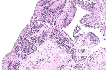
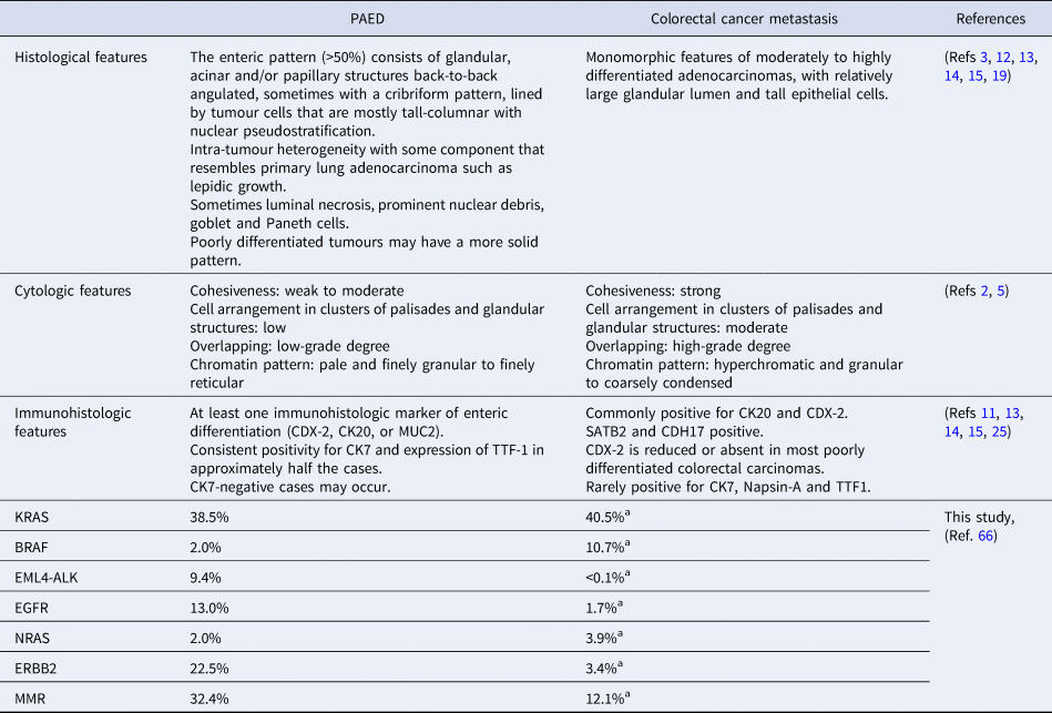
Microscopy examination shows tumour cells resembling the colorectal cancer with back to back angulated acinar structure (Fig. 1). Also, tumour cells appear cuboidal or tall columnar, exhibiting nuclear pseudo-stratification without significant globet cell differentiation. Necrotic debris and central necrosis may be occasionally found, whereas poorly differentiated tumours may have a more solid pattern (Refs Reference Inamura3, Reference Chen20) (Table 1).
Representative image of a moderately differentiated enteric adenocarcinoma infiltrating the bronchial mucosa (EE 20x).

Main differences between PAED and colorectal metastasis

a Data extrapolated from cBioPortal for Cancer Genomic (https://www.cbioportal.org/) analysing 533 colorectal cancer metastases samples from the ‘Metastatic Colorectal Cancer (MSKCC, Cancer Cell 2018)’ dataset.
PAED cells might be distinguished from MCC and conventional lung adenocarcinoma even at the cytological observation. Indeed, Satoh et al., by evaluating cytological samples deriving from seven cases of PAED, 10 pulmonary adenocarcinomas and 10 patients with MCC, found that the presence of specific tumour cell features such as cluster formation, tendency to overlap and adhesion, together with typical nuclear characteristics (i.e. irregular shape, thick nuclear membrane, presence of nucleoli and intranuclear cytoplasmic inclusion) could facilitate the differential diagnosis (Ref. Reference Satoh5). Indeed, the authors found that PAED was characterised by a low degree of cell stratification, weak adhesiveness and low frequency of palisade or gland structures. On the other hand, pulmonary adenocarcinoma samples mainly include cuboidal or round cells, whereas MCC exhibited cells with hyperchromatic and condensed nuclear chromatin, organised into palisade or gland structures (Ref. Reference Satoh5) (Table 1).
Although PAED characterisation by IHC has been largely investigated, no univocal features have been described to date. At present, positive staining for at least one of the typical colon markers (CDX2, CK20 and MUC2) is necessary to confirm PAED diagnosis. Markers of lung differentiation, such as Cytokeratin 7, Transcriptional Thyroid Factor 1 and Napsin-A, are expressed in approximately 50% of cases, but their negativity does not exclude PAED diagnosis (Refs Reference Travis13, Reference Chen20, Reference de Castria and de Azevedo Souza31, Reference Hatanaka34) (Table 1).
Chen et al. reviewed 129 cases of PAED describing that the rate of positive staining for CDX2 (79.1%) was higher than CK20 one (48.1%), whereas the pneumocyte markers CK7 and TTF1 were found expressed in 89.9 and 40.3% of cases, respectively. Moreover, the authors demonstrated, by comparing 129 PAED samples with 50 colorectal cancers, that the concomitant positive IHC staining for CK7 and CDX2 was both highly sensitive (71.3%) and specific (82%) to differentiate PAED from colorectal malignancies (Ref. Reference Chen20).
Interestingly, Feng et al. identified CDX2 and CEA as the frequently expressed markers (86.6 and 93.3% respectively), whereas CK20 was found only in 30.0% of cases. In addition, a statistically significant correlation between CK20-positive/CDX2-negative staining and longer overall survival was found (Ref. Reference Feng24).
On the other hand, Zhao et al. analysed 28 cases of PEAC and showed Villin as the most frequently expressed markers (89.2%), followed by CK7 (66.6%), CDX2 (57.1%), CK20 (36.0%), TTF1 (35.7%) and Napsin A (23.0%) (Ref. Reference Zhao19). Moreover, Lin et al. confirmed the frequent expression of Villin (80.0%), but found discrepant data for the other markers (CDX2: 90.0% of expression rate, CK20: 70.0%; CK7: 30.0%; TTF1: 30.0%; Napsin A: 25%) over a series of 11 cases (Ref. Reference Lin23).
Further IHC markers have been recently investigated, including Cadherin-17 (CDH17) and SATB homeobox 2 (SATB2), that were described as having a high specificity and sensitivity to distinguish PAED from MCC (Ref. Reference Bian11).
Meanwhile, another study including eight samples of PAED detected a low positivity for SATB2 (12.5%) in the absence of β-Catenin expression, whose levels were found contrariwise high in MCC (100 and 55%, respectively) (Ref. Reference Matsushima35).
The biggest series of PAED has been described by Nottegar et al., who found the co-expression of CK7 and CDX2 in all the analysed samples (N = 46), even if on small biopsies. Once a score was attributed to CDX2, an inverse relationship was observed between this marker and the presence of pneumocyte markers TTF1, Napsin A and surfactant protein-A (SP-A), which were detected in about 50% of cases (Ref. Reference Nottegar12).
Hence, despite the increasing incidence of PAED, there are still concerns associated with its differential diagnosis from MCC. In this regard, five senior pathologists have been asked in a recent study to formulate a blinded diagnosis of 15 PAED and four MCC samples. In all cases, at least one of the pathologists formulated a wrong diagnosis, with error rates ranging from 20 to 60%. It is of note that one single case of lung metastasis from colorectal cancer was erroneously classified by all pathologists as a primary pulmonary tumour (Ref. Reference Jurmeister26).
Genomic assessment
Although a genomic assessment of PAED has not been extensively performed, current evidence suggests that a high degree of molecular heterogeneity characterises these tumours, as compared with conventional lung adenocarcinoma.
In most case reports, the authors focused on those genes, such as EGFR, KRAS and EML4-ALK, that are commonly investigated in lung adenocarcinoma patients (Refs Reference Stojsic21, Reference Qureshi and Furrukh36, Reference Laszlo37). For instance, in 2014, Wang et al. evaluated the mutational status of EGFR and KRAS genes, as well as EML4-ALK translocations, in nine PAED that were compared with 20 MCC and 20 typical primary adenocarcinomas. Although PAED samples did not harbour EGFR mutations, the latter were detected in 30% of typical adenocarcinomas, whereas KRAS mutations were found only in two MCC samples and one case of typical adenocarcinoma, confirming the different pathogenesis of these malignancies (Ref. Reference Wang16).
Conflicting data came also from subsequent studies. Nottegar et al. identified KRAS mutations in four out of eight PAED samples, with a concomitant PIK3CA gene mutation and EML4-ALK translocation in one case; no mutations were detected in EGFR, BRAF and NRAS (Ref. Reference Nottegar38).
Zhao et al. described a different incidence of EGFR and KRAS mutations (10 and 12.5%, respectively) over a case series including 28 PAED (Ref. Reference Zhao19). Canney et al. reported the case of a patient carrying a germline mutation of the hMSH2 who developed a right colon adenocarcinoma and two metachronous lung lesions. One of the lung nodules was histologically analysed and turned out as an intestinal-type lung adenocarcinoma with the loss of MSH2 and MSH6 proteins, leading the authors to hypothesise that it could represent a rare tumour variant in the HNPCC spectrum (Ref. Reference Canney8). Several subsequent studies confirmed the presence of somatic MMR genes mutations in PAED lesions (MSH2, PMS2, MSh6 and MLh1) (Refs Reference Chen20, Reference Lin23, Reference Zhang25).
A recent work by Chen et al. (Ref. Reference Chen20) analysed 129 cases of PAED, including 111 from previous literature; the authors described KRAS as the most commonly mutated gene (48%), whereas no mutations in EGFR and BRAF were detected, in agreement with previous data from Nottegar et al. (Refs Reference Nottegar12, Reference Nottegar38). Four out of five patients analysed (80%) harboured MMR alterations, whereas two out of five patients exhibited amplification and/or mutations of ERBB2. The authors also detected a higher TMB in PAED samples, as compared with conventional lung adenocarcinoma (Ref. Reference Chen20), suggesting a potentially higher sensitivity to immunotherapy of the former (Ref. Reference Planchard39).
Thanks to the advent of advanced nucleic acid sequencing technologies, new opportunities emerged to delineate the molecular profiles of most malignancies, which provide useful for differential diagnoses as well as to define prognosis and susceptibility to therapy (Ref. Reference Savonarola40).
In 2017, Lin et al. performed, by using a panel of 170 oncogenes and anti-oncogenes, a targeted next-generation sequencing analysis on seven PAED samples, and identified frequent mutations in ALK/ROS1 (71%), MMR genes MSH2/MSH6 (42%) and TP53 (57%) (Ref. Reference Lin23).
A similar approach was employed by Zhang et al. by applying a larger panel of 259 genes on 13 cases of PAED. Interestingly, the authors described in about 75% of cases the presence of typical NSCLC driver mutations in EGFR, ALK and ERBB2 genes. The same gene panel was applied to 15 pulmonary metastases from colorectal carcinoma and five primary colorectal tumours, showing in 91% of cases typical driver mutations of this neoplasm (in APC, KRAS, NRAS). These findings allowed to draw up a possible algorithm suitable for differential diagnosis (Ref. Reference Zhang25).
More recently, Jurmeister et al. have employed a commercial panel, including 22 genes associated with colorectal and lung cancer, together with a methylation and a copy number analysis, in 15 PAEDs. They showed that KRAS and TP53 were the most frequently mutated genes, at a frequency of 60 and 33%, respectively, whereas the gain of 1p (53%) and 20q (53%) and the loss of 3p (66%) and 1q (53%) were the main chromosomal alterations. Furthermore, the authors demonstrated that the study of methylation patterns, mainly involving CACNB2, HOXA9, HOXD1, HOXD8, RNLS, FAD and KRT7 genes, may differentiate molecular alterations of PAED from colorectal adenocarcinomas ones (Ref. Reference Jurmeister26). In a further interesting recent study, the same research group Jurmeister et al., using a gene panel covering 404 cancer-related genes on seven PAED samples, observed TP53 and KRAS mutations in six and three specimens respectively, whereas all the samples showed a high TMB (16.8 mutations per megabase). Furthermore, the immunohistochemical analysis indicated that all six samples showed high proliferation rates in Ki-67 and three samples showed membranous PD-L1 staining in tumour cells. These data, although preliminary, indicated for the first time the possibility of a treatment with immune checkpoint inhibitors for patients with PAED. Moreover, the finding in two samples of a RICTOR gene amplification induces the authors to hypothesise a potential therapeutic use of mTORC1/2 inhibitors in these patients (Ref. Reference Jurmeister33).
Finally, using a 47-microRNA (miRNA) cancer-specific array on a PAED patient, Garajová et al. found a signature which turned out similar to that expressed by NSCLC, but different from that of colorectal cancer. Interestingly, some miRNAs associated with tumour aggressiveness (miR-31*, miR-126*, miR-506, miR-508-3p and miR-514) showed a partial overlapping with the profile expressed by the pancreatic ductal adenocarcinoma (Ref. Reference Garajova30).
Analysis of literature data
The review of the literature allowed to identify 295 patients (116 males, 90 females and 89 subjects without gender information) from 58 published articles. For each patient or group of patients, when possible, demographic data, ethnicity, IHC and mutational analysis results were extrapolated, transcribed into excel sheet and categorised. The patient mean age was 63.24 years (range: 25–81).
Most studies were single-case reports or small patient series (Refs Reference Tsao and Fraser1, Reference Weidner2, Reference Inamura3, Reference Yousem4, Reference Satoh5, Reference Maeda6, Reference Li7, Reference Canney8, Reference Wei9, Reference Tominaga10, Reference Wang16, Reference El Hammoumi, El Ochi and and Kabiri17, Reference Stojsic21, Reference Todisco22, Reference Handa29, Reference Garajova30, Reference de Castria and de Azevedo Souza31, Reference Lin32, Reference Hatanaka34, Reference Matsushima35, Reference Qureshi and Furrukh36, Reference Laszlo37, Reference Nottegar38, Reference Fujita41, Reference Lin42, Reference Ishida43, Reference Metro44, Reference Shina45, Reference Fujiwara, Iwashiro and Kimura46, Reference Gomez-Hernandez47, Reference Matsubara48, Reference Prakobkit49, Reference Shomura50, Reference Sun51, Reference Hayama52, Reference Miura and Sakamoto53, Reference Miyaoka54, Reference Suzuki55, Reference Kimura56, Reference Toyazaki, Yamamoto and Shinji57, Reference Mizoguchi58, Reference Nojima59, Reference Saito60, Reference Ogihara61) whereas sporadic cases were also described within larger studies on lung cancer, from which it was not always possible to retrieve demographic and clinical information (Refs Reference Lee, Kim and Shim62, Reference Suzuki63, Reference Calio64, Reference Yang65).
The studies involving more than 10 patients are relatively recent and date back to the last 3 years, suggesting the increasing interest towards this clinical entity (Refs Reference Bian11, Reference Nottegar12, Reference Zhao19, Reference Chen20, Reference Lin23, Reference Feng24, Reference Zhang25, Reference Jurmeister26), whereas the largest study included 46 subjects (Ref. Reference Nottegar12).
According to the published studies, most patients with PAED diagnoses were Asian (185/295 subjects, among whom 132 came from China, 43 from Japan and 10 from Korea) followed by European + North America patients (103/295, including 63 from Italy), whereas only seven cases were described in Africa and Brazil (Supplementary Table 1).
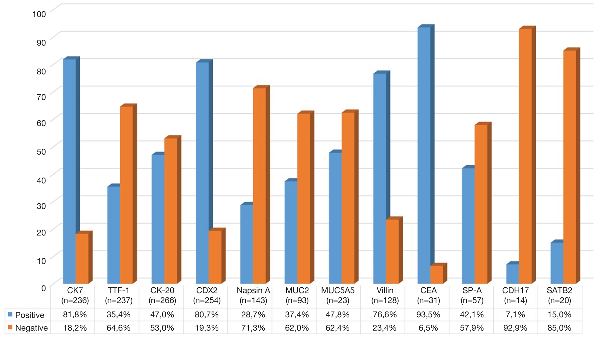
We found that the most represented IHC markers were CEA (93.5%), CK7 (81.8%), CDX2 (80.7%) and Villin (76.6%), whereas a minor expression was observed for MUC5A5 (47.8%), CK20 (47.0%), SP-A (42.1%), MUC2 (37.4%), TTF1 (35.4%) and Napsin A (28.7%) (Fig. 2).
Graphical representation of the percentages of the positive (light blue bars) and negative (orange bars) immunostaining in PAED patients for the various markers used in all the studies considered analysed in this review. In the lower portion of the graphic, the absolute number of analysed samples and the percentages of positive and negative samples are reported.

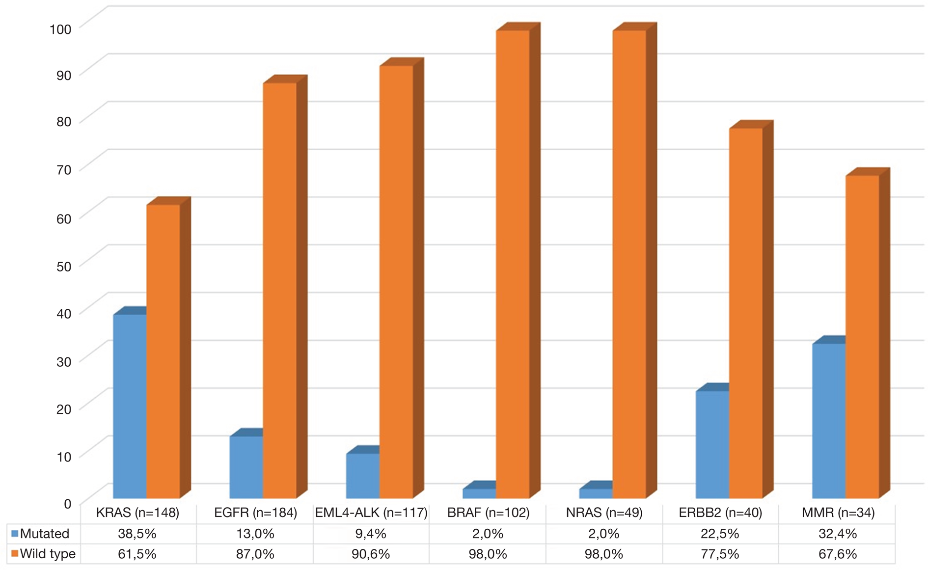
We also recorded the majority of mutations in KRAS (38.5%, N = 148), MMR (32.4%, N = 34), ERBB2 (22.5%, N = 40) and EGFR (13.3%, N = 184) genes, whereas the translocation of EML4-ALK was detected in 9.4% of cases (N = 117). Less common were BRAF (2.0%, N = 102) and NRAS (2.0%, N = 49) mutations (Fig. 3).
Graphical representation of the percentages of the mutated (light blue bars) and wild-type (orange bars) results of the mutational analysis in PAED patients for the various analysed genes in all the studies considered in this review. In the lower portion of the graphic, the absolute number of analysed samples and the percentages of mutated and wild-type samples are reported.

Using cBioportal for Cancer Genomic (https://www.cbioportal.org/), we have subsequently compared these latest results with data extrapolated from 533 colorectal cancer metastases samples from the dataset ‘Metastatic Colorectal Cancer (MSKCC, Cancer Cell 2018)’ (Refs Reference Yaeger66, Reference Gao67).
The analysis showed a higher frequency of alterations in PAEDs compared with colorectal cancer metastases for the EGFR (13.0 versus 1.7%), ERBB2 (22.5 versus 3.4%), EML4-ALK (9.4 versus <0.1%) genes and MMR (32.4 versus 12.1%) genes. The mutation frequencies were relatively similar for KRAS (38.5 versus 40.5%) and NRAS (2.0 versus 3.9%) and lower for BRAF (2.0 versus 10.7%) genes (Table 1).
Recent evidence supports the association between patient ethnicity and NSCLC features (Refs Reference Schabath, Cress and Munoz-Antonia68, Reference Araujo and Carbone69, Reference Heath70), suggesting that both racial and environmental factors may drive cancer molecular alterations, and justify its heterogeneous clinical evolution.
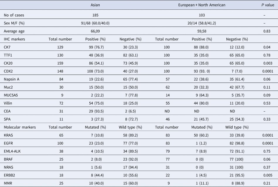
In this context, we analysed the potential differences between PAEDs diagnosed in Asia and those reported in European/North American countries. As shown in Table 2, most PAED cases (185/295) were described in Asia, whereas gender distribution was identical in the two groups. The mean age at the time of diagnosis was 66.09 years in Asian patients and 59.58 years in European/North American ones.
Distributions of immunostaining and mutational analysis frequencies in PAED patients from the studies considered in this review

When focusing on IHC features, we observed a high similarity between the two groups, in terms of TTF1, MUC2, MUC5A5, Villin, Napsin A and SP-A expression. On the other hand, we found a significantly higher rate of CK20-positive tumours in the Asian population (54.1 versus 35.0% of European/North American ones, P = 0.003), whereas CK7 and CDX2 were significantly more expressed in PAED samples from Europe and North America, as compared with those reported in Asia (P = 0.04 for CK7, P < 0.0001for CDX2).
We next sought to determine the molecular differences between the two groups, focusing on the most common gene mutations identified in NSCLC. In particular, we observed a significantly higher rate of EGFR mutations in Asian patients as compared with those from Europe and North America (23.0 versus 1.2%, P < 0.0001), whereas an opposite trend was found for KRAS molecular alterations (10.8 versus 60.2% respectively, P < 0.0001). In this regard, recent reports on NSCLC described a similar geographical distribution of EGFR and KRAS mutations, which were also mutually exclusive (Refs Reference Soh71, Reference Graham72).
Moreover, no significant differences between the groups were detected with respect to EML4-ALK rearrangements, NRAS and BRAF mutations and MMR deficiency. On the other hand, PAED from Asian countries harboured more ERBB2 mutations, as compared with those diagnosed in Europe and North America (ERBB2 mutations: 44.4 versus 4.5%, P = 0.005). However, because of the rarity of these molecular alterations in NSCLC (Refs Reference Schabath, Cress and Munoz-Antonia68, Reference Dearden73, Reference Garrido-Castro and Felip74), no definitive conclusions can be drawn on their inter-racial variability.
Discussion
Lung adenocarcinoma with enteric differentiation has been long considered a rare clinical-pathological entity, with most reports being anecdotal for decades. However, an increasing interest has been addressed by both pathologists and clinicians towards this NSCLC variant, as proved by the numerous cases described in the last 3 years (Refs Reference Bian11, Reference Nottegar12, Reference Zhao19, Reference Chen20, Reference Lin23, Reference Feng24, Reference Zhang25, Reference Jurmeister26). At present, there is the lack of consensus about the diagnostic criteria to be employed, making the differential diagnosis between PAED and MCC often complicated and mainly based on the exclusion of synchronous or metachronous gastrointestinal malignancies (Refs Reference Canney8, Reference El Hammoumi, El Ochi and and Kabiri17, Reference Travis18, Reference Chen20, Reference Stojsic21, Reference Handa29, Reference Garajova30).
Indeed, both clinical presentation and radiological features of PAED are almost identical to lung adenocarcinoma (Refs Reference Wang16, Reference Zhao19), although the main nodule diameter is often higher than 3 cm, differently from other NSCLC histotypes (Ref. Reference Zhao19). Serum CYFRA 21.1 within the normal ranges may help in distinguishing this variant from typical adenocarcinomas, whereas the frequent rising of CEA and CA19.9 may complicate its differentiation from MCC (Refs Reference Zhao19, Reference Chen20).
IHC features of PAED have been described by several authors, but data are often conflicting and inconclusive (Refs Reference Travis13, Reference Zhao19, Reference Chen20, Reference Lin23, Reference Feng24, Reference de Castria and de Azevedo Souza31, Reference Hatanaka34). However, scientific societies agree in considering, as major pathological characteristics of PAED, the IHC positivity for at least one marker of colonic differentiation, among CDX2, CK20 and MUC2, as well as the presence of an entero-like tumour cell morphology in more than half sample (Travis 2013, Truini 2015). Interestingly, Matsushima et al. showed the lack of β-Catenin in PAED samples, compared with the 100% positivity rate of MCCs (Ref. Reference Matsushima35); however, the exiguous number of analysed cases does not allow to consider this as an unequivocal criterion for differential diagnosis, for which further investigation is needed.
Molecular characterisation of PAED has been attempted by several authors, who investigated the mutational state of several genes involved in lung and colorectal cancer pathogenesis (Refs Reference Wang16, Reference Stojsic21, Reference Qureshi and Furrukh36, Reference Laszlo37). Although some discrepancies exist among different reports (Refs Reference Zhao19, Reference Nottegar38), we observed a strong correlation between EGFR and KRAS mutational status and ethnicity, as previously reported for other NSCLC variants (Refs Reference Soh71, Reference Graham72). This suggests that, besides the morphological analogy between intestinal malignancies and PAEDs, the latter exhibit a higher degree of molecular similarity to NSCLC, which might influence their clinical management. In agreement with this observation, treatment of PAED with colorectal cancer chemotherapy regimens was not effective (Refs Reference de Castria and de Azevedo Souza31, Reference Lin32), whereas the administration of taxanes, platinum derivatives and gemcitabine resulted in higher response rates (Refs Reference Handa29, Reference Lin32).
However, in the ‘personalised medicine’ era, further clinical investigation is desirable to investigate the potential therapeutic role of targeted therapies and immunotherapy in PAED. In this regard, the identification of ERBB2 amplification in 44.4% of the Asian patients included in our analysis may pave the way to potential targeted anti-ERBB2 treatments. On the other hand, both the frequent alteration of MMR and the high TMB reported in the literature, as well as the expression of immune checkpoint regulators, may suggest a potential sensitiveness of PAED to immunotherapy (Ref. Reference Jurmeister33), whose introduction in the clinical practice may significantly improve the outcome of such malignancies.
Supplementary material
The supplementary material for this article can be found at https://doi.org/10.1017/erm.2020.2.
Acknowledgements
This work was supported by the AIRC Associazione Italiana per la Ricerca sul Cancro [grant number 17536] and from the Apulia Region (Oncogenomic Project and Jonico-Salentino Project).
Conflict of interest
All the authors confirm that there are no conflicts of interest.