Introduction
Isolated sphenoid fungal sinusitis is a relatively rare condition, as is isolated sphenoid sinusitis in general. As a result, management of isolated sphenoid fungal sinusitis is generally non-standardised, but is frequently similar to treatment for isolated sphenoid bacterial sinusitis. Patients may have no symptoms during the early stages of the disease, and may later develop symptoms such as localised pain and bloody nasal discharge when localised inflammation becomes more serious.
Many important structures lie adjacent to the sphenoid sinus (e.g. the cavernous sinus, pituitary, internal carotid artery, and cranial nerves II, III, IV, V1, V2 and VI).Reference Lawson and Reino1 However, it is often difficult to make an early diagnosis of isolated sphenoid fungal sinusitis, based on the typical clinical picture of slight nasal symptoms plus nonspecific central neurological symptoms such as headache, visual disturbances and cranial nerve palsies. An isolated, opacified sphenoid sinus on computed tomography (CT) or magnetic resonance image scanning will often be the only pathological finding.
Sphenoid sinus mycetoma or ‘fungal ball’ is rare and has attracted little research, especially regarding associated vision loss. There have been only three reported cases of sphenoid fungal ball in conjunction with vision loss.Reference Lawson and Reino1–Reference Müller, Wolf and Velten3 We recently treated two patients with sphenoid sinus fungal ball and visual loss; here, we summarise our findings.
Case reports
Case one
An 81-year-old woman was referred with a five-month history of right eye pain, recalcitrant headache and vision loss. She had suffered cholecystitis for 20 years, and also had a history of previous ovariotomy, hysterectomy, radical resection of sigmoid flexure carcinoma, transverse colostomy and six months' chemotherapy. She had initially been treated with antibiotics and had recovered partially. However, the aforementioned symptoms had persisted and new symptoms, including hyposmia and bloody rhinorrhoea, had developed four months later.
On presentation, the patient was afebrile and reported no diplopia or emesis. On examination, a partial right IIIrd cranial nerve palsy with sectional ptosis was observed. The nose appeared normal.
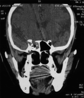
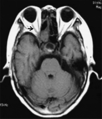
The patient's blood serum eosinophil count was normal. A CT scan showed a right sphenoid sinus mass of variable density, together with erosion of the right petrous apex and cavernous sinus (Figure 1). Magnetic resonance imaging showed inflammation of the right optic nerve (Figure 2).
Coronal computed tomography scan of case one, showing a right sphenoid sinus mass of variable density together with erosion of the right petrous apex and cavernous sinus. The fungal ball (arrow) shows high density, a typical feature.

Axial magnetic resonance imaging scan of case one, showing an inflamed right optic nerve (arrow).

Based on these radiological findings, we suspected invasive fungal sinusitis. The patient was treated with 0.2 g once daily fluconazole four times daily, prior to surgery. An endoscopic sphenoidotomy and extirpation of sphenoid fungal masses were performed. Because the mucosa was grossly inflamed and oedematous, we continued using fluconazole post-operatively (0.4 g once daily for seven days and then 0.2 g once daily for two days) as antifungal treatment.
Histopathological examination of an intra-operative biopsy specimen revealed chronic inflammation of the sphenoid sinus mucosa, with a few fungal mycelia. Although fungal elements were seen during microscopy, neither fungi nor bacteria grew on culture.
Three days after surgery, the patient's headache eased, and six days later her eyesight improved. Six weeks after surgery, all the patient's symptoms had resolved and sphenoid sinus (It means that the natural drainage mechanism of the sinus) drainage was unblocked.
Case two
A 50-year-old woman was referred with a 10-day history of headache and a one-week history of left vision loss with a partial visual field defect.
On examination, the patient's vision was limited to light perception. The nose appeared normal.
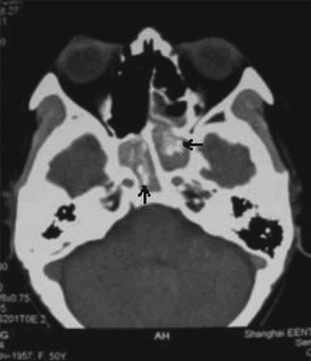
The patient's blood serum eosinophil count was normal. Computed tomography indicated a sphenoid sinus mass of variable density present in the left spheno-ethmoidal recess (Figure 3).
Axial computed tomography scan of case two, showing bilateral sphenoid sinus masses of variable density, typical of a fungus ball the arrow shows high density.

Bilateral sphenoidotomy was performed as an emergency procedure. The mucosa was grossly inflamed and oedematous; the patient also exhibited partial absence of the inner wall of the left optic canal. The patient was treated with 400 mg methylprednisone four times daily to reduce inflammation of the optic nerve.
As in case one, histopathological examination of this patient's biopsy specimen also revealed chronic inflammation of the sphenoid sinus mucosa, with fungal mycelia.
Three days post-operatively, the patient's visual field dramatically recovered. Eight days later, she recovered normal eyesight. Three weeks later, all symptoms had resolved and the sphenoid sinus drainage was unblocked.
Both patients were followed up for nearly four months without recurrence of disease.
Discussion
Isolated sphenoid sinus disease is comparatively rare; Hnatuk et al. Reference Hnatuk, Macdonald and Papsin4 estimated that the condition constitutes less than 1 per cent of all sinus disease. The incidence of isolated fungal sphenoid sinusitis is even lower. Friedman et al. Reference Friedman, Batra, Fakhri, Citardi and Lanza5 found that fungal sinusitis, most cases of which were characterised by a fungal ball, accounted for 31 per cent of inflammatory sphenoid disease incidence. However, Lawson and ReinoReference Lawson and Reino1 found the incidence to be only 7.5 per cent. Sphenoid fungal sinusitis is difficult to diagnose and treat in its early stages, due in part to the inaccessible position of the sphenoid sinus and the unremarkable inflammatory presentation with nonspecific symptoms. Common symptoms of sphenoid sinus fungal ball include bloody rhinorrhoea and post-nasal drainage, which in conjunction with vision loss are rare and have not been adequately reported. Both our cases developed headaches and vision loss as primary symptoms; case one presented with a IIIrd cranial nerve palsy and case two with a partial visual field defect.
Grillone and KasznicaReference Grillone and Kasznica6 stated that the optic nerve is affected by sphenoid sinus fungal balls more often than any other cranial nerve. Lee et al. Reference Lee, Huang and Lee2 reported that the cranial nerve most affected was the optic nerve (54 per cent) followed by the VIth (40 per cent). The preponderance of optic nerve involvement is probably attributable to the local anatomy of the region.Reference Dale and Mackenzie7 In 79 per cent of cadavers, the inner wall of the optic canal is less than 5 mm thick, and it is completely absent in 8 per cent.Reference Unlu, Aslan, Goktan and Egrilmez8 The mechanisms responsible for visual disturbance due to sphenoid sinus disease may involve cranial neuritis (resulting from adjacent or nearby infection), pressure ischaemia (attributable to compression by an expansile lesion) or ischaemic infarction (resulting from thrombophlebitis or vasculitis).Reference Lee, Huang and Lee2
In our first case, the right optic nerve was impaired by inflammation and compression because of bony absorption. In case two, the left optic nerve was affected by lack of a complete bony wall outside the nerve, compromising its protection. There appeared to be no histopathological evidence of invasive fungal sinusitis, eliminating the possibility of ischaemic infarction resulting from thrombophlebitis or vasculitis. In case one, the bony absorption seen in the CT scan, which was previously suspected due to age and an immunocompromised state, cannot be considered diagnostic for invasive fungal sinusitis. DeShazo et al. Reference deShazo, O'Brien, Chapin, Soto-Aquilar, Gardner and Swain9 suggested that bony absorption may be induced by ischaemic infarction resulting from thrombophlebitis, vasculitis or osteolysis attributable to compression. At the same time, bony destruction is a common feature of carcinoma, which should be kept in mind during the initial management and follow-up care of patients. In our case two, osteolysis was suspected to be the cause of compression because the bony wall was congenitally complete. A possible diagnosis of allergic fungal sinusitis was excluded by the normal blood serum eosinophil count, lack of allergic disease history, inconclusive CT results, surgical findings and post-operative pathology.
Visual disturbance is not an uncommon complication in cases of sphenoid sinus disease and must be managed properly to prevent permanent neuropathy.Reference Lee, Huang and Lee2 Primary treatment consists of surgically opening the sphenoid sinus (this also enables post-operative endoscopic visualisation of the sphenoid). Endoscopy is best suited for this task, and the use of angled lenses facilitates complete extirpation of fungal material from the sphenoid sinus.Reference Metson and Gliklich10 In the case of sphenoid sinus disease with ophthalmic complications, a more radical resection of the anteroinferior wall of the sphenoid sinus should be performed in order to enable extensive drainage and optimal decompression; this is very different from the minimally invasive and conventional functional endoscopic sinus surgery.Reference Lee, Huang and Lee2 Additional medical treatment of the fungus will depend upon the degree of invasiveness. Adequate treatment of a sphenoid sinus fungal ball can be achieved with surgical debridement of the infectious material to maintain adequate aeration of the sinus.Reference Blitzer and Lawson11 Both our cases were thus treated, with the addition of antifungal therapy in case one to prevent invasive fungal sinusitis, and a hormone to alleviate optic nerve inflammation in case two.
What is the efficacy of endoscopic surgery for patients with sphenoid sinus disease in conjunction with visual disturbances? Heretofore, little information has been available. A 75 per cent complete recovery from abducens palsy (lasting more than 96 hours) in four patients with isolated sphenoid sinus disease was previously reported.Reference Lee, Lee and Huang12 Lee et al. Reference Lee, Huang and Lee2 found that visual improvement occurred in patients in whom visual disturbance had persisted pre-operatively for less than six months. A shorter duration between symptom onset and surgery did not necessarily guarantee an earlier recovery; the recovery of vision also depended on the severity of visual disturbance and the severity of sphenoid sinus disease.Reference Lee, Huang and Lee2 Lee et al. Reference Lee, Huang and Lee2 also found that patients with sphenoid sinus fungal ball and diplopia recovered completely after surgery, while those with vision loss did not. To our knowledge, no literature exists addressing the question of the timing of surgery for sphenoid sinus fungal ball in conjunction with vision loss; however, Deans and Welch have proposed that vision loss in conjunction with sphenoiditis be considered an emergency.Reference Deans and Welch13 These same authors have stated that the prognosis is good if patients had residual vision before surgery, but poor for those with no light perception pre-operatively. Post-operative management is critical because recovery from visual disturbance is frequently slow and long-term follow up is needed.Reference Lee, Huang and Lee2 Our two cases had a five-month and a one-week history of vision loss, exacerbated by delayed diagnosis of sphenoid sinus fungal ball. Both cases presented with some residual vision, and both recovered completely. All other symptoms were resolved by timely surgery, and the disease did not recur.
Conclusion
Our report of the management of two patients with sphenoid sinus fungal ball in conjunction with vision loss indicates that the otolaryngologist should keep this disease in mind even when symptoms are nonspecific. A diagnosis should be made as early as possible through advanced imaging, as timely surgery may prevent permanent visual disturbance. Endoscopic surgery can be a safe and viable therapeutic measure for patients with sphenoid sinus fungal ball in conjunction with vision loss. Post-operative follow up and management are also important. The prognosis is good if the patient has residual vision before surgery.