Introduction
Kawasaki disease is an acute systemic vasculitis of childhood characterized by prolonged high fever ( ≥ 5 days), mucocutaneous manifestations, cervical lymphadenopathy, and predominant medium-sized artery inflammation, particularly of the coronary arteries. Although its aetiology remains incompletely understood, current evidence implicates dysregulated innate and adaptive immune responses to infectious and environmental triggers in genetically susceptible hosts. The disease primarily affects children under 5 years of age and is classified by the 2012 International Chapel Hill Consensus Conference on the Nomenclature of Vasculitides as an arteritis associated with mucocutaneous lymph node syndrome. Reference Jennette1–Reference McCrindle, Rowley and Newburger7 Since its initial description by Tomisaku Kawasaki in the 1960s, Kawasaki disease has been recognized worldwide, with marked geographic variability and the highest incidence reported in East Asia, particularly Japan and Korea (>200 per 100,000 children <5 years). Reference Kawasaki8–Reference Kang, Lee and Choe11
The diagnosis of Kawasaki disease can be challenging, particularly in infants, older children, and cases with incomplete or atypical presentations, as its clinical features often overlap with common viral infections and other febrile illnesses. Reference Pilania, Bhattarai and Singh12 For over four decades, intravenous immunoglobulin combined with aspirin has been the standard first-line therapy for Kawasaki disease. Early diagnosis and prompt treatment significantly reduce the risk of coronary artery aneurysms from approximately 25% in untreated cases to 4%. However, intravenous immunoglobulin resistance occurs in 7.5–26.8% of patients, leading to persistent inflammation and increased risk of coronary complications. Reference Li, Cai and Lu13–Reference Yan, Zhang, Xiong, Cheng, Chen and Zhang16 Coronary involvement determines the long-term prognosis, as affected children may develop dilatation or aneurysms, thrombosis, myocardial ischaemia or infarction, and, less commonly, valvular disease or myocardial fibrosis. Reference Dedeoglu, Barut and Oztunc17 Although current management has significantly improved outcomes, mortality remains low (0.1–0.8%), with most fatal cases attributable to coronary artery occlusion and myocardial infarction. Reference Kato, Sugimura and Akagi18–Reference Dahdah21 Kawasaki disease is now considered the leading cause of acquired heart disease in children in many developed countries. Reference Pilania, Tremoulet, Prinja, Dahdah and Singh22
Despite extensive international research, regional differences in clinical presentation, intravenous immunoglobulin response, and coronary outcomes suggest that genetic and environmental factors modulate disease expression. Data from the Turkish paediatric population, however, remain limited. A clearer understanding of clinical, laboratory, and echocardiographic predictors of severe disease and persistent coronary abnormalities is needed to improve early risk stratification. This study aimed to determine clinical, laboratory, and echocardiographic predictors of severe Kawasaki disease in a Turkish paediatric cohort.
Materials and methods
Study design
This retrospective, single-centre cohort study was conducted at the Cerrahpaşa Pediatric Rheumatology and Pediatric Cardiology Departments and reflects a long-term, integrated paediatric rheumatology–cardiology experience in the management of Kawasaki disease. Children diagnosed with Kawasaki disease between January 2009 and April 2025 were followed longitudinally for clinical, laboratory, treatment-related, and cardiovascular outcomes.
Patient population
The study included children younger than 18 years at diagnosis with at least 6 months of follow-up and complete clinical, laboratory, treatment, and echocardiographic data. Patients with incomplete medical records or alternative diagnoses, including other vasculitides, infectious diseases, or systemic inflammatory diseases, were excluded.
Kawasaki disease was classified as complete or incomplete according to the American Heart Association diagnostic criteria applicable at the time of diagnosis. Reference McCrindle, Rowley and Newburger7 Complete Kawasaki disease was defined as fever lasting at least 5 days accompanied by four or more principal clinical features, whereas incomplete Kawasaki disease was defined as prolonged fever with fewer than four principal clinical features in the presence of supportive laboratory and/or echocardiographic findings.
Data collection
Data were extracted from hospital paper charts and electronic records. Baseline demographic and clinical variables included age at diagnosis (months), sex, duration of fever before diagnosis, time from symptom onset to first intravenous immunoglobulin administration, length of hospital stay, and ICU requirement. Detailed clinical features and comorbid conditions were recorded, including bilateral non-exudative conjunctivitis, oral mucosal changes, extremity findings, and cervical lymphadenopathy.
Baseline laboratory parameters collected at diagnosis prior to intravenous immunoglobulin initiation included C-reactive protein, erythrocyte sedimentation rate, complete blood count, serum sodium, albumin, ferritin, aspartate aminotransferase, alanine aminotransferase, urea, creatinine, bilirubin, D-dimer, fibrinogen, international normalized ratio, activated partial thromboplastin time, prothrombin time, procalcitonin, and cardiac biomarkers (troponin, pro-B-type natriuretic peptide, and creatine kinase–myocardial band), as well as blood culture and viral respiratory panel results.
Hypoalbuminaemia was defined as a serum albumin level <3.5 g/dL. Thrombocytosis was defined as a platelet count >450,000/mm3. Anaemia was determined using age-specific thresholds: haemoglobin <9.5 g/dL for infants <6 months, <11 g/dL for children aged 6 months to 5 years, and <11.5 g/dL for those aged 5–11 years. Elevated acute-phase reactants were defined as C-reactive protein >5 mg/L and/or erythrocyte sedimentation rate >20 mm/h. Laboratory normalization was defined as C-reactive protein and erythrocyte sedimentation rate returning to normal laboratory reference ranges (for C-reactive protein < 5 mg/L, for erythrocyte sedimentation rate < 20 mm/h). The time to normalization (in months) of initially abnormal inflammatory markers (C-reactive protein/erythrocyte sedimentation rate) was recorded for longitudinal analyses.
Treatment-related variables included the use and timing of intravenous immunoglobulin administration and aspirin therapy (initial anti-inflammatory and subsequent antiplatelet regimens). The use of corticosteroids (including pulse methylprednisolone and maintenance steroid therapy), biologic agents (anakinra, infliximab, and canakinumab), and anticoagulation therapies (low-molecular-weight heparin or other agents) were recorded. Intravenous immunoglobulin resistance and the need for re-treatment or adjunctive therapies were also documented. Therapeutic decisions were interpreted in the context of the contemporaneous American Heart Association guidelines applicable at the time of diagnosis, acknowledging the evolution of Kawasaki disease management recommendations across successive guideline updates during the study period (including the 2004, 2017, and 2024 statements). Reference McCrindle, Rowley and Newburger7,Reference Newburger, Takahashi and Gerber23,Reference Jone, Tremoulet and Choueiter24 Accordingly, variations in adjunctive immunomodulatory therapy, anticoagulation strategies, and treatment escalation reflect era-appropriate, guideline-adherent real-world clinical practice rather than protocol-driven inconsistencies. No attempt was made to retrospectively standardize treatment approaches across diagnostic eras. All patients received standard first-line therapy consisting of intravenous immunoglobulin (2 g/kg administered over 10–12 hours). Intravenous immunoglobulin resistance was defined as persistent or recrudescent fever ≥36 hours after completion of the initial intravenous immunoglobulin infusion. Reference Jone, Tremoulet and Choueiter24
Echocardiography, coronary artery assessment, and disease severity classification
Echocardiographic evaluations were performed at diagnosis and during follow-up at 2 weeks, 6 weeks, 3 months, 6 months, 12 months, and at the last available visit. To ensure consistency in coronary artery measurements, all echocardiographic assessments for each patient throughout follow-up were conducted by the same paediatric cardiologist who performed the initial examination. Recorded echocardiographic parameters included left main coronary artery, left anterior descending artery, and right coronary artery Z-scores, presence and severity of coronary dilatation or aneurysms, progression or regression of coronary involvement, and the occurrence of coronary thrombosis or stenosis. Additional cardiac findings such as myocardial dysfunction, pericardial effusion, and valvular abnormalities were also documented.
Coronary artery dimensions were evaluated by transthoracic echocardiography, and Z-scores for the left main coronary artery, left anterior descending artery, and right coronary artery were calculated using the Dallaire and Dahdah reference equations. Reference Dallaire and Dahdah25 In patients diagnosed before 2017, coronary artery measurements had originally been recorded as absolute internal diameters (mm) in accordance with contemporaneous American Heart Association guidelines. Reference Newburger, Takahashi and Gerber23 For the purposes of standardized severity assessment and cross-era comparability, baseline coronary artery Z-scores were retrospectively calculated for all patients using recorded height and weight at diagnosis. Using these calculated Z-scores, coronary artery involvement across the cohort was classified according to the 2017 American Heart Association Z-score–based definitions as normal (Z < 2), dilatation (Z 2–<2.5), small aneurysm (Z 2.5–<5), medium aneurysm (Z 5–<10), or large/giant aneurysm (Z ≥10). Reference McCrindle, Rowley and Newburger7 This reclassification was performed exclusively for analytical and research purposes and did not influence clinical management at the time of diagnosis.
Severe Kawasaki disease was defined as a maximum baseline coronary artery Z-score ≥ 5.0 in any major coronary segment at diagnosis, consistent with moderate-to-large aneurysm criteria. Treatment escalation variables were excluded from the primary outcome definition to minimize incorporation bias. As a sensitivity analysis, persistent coronary abnormality at 12 months (Z-score ≥ 2.0) was also evaluated. Comparative analyses were conducted to examine differences between severity groups with respect to demographic characteristics, clinical presentation, inflammatory profile, treatment requirements, and short- and long-term cardiovascular outcomes.
Ethical considerations
The study was approved by the Istanbul University-Cerrahpasa Institutional Review Board on 12.03.2025 (protocol number: 2025/152). Written consent was obtained from the legal representatives of the patients in compliance with the Declaration of Helsinki.
Statistical analysis
All statistical analyses were performed using IBM SPSS Statistics (version 26). Continuous variables were assessed for normality using the Kolmogorov–Smirnov test and visual inspection of histograms. Normally distributed variables were presented as mean ± standard deviation, whereas non-normally distributed variables were summarized as median with interquartile ranges (Q1–Q3). Categorical variables were expressed as frequencies and percentages. Comparisons between severe and non-severe Kawasaki disease groups were conducted using the independent samples t-test or Mann–Whitney U test for continuous variables and Pearson’s chi-square or Fisher’s exact test for categorical variables, as appropriate. Univariable logistic regression analyses were conducted to identify candidate predictors of severe disease. Variables with p < 0.10 in univariate analysis were considered for inclusion in multivariable models. Due to the limited number of severe cases, parsimonious multivariable models were constructed to minimize overfitting. Collinear delay-related variables were not included in the same model. Sensitivity analyses using alternative model specifications were performed to assess model robustness. Platelet counts were scaled per 100,000/µL for regression analyses. Hypoalbuminaemia was defined as a serum albumin level <3.5 g/dL. Receiver operating characteristic curve analyses were restricted to baseline variables available at initial presentation. Time-to-event analyses were not performed because of treatment-related confounding and the limited number of outcome events. A two-sided p-value < 0.05 was considered statistically significant.
Results
Baseline demographic, clinical, laboratory, and treatment characteristics of the study cohort
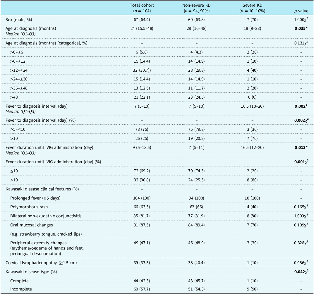
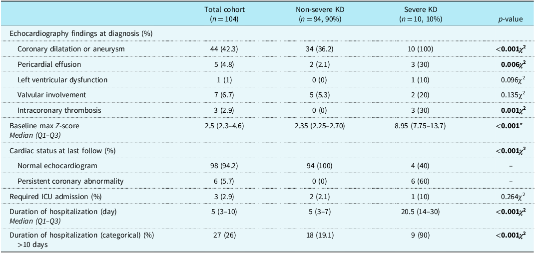
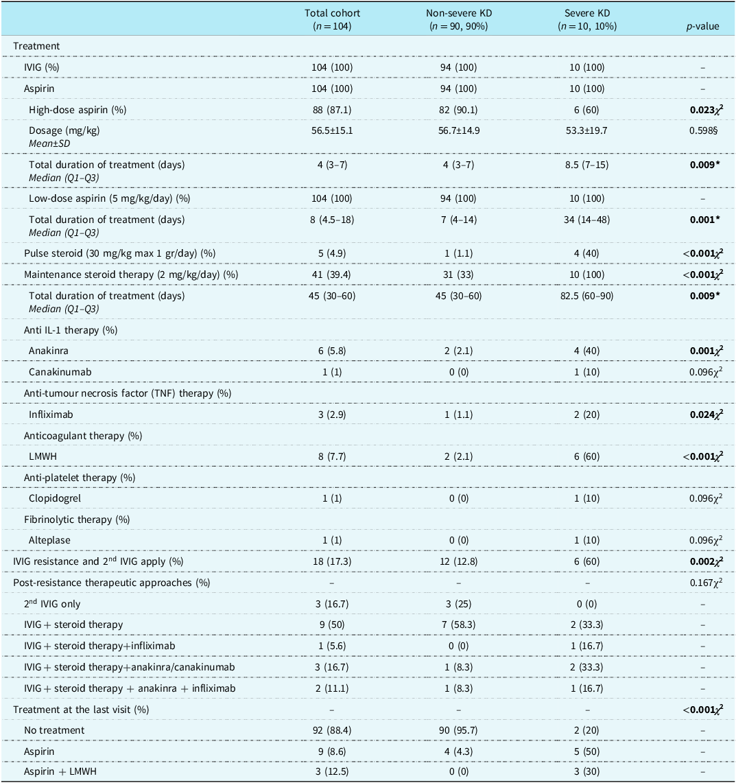
The study included 104 children with Kawasaki disease, of whom 64.4% (n = 67) were male, with a median age at diagnosis of 24 months (Q1–Q3: 15.5–48). All patients presented with ≥5 days of fever, and common clinical features included oral mucosal changes (n = 91, 87.5%), bilateral non-exudative conjunctivitis (n = 85, 81.7%), and polymorphous rash (n = 66, 63.5%). The median time from symptom onset to diagnosis was 7 days (Q1–Q3: 5–10), and 78 patients (65%) were diagnosed within this interval. In addition, 60 patients (57.7%) met the criteria for incomplete Kawasaki disease. The median duration of fever before intravenous immunoglobulin administration was 9 days (Q1–Q3: 5–13.5) (Table 1). Coronary involvement at diagnosis was identified in 42.3% (n = 44) of patients, with most affected children showing coronary dilatation; 10 patients (10%) had moderate-to-large aneurysm with baseline Z-score >5 (Table 2). Laboratory evaluation showed mean haemoglobin of 10.2 ± 1.7 g/dL, median platelet count of 497 (360–708) x 109/L, and mean albumin level of 3.6 ± 0.5 g/dL; inflammatory markers (C-reactive protein, erythrocyte sedimentation rate, ferritin, and fibrinogen) were elevated as shown in the Table S1. All patients received intravenous immunoglobulin and aspirin, and 39.4% (n = 41) were treated with maintenance corticosteroids. Intravenous immunoglobulin resistance developed in 18 patients (17.3%). Small proportions required biologic therapy (anti-IL-1, 7 (6.8%); anti-tumour necrosis factor (TNF), 3(2.9%)), and 7.7% (n = 8) received anticoagulation (Table 3).
Clinical and demographic characteristics of the overall Kawasaki disease cohort and comparison between patients with severe and non-severe Kawasaki disease

Data are presented as median (Q1–Q3) or %.
Bold values indicate statistical significance (p < 0.05).
IVIG = intravenous immunoglobulin; KD = Kawasaki disease.
*Mann–Whitney U test.
χ² Chi-square test.
Comparison of echocardiographic characteristics and coronary artery involvement between severe and non-severe Kawasaki disease (KD) groups

Data are presented as median (Q1–Q3) or %.
Bold values indicate statistical significance (p < 0.05).
*Mann–Whitney U test.
χ2 Chi-square test.
Comparison of treatment modalities between severe and non-severe Kawasaki disease groups

Data are presented as mean ± SD, median (Q1–Q3), or %.
Bold values indicate statistical significance (p < 0.05).
IVIG = intravenous immunoglobulin; KD = Kawasaki disease; LMWH = low-molecular-weight heparin.
§ Student’s t-test.
*Mann–Whitney U test.
χ2 Chi-square test.
Clinical, laboratory, treatment, and outcome differences between severe and non-severe Kawasaki disease
Of the total cohort, 10 patients (9.6%) met the criteria for severe Kawasaki disease. Severe cases were significantly younger (median 18 vs. 28 months, p = 0.035) and had longer fever-to-diagnosis intervals (median 16.5 vs. 7 days, p = 0.003) and longer duration of fever before intravenous immunoglobulin administration (median 16.5 vs. 7 days, p = 0.013). Disease severity was not significantly associated with age at diagnosis (p = 0.165), although higher severity rates were observed in younger infants. In contrast, fever duration before intravenous immunoglobulin administration showed a strong association with disease severity. The proportion of severe cases increased progressively with longer fever duration, from 2.8% in patients treated within 5–10 days to 35.7% in those treated after 15–20 days (p = 0.001, linear-by-linear association p = 0.001). Severe disease was approximately nine times more frequent in patients who received intravenous immunoglobulin after day 10 compared with those treated within the first 10 days (25.0% vs. 2.8%, p < 0.001). No statistically significant differences were observed between the severe and non-severe groups in terms of classical Kawasaki disease manifestations (all p > 0.05). Detailed comparisons of clinical characteristics are presented in Table 1. The frequency of incomplete Kawasaki disease was significantly higher in the severe group compared with the non-severe group (p = 0.042).
At baseline echocardiography, thrombosis and pericardial effusion were observed significantly more frequently in the severe group (p = 0.001 and p = 0.006, respectively), while left ventricular dysfunction and valvular involvement did not differ significantly between groups. Baseline maximum coronary Z-scores were markedly higher in the severe group compared with the non-severe group (median [IQR[: 8.95 [7.75–13.7] vs. 2.35 [2.25–2.70], p < 0.001) (Table 2). In the severe group, median maximum coronary Z-scores initially increased during the first 3 months, peaking at 3 months (16.0 [IQR: 8.55–22.15]), followed by a gradual decline thereafter. Median Z-scores decreased to 5.5 (4.95–17.6) at 6 months, 4.4 (2.85–13.55) at 12 months, and 4.0 (3.4–13.0) at the last follow-up. However, the overall change over time did not reach statistical significance (Friedman test, p = 0.098) (Figure 1).
Longitudinal changes in maximum coronary Z-scores in the severe group. Median values with interquartile ranges are shown at baseline and during follow-up.

Figure 1. Long description
A line graph showing longitudinal changes in maximum coronary Z-scores in the severe group. The x-axis represents follow-up time with labels including Baseline, 2 weeks, 6 weeks, 3 months, 6 months, 12 months, and Last visit. The y-axis represents maximum coronary Z-score ranging from 2.5 to 22.5. The graph shows median values with interquartile ranges at each follow-up point. The data points indicate an initial Z-score around 10 at baseline, a slight increase at 2 weeks, a more significant rise at 6 weeks, peaking at 3 months, followed by a decline at 6 months, and further decreases at 12 months and the last visit. All values are approximated.
Patients with severe disease had significantly lower serum albumin levels compared with non-severe patients (mean ± SD: 3.3 ± 0.4 vs. 3.7 ± 0.5 g/dL, p = 0.015) and lower haemoglobin levels (9.1 ± 1.2 vs. 10.4 ± 1.7 g/dL, p = 0.026). Platelet counts were significantly higher in the severe group (median [IQR]: 744.000 [670.900–810.000] vs. 469.000 [350.000–612.000], p = 0.001). Hypoalbuminaemia (<3.5 g/dL) was more frequent in patients with severe disease than in those with non-severe disease (31.8% vs. 2.5%, p = 0.002). All patients with severe disease were anaemic, whereas no severe cases were observed among patients without anaemia (p = 0.026). Severe disease was observed only in patients with thrombocytosis (>450 × 109/L) (p = 0.030). The time to normalization of inflammatory laboratory markers (C-reactive protein and erythrocyte sedimentation rate) was significantly longer in patients with severe disease compared with those with non-severe disease (median (IQR): 6 [3–6] vs. 2 [1–3] months, p = 0.014) (Table S1).
Severe cases also required more intensive treatment, including pulse steroids (n = 4, 40% vs. n = 1, 1.1%, p < 0.001), maintenance steroids (n = 10, 100% vs. n = 31, 33%, p < 0.001), anti-IL-1 therapy (anakinra) (n = 4, 40% vs. n = 2, 2.1%, p = 0.001), anti-tumour necrosis factor (TNF) therapy (infliximab) (n = 2, 20% vs. n = 1, 1.1%, p = 0.024), intravenous immunoglobulin resistance and re-treatment (n = 6, 60% vs. n = 12, 12.8%, p = 0.002), and anticoagulant therapy (ow-molecular-weight heparin) (n = 6, 60% vs. n = 2, 2.1%, p < 0.001). Severe patients had longer hospital stays (median 20.5 vs. 5 days, p < 0.001) (Table 3).
Outcome analysis in the severe subgroup: normalization versus persistent coronary abnormalities
Among patients with severe disease (n = 10), longitudinal echocardiographic follow-up revealed substantial heterogeneity in coronary outcomes. Although median maximum coronary Z-scores showed an overall declining trend over time, no statistically significant difference was observed across follow-up time points (Friedman test, p = 0.098). At the last follow-up, coronary normalization occurred in 4 patients (40%), whereas persistent abnormalities remained in 6 (60%). No significant differences in clinical, laboratory, or treatment-related variables were identified between patients with normalized and persistent disease. Overall, no individual factor reliably predicted coronary recovery within the severe subgroup (Table S2).
Predictors of severe Kawasaki disease: univariate and multivariate logistic regression findings
In univariate analysis, diagnostic delay, prolonged fever duration before intravenous immunoglobulin administration, delayed IVIG treatment (>10 days), baseline pericardial effusion, hypoalbuminaemia, and higher platelet counts were significantly associated with severe disease (Figure 2(a)). Delayed intravenous immunoglobulin administration was associated with an approximately 12-fold increase in risk, and baseline pericardial effusion with nearly a 20-fold increase. Each additional day of diagnostic delay increased the risk by approximately 20%, whereas each 1 g/dL increase in serum albumin reduced the risk by nearly 90%. In multivariable analyses, delayed intravenous immunoglobulin administration and baseline pericardial effusion remained the most robust independent predictors of severe disease across different models, conferring more than a tenfold increased risk. Diagnostic delay also remained independently associated with severity in alternative models. In sensitivity analysis, higher serum albumin levels remained strongly protective after adjustment for treatment delay (Figure 2(b)).
Forest plots of univariate ( a ) and multivariable ( b ) logistic regression analyses identifying predictors of severe Kawasaki disease. Odds ratios with 95% confidence intervals are displayed on a logarithmic scale.

Figure 2. Long description
The table compares research and engagement processes across several key aspects. It has two columns labeled ‘Research’ and ‘Engagement,’ and six rows detailing different elements. The first row discusses the goals, with research aiming to collect data and produce generalizable knowledge, while engagement focuses on collaborative work to enhance specific research projects. The second row describes participant roles, noting that participants are studied in research but are partners in engagement. The third row covers consent processes, indicating that research participants complete informed consent, whereas engagement generally does not require it. The fourth row addresses compensation, stating that research participants receive incentives, while engagement partners are compensated for their time and expertise. The fifth row outlines data collection methods, mentioning surveys, focus groups, and interviews for research, and feedback sessions and advisory board membership for engagement. The final row discusses approval requirements, noting that research projects must be approved by the Institutional Review Board, while engagement is not considered research but may seek a ‘Not Human Subjects Research’ determination.
Discussion
In this single-centre cohort, delayed intravenous immunoglobulin administration and baseline pericardial effusion emerged as the strongest independent predictors of severe Kawasaki disease. Hypoalbuminaemia and elevated inflammatory markers further characterized a high-risk inflammatory phenotype. Severe cases exhibited markedly higher baseline coronary Z-scores and a prolonged inflammatory course.
Younger age at diagnosis emerged as an important differentiating factor for severe Kawasaki disease in our cohort. Large studies from Japan and Korea, settings with the highest Kawasaki disease incidence, have repeatedly demonstrated that infants and toddlers carry the greatest risk for coronary artery aneurysms, intravenous immunoglobulin resistance, and treatment-refractory inflammation. Reference Kim, Choi and Woo26–Reference Yamashita, Ae and Yashiro29 Similar patterns have been reported in contemporary European cohorts from Italy, Spain, and the United Kingdom, all highlighting the vulnerability of children under two years of age and the heightened likelihood of adverse coronary outcomes in this group. Reference Morana, Guida and Andreozzi30–Reference Tulloh, Mayon-White and Harnden32
Recent multicentre and single-centre Turkish studies have shown that younger age, prolonged fever before diagnosis, delayed intravenous immunoglobulin administration, and elevated inflammatory markers significantly increase the risk of coronary involvement and treatment resistance. Reference Türkuçar, Kaya and Çakmak33,Reference Kaya Akca, Arslanoglu Aydin and Aykan34 Consistent with prior reports, we observed that diagnostic delay (>10 days; p = 0.002), particularly beyond 10–20 days of fever, along with prolonged fever before intravenous immunoglobulin (median 16.5 vs. 7 days; p = 0.013), was strongly associated with severe disease, supporting the established concept that sustained untreated inflammation amplifies vascular injury and promotes aneurysm formation. Collectively, the alignment between our cohort, regional Turkish data, and large Asian, European, and Arabian studies reinforces the global relevance of age and treatment timing as central, clinically relevant determinants of Kawasaki disease severity. Reference Xia, Qiu and Wen35–Reference Kenza, Maryam, Asmaa, Ghita, Hanan and Abdenasser40
In the present study, pericardial effusion was significantly more frequent at initial echocardiography in the severe group and remained an independent risk factor for disease severity in multivariate analysis. This finding suggests that pericardial effusion may serve as an early indicator of more extensive cardiovascular involvement and an unfavourable disease trajectory in patients with severe Kawasaki disease, consistent with previous reports by Chbeir et al. and Lega et al. Reference Chbeir, Gaschignard and Bonnefoy41,Reference Lega, Bozio and Cimaz42
In the present study, serum albumin emerged as a key laboratory marker associated with disease severity. Although both lower albumin levels and higher platelet counts were significantly associated with severe disease in univariate analyses, only hypoalbuminaemia remained an independent predictor in multivariable models. Each 1 g/dL increase in serum albumin was associated with a substantial reduction in the risk of severe disease, highlighting its value for early risk stratification. This finding does not imply a causal role of albumin but rather reflects the combined effects of systemic inflammation, endothelial dysfunction, and increased vascular permeability in Kawasaki disease. While other inflammatory markers lost significance after adjustment, likely due to shared biological pathways, serum albumin appears to function as an integrative surrogate marker. Consistent with previous reports, our findings support the role of hypoalbuminaemia as a marker of high-risk disease and adverse coronary outcomes. Reference Xia, Qiu and Wen35,Reference Jiang, Li and Xu37,Reference Soriano-Ramos43–Reference Liu, Wang and Du45
In our cohort, baseline maximum coronary Z-scores were markedly higher in the severe group than in the non-severe group, supporting the validity of our severity definition based on early coronary involvement. Longitudinal analysis within the severe subgroup revealed a distinct trajectory, with Z-scores initially increasing during the first 3 months and subsequently declining over time, reflecting ongoing vascular inflammation and delayed remodelling in the early disease phase. Similar early progression followed by gradual regression has been reported in previous longitudinal studies of high-risk Kawasaki disease, particularly in cohorts with large and giant coronary aneurysms. Reference Friedman, Gauvreau and Hamaoka-Okamoto46–Reference Tsuda and Hashimoto48
Despite overall improvement, substantial heterogeneity in coronary outcomes was observed within the severe subgroup, with persistent coronary abnormalities in 6 of 10 patients (60%) at last follow-up. No significant differences in baseline clinical, laboratory, or treatment-related variables were identified between patients with persistent and regressed disease, likely reflecting limited statistical power due to the small sample size. In contrast, Guida et al. reported that patients with persistent coronary artery lesions were younger, more likely to be intravenous immunoglobulin-resistant and late-treated, and had lower haemoglobin and albumin levels and higher C-reactive protein values. Reference Guida, Morana and Tarì49 The discrepancy between these findings underscores the clinical importance of early identification of high-risk patients and highlights the need for larger, multicentre studies to refine prognostic stratification and optimize long-term management in severe Kawasaki disease.
Our treatment patterns closely mirror contemporary high-risk Kawasaki disease management. International guidelines and major trials, including the 2017 American Heart Association statement and the RAISE regimen, support early corticosteroids in children with high inflammatory burden or elevated baseline Z-scores, and increasing real-world data similarly endorse infliximab or IL-1 blockade for refractory disease. Reference Jone, Tremoulet and Choueiter24, Reference Buda, Friedman-Gruszczyńska and Książyk50–Reference Marchesi, Tarissi de Jacobis and Rigante55 In this context, the reduced use of high-dose aspirin and the substantially greater reliance on prolonged low-dose aspirin, corticosteroids, anakinra, infliximab, and anticoagulation in our severe cases are fully consistent with these evidence-based “intensified care” approaches. These findings emphasize that severe Kawasaki disease not only presents with a more aggressive inflammatory profile but also requires a distinctly escalated therapeutic strategy.
This study has several limitations. Its retrospective, single-centre design and relatively small sample size, particularly within the severe subgroup, limited statistical power and reduced our ability to identify reliable predictors of coronary persistence and perform comprehensive risk stratification. In addition, variability in follow-up duration and incomplete echocardiographic data in the non-severe group precluded fully standardized temporal analyses across severity groups. Nevertheless, our cohort was characterized by detailed longitudinal echocardiographic assessment in patients with severe disease and comprehensive clinical and laboratory profiling. This enabled the identification of clinically meaningful patterns in coronary Z-score trajectories and early risk markers. Despite inherent constraints, our findings provide valuable real-world evidence and generate testable hypotheses for future large-scale, prospective, multicentre studies aimed at improving long-term risk stratification and individualized management in Kawasaki disease.
Conclusion
In this study, pericardial effusion on initial echocardiography, baseline hypoalbuminaemia, and delayed intravenous immunoglobulin administration emerged as key early markers of severe coronary involvement in Kawasaki disease. Baseline coronary Z-scores reliably identified high-risk patients, while longitudinal analyses revealed a heterogeneous disease course with frequent persistence of coronary abnormalities. Current clinical and laboratory parameters remain insufficient for precise long-term risk prediction, underscoring the need for early immunomodulatory intervention and large, prospective, multicentre studies integrating advanced biomarkers and standardized imaging to optimize long-term cardiovascular outcomes.
Supplementary material
The supplementary material for this article can be found at https://doi.org/10.1017/S1047951126113304.
Data availability statement
All data generated or analysed during this study are included in this published article.
Acknowledgements
We would like to thank all specialists, residents, and faculty members of the Cerrahpaşa Department of Pediatrics for their valuable contributions.
Authors contribution
Nergis Akay: Writing – original draft, Writing – review & editing, Conceptualization, Methodology, Investigation, Formal analysis, Data curation. Zeynep Torunoglu: Methodology, Investigation. Ece Aslan: Methodology, Investigation. Umit Gul: Methodology, Investigation. Elif Kilic Konte: Methodology, Investigation. Nida Gulderen Kalay Senturk: Methodology, Investigation, Data curation. Mehmet Yildiz: Writing – review & editing, Conceptualization. Sezgin Sahin: Writing – review & editing, Conceptualization. Kenan Barut: Writing – review & editing, Conceptualization. Reyhan Dedeoglu: Writing – review & editing, Conceptualization, Project administration. Ozgur Kasapcopur: Writing – review & editing, Conceptualization, Project administration. All authors reviewed and revised the manuscript and approved the final version of the manuscript.
Financial support
This research did not receive any specific grant from funding agencies in the public, commercial, or not-for-profit sectors.
Competing interests
The authors declare that they have no known competing financial interests or personal relationships that could have appeared to influence the work reported in this paper.
Ethical standard
The study was approved by the Istanbul University-Cerrahpasa Institutional Review Board on 12.03.2025 (protocol number: 2025/152). Written consent was obtained from the legal representatives of the patients in compliance with the Declaration of Helsinki.