Introduction
The oviduct is a complex and organized thin tubular structure connecting the ovary with the uterus in which the earliest reproductive events occur (Maillo et al., Reference Maillo, Lopera-Vásquez, Hamdi, Gutiérrez-Adán, Lonergan and Rizos2016a). Together with other molecules derived from plasma and follicular fluid, the oviduct provides an optimal microenvironment for reproductive functions (Hunter, Reference Hunter2012). The oviductal fluid contains simple and complex carbohydrates, ions, lipids, phospholipids and proteins that influence sperm capacitation, final oocyte maturation, fertilization and early embryonic development (Killian, Reference Killian2004). Also, in the oviductal fluid, the activity of proteolytic enzymes involved in multiple events that take place in the oviduct was demonstrated (Jiménez-Díaz et al., Reference Jiménez-Díaz, Giunta, Valz-Gianinet, Pereyra-Alfonso, Flores and Miceli2000; Gabler et al., Reference Gabler, Killian and Einspanier2001; Roldán-Olarte et al., Reference Roldán-Olarte, García, Jiménez-Díaz, Valdecantos and Miceli2012). The urokinase plasminogen activator system (uPAS) promotes extracellular proteolysis by regulating plasminogen activation in the vicinity of cells and activates intracellular signalling pathways (Hildenbrand et al., Reference Hildenbrand, Allgayer, Marx and Stroebel2010). The urokinase plasminogen activator (uPA) is a member of the uPAS, which can bind to its receptor (uPAR) and initiate a proteolytic cascade resulting in the conversion of plasminogen to plasmin (Baldwin et al., Reference Baldwin, Sood, Elfline, Luke, Dewyer, Díaz, Myers, Wakefield and Henke2012). Plasmin has a proteolytic function that leads to the degradation of a number of extracellular basement membrane components. In addition, uPA-mediated plasmin can act over the oocyte and cumulus cell surface (García et al., Reference García, Miceli, Rizo, García, Valdecantos and Roldán-Olarte2016) and the proteolytic activity of uPA is needed for successful oocyte maturation in bovine (Roldán-Olarte et al., Reference Roldán-Olarte, Maillo, Sánchez-Calabuig, Beltrán-Brena, Rizos and Gutiérrez-Adán2017). uPA/uPAR binding is involved in signalling pathways that activate transcription factors and regulate the synthesis of molecules concerned with the arrangement of a particular oviductal microenvironment (García et al., Reference García, Miceli, Valdecantos, García and Roldán-Olarte2014). Plasmin activation and uPA also result in the release of growth factors, including members of the vascular endothelial growth factor (VEGF) family (Plouët et al., Reference Plouët, Moro, Bertagnolli, Coldeboeuf, Mazarguil, Clamens and Bayard1997; Ferrara, Reference Ferrara2010; Salajegheh, Reference Salajegheh2016). Vascular endothelial growth factors and their receptors are positive regulators of angiogenesis (Lee et al., Reference Lee, Jeong, Han and Baek2015). The VEGF family includes VEGF glycoproteins members (VEGF-A, VEGF-B, VEGF-C, VEGF-D, and VEGF-E) and placental growth factors 1 and 2 (PLGF) (Salajegheh, Reference Salajegheh2016). VEGFR-1, VEGFR-2 and VEGFR-3 are known as the primary receptors for VEGF (Lee et al., Reference Lee, Jeong, Han and Baek2015). The binding of the VEGF ligand to VEGFR causes dimerization and activation via transphosphorylation and signal transduction. VEGFR-2 is predominantly expressed by activated endothelia of blood vessels and VEGFR-3 by lymphatic endothelia (Shibuya, Reference Shibuya2014). VEGF-D, known as c-fos-induced growth factor, has a putative role in angiogenesis and lymphangiogenesis via VEGFR-2 and VEGFR-3 (Achen et al., Reference Achen, Jeltsch, Kukk, Makinen, Vitali, Wilks, Alitalo and Stacker1998). VEGF actions include the mitogenic activation of endothelial cells and the necessary expression and induction of serine proteases and collagenases, increasing endothelial chemotaxis and vasodilation (Vempati et al., Reference Vempati, Popel and MacGabhann2014). VEGF–VEGFR systems act in an autocrine and paracrine manner and are thought to be involved in ovarian follicular development, oocytes maturation, luteal angiogenesis, endometrial changes, embryo implantation, formation of placental blood vessels and embryo development (Yan & Shi, Reference Yan and Shi2016). In addition, members of the VEGF family are able to modify the activation and expression of uPAS components. It has been demonstrated that the binding of VEGFs to their receptors on endothelial cells led to increased expression and activity of uPAS, suggesting a role for VEGFs in the regulation of extracellular matrix degradation, cell adhesion, and migration (Olofsson et al., Reference Olofsson, Korpelainen, Pepper, Mandriota, Aase, Kumar, Gunji, Jeltsch, Shibuya, Alitalo and Eriksson1998; Behzadian et al., Reference Behzadian, Windsor, Ghaly, Liou, Tsai and Caldwell2003; Prager et al., Reference Prager, Mihaly, Brunner, Koshelnick, Hoyer-Hansen and Binder2009).
The main objective of this work was to analyze the effect of VEGF-D in the regulation of the expression of components of uPAS in bovine oviductal epithelial cells (BOEC). In the first place, the expression of VEGF-D and its receptors VEGFR-2 and VEGFR-3 in cells obtained from BOEC was studied before and after in vitro conditions. Then the effect of exogenous VEGF-D on the expression levels of uPA, uPAR, and PAI-1 in BOEC cultures was analysed and also the effect of VEGF-D and uPA on cell migration in monolayer cell cultures.
Materials and Methods
Tissue preparation
Bovine oviductal epithelial cells (BOEC)
Genital tracts from young beef cows (Bos taurus) were collected from a local abattoir within 20 min of death and transported to the laboratory in sodium phosphate buffer (PBS), pH 7.4 at 4°C for processing within 3 h after collection. Ipsilateral oviducts to the active ovary, corresponding to the mid-luteal stage (presence of corpus luteum, days 6–12), were used. We had previously studied the expression of genes involved in uPAS, which could be relevant to understanding the changes in oviductal physiology (Roldán-Olarte et al., Reference Roldán-Olarte, García, Jiménez-Díaz, Valdecantos and Miceli2012; García et al., Reference García, Miceli, Valdecantos, García and Roldán-Olarte2014). Oviducts were ligated, dissected, washed in fresh PBS and then quickly dipped in 70% ethanol and washed again in PBS. After discarding infundibulum and utero tubal junction, oviducts were gently squeezed in a stripping motion with forceps to obtain epithelial cells (Rottmayer et al., Reference Rottmayer, Ulbrich, Kolle, Prelle, Neumueller, Sinowatz, Meyer, Wolf and Hiendleder2006).
BOEC suspension cultures
In each experiment, epithelial cells collected from six to eight oviducts from different animals were pooled and placed in culture medium TCM-199 (Gibco, Life Technology, Burlington, ON, Canada) supplemented with 10% bovine fetal serum (Internegocios, Buenos Aires, Argentina), 25 mM HEPES (Gibco, Life Technology, Burlington, ON, Canada), 0.2 mM sodium pyruvate, 100 IU/ml of penicillin, 100 µg/ml of streptomycin and 0.25 µg/ml of fungizone (Gibco, Life Technology, Burlington, ON, Canada). The cell suspension was pipetted 15 times through a 21-gauge syringe needle. Three washing steps were performed, each followed by 25 min sedimentation in culture medium in the cell culture incubator. Cell viability at seeding was analyzed using trypan blue (Sigma) staining and microscopic observation of beating cilia. Cells were grown in 25 mm2 bottle cultures (Nunclon, Roskilde, Denmark) in 5 ml TCM-199 supplemented as described previously. Suspension cultures were performed at 38.5°C in a humidified atmosphere and in 5% CO2 in air. After 48 h, cell cultures were washed twice with PBS. The epithelial nature of the cultured cells was confirmed by immunocytochemical analysis, using monoclonal anti-pan keratin (AE1/AE3/PCK26) primary antibody (Ventana Medical Systems, Inc., 760- 2595); more than 90% of the cells were stained positively for cytokeratin (Valdecantos et al., Reference Valdecantos, Bravo-Miana, García, García, Roldán-Olarte and Miceli2017). For the VEGF-D stimulation assays, 20 mg of cell suspension culture were placed in each well of a 24-well culture plate (Nunclon, Roskilde, Denmark) in 500 µl of serum-free medium containing 1% bovine serum albumin (Sigma) for 2 h. After this step, the culture medium was replaced by another medium supplemented with recombinant VEGF-D at 1 or 10 ng/ml (Sigma, V6012) as previously described by others authors (Wijayagunawardane et al., Reference Wijayagunawardane, Kodithuwakku, Yamamoto and Miyamoto2005). Suspension cell cultures were collected at 0, 12, 24 or 48 h after incubation in the presence of VEGF-D. Cells from each sample were centrifuged at 10,000 g and supernatants discarded. Finally, the cells were washed twice with PBS and then used for total RNA isolation.
Total RNA isolation
Samples of BOEC obtained from oviducts in mid-luteal phase by mechanical processing (n = 4), were used to analyse the expression of VEGF-D, VEGFR2 and VEGFR3. Cell suspension cultures of BOEC before and after VEGF-D stimulation (n = 4 from each condition and time) were also used. Total RNA was isolated by using Tri-Reagent (Molecular Research Center Inc., Cincinnati, OH, USA). Total RNA yield was determined spectroscopically at 260 nm. RNA quantity and quality were verified by electrophoresis on 1% (w/v) formaldehyde agarose gel containing 0.06 μg/ml SYBR ® Safe DNA Gel Stain (Invitrogen, Burlington, ON, Canada).
Reverse transcription-polymerase chain reactions (RT-PCR)
Total RNA (1 μg) was reverse transcribed using Moloney murine leukemia virus enzyme (Promega, Madison, WI, USA) and oligo(dT)17 (García et al., Reference García, Miceli, Valdecantos, García and Roldán-Olarte2014). A 25 μl final volume of the reaction mixture containing 50 mM Tris–HCl (pH 8.3), 75 mM KCl, 3 mM MgCl2, 0.5 mM of each dNTP, 25 pM oligo(dT), 10 mM dithiothreitol and 200 units of reverse transcriptase was incubated at 42°C for 1 h followed by reverse transcriptase inactivation at 94°C for 5 min.
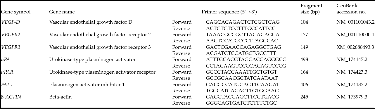
Aliquots of 1 µl of the RT reaction were amplified by end-point PCR with 2.0 U of Taq DNA polymerase (Invitrogen, Burlington, ON, Canada) in a 20 µl reaction volume containing 10 mM Tris–HCl (pH 9.0), 50 mM KCl, 0.1% Triton X-100, 2.5 mM MgCl2, 0.2 mM of each dNTP and 1 µM of each oligonucleotide specific primer for the gene of interest: VEGF-D, VEGFR2, VEGFR3, uPA, uPAR, PAI-1 and β-ACTIN (see Table 1). PCR conditions were previously assayed to determine the appropriate number of cycles for the amplification of each fragment. PCR cycles consisted of: (1) an initial denaturing step at 94°C for 1 min; (2) 30 cycles (VEGF-D, VEGFR2, VEGFR3, PAI-1 and β-ACTIN) or 22 cycles (uPA, uPAR and β-ACTIN) of denaturation for 30 s at 94°C, annealing for 30 s at the appropriate annealing temperature for each pair of primers, extension for 50 s at 72°C; (3) a final extension step at 72°C for 7 min. Each PCR assay was carried out at least twice for each sample. Primer pairs were designed with primer3 software from published Bos taurus cDNA sequences.
Primers used to amplify specific bovine transcripts from oviductal epithelial cells

GenBank accession numbers, primer sequences, and amplification fragment sizes are shown in Table 1. β-ACTIN mRNA was used as a control for RNA semi-quantification and PCR reaction efficiency (Soutar et al., Reference Soutar, Dillon and Ralston1997). A PCR reaction with specific primers for β-ACTIN was carried out in the same amplification conditions (number of the cycles) as the genes of interest. Non-template controls were included in each experiment. PCR products were resolved on 1.5% agarose gel containing 0.06 µg/ml SYBR ® Safe DNA Gel Stain (Invitrogen, Burlington, ON, Canada). The nature of the PCR products obtained was confirmed by sequencing. The gels were photographed with a digital camera (Olympus C-5060). The intensities of the bands were analyzed with the NIH ImageJ software for the semi-quantitative determination of the expression levels of VEGF-D, VEGFR2, VEGFR3, uPA, uPAR and PAI-1 in relation to intensity of the corresponding β-ACTIN fragment for each sample and condition of the assays.
Wound healing assay
Oviductal epithelial monolayer cell cultures
For each experiment, four ipsilateral oviducts at the mid-luteal stage were used to obtain oviductal epithelial cells for primary cultures. The collected cells were incubated at 38.5°C, in 5% CO2 in air and 100% humidity for 48 h to obtain cell suspension cultures. Later incubation of these cell cultures for 5 days allowed the formation of monolayer cell cultures. Then, cultures were washed twice with PBS, resuspended in 2 ml of trypsin–EDTA (Gibco BRL) and incubated for 3 min at 37°C. Trypsin action was blocked with 2 ml culture medium (Valdecantos et al., Reference Valdecantos, Bravo-Miana, García, García, Roldán-Olarte and Miceli2017). The cells were recovered in a sterile Petri dish and disaggregated mechanically with a pipette. Cells were collected in a 50 ml sterile conical tube with 15 ml medium TCM-199 and washed twice at room temperature. Material was collected in culture medium TCM-199 supplemented with 10% bovine fetal serum, 25 mM HEPES, 0.2 mM sodium pyruvate, 100 IU/ml penicillin, 100 µg/ml streptomycin and 0.25 µg/ml fungizone. The cells (2 × 10⁵/ml) were incubated at 38.5°C, in 5% CO2 in air and 100% humidity in 24-well culture plates until confluency. Finally, monolayer cell cultures were incubated with 500 µl of serum-free medium containing 1% bovine serum albumin for 2 h.
Wound healing assay
Wound healing assays were performed to study cell migration capacity, as previously described (García et al., Reference García, Valdecantos, Miceli and Roldán-Olarte2017). Briefly, artificial gaps were made in each monolayer using a 200 µl pipette tip. Immediately after creating a scratch of cultures, cells were washed and incubated in medium supplemented with VEGF-D 10 ng/ml or uPA 10 nM (García et al., Reference García, Miceli, Valdecantos, García and Roldán-Olarte2014). Three experiments from each treatment were carried out. Images were captured at different intervals (0, 6, 12 or 24 h) using an inverted light microscope (Zeiss Axiovert 25) and photographed (n = 5) for each treatment and time with a digital camera (Arcano). To obtain the same field of interest during the observations, etches were made on the bottom of each well and used as microscopic reference points to make sure that measurements were always carried out at exactly the same spot (Jordaens et al., Reference Jordaens, Arias-Alvarez, Pintelon, Thys, Valckx, Dezhkam, Bols and Leroy2015). Measurements of each scratch area were obtained using ImageJ software at every time point and the percentage of wound region (% WR) was determined (wound region/total size of image per 100) (Cardona, Reference Cardona2015).
Statistical studies
RT-PCR and wound closure data are expressed as the mean ± standard error of the mean (SEM). Statistical analysis of the data was performed using the InfoStat Statistical Package (InfoStat 2015, http://www.infostat.com.ar). Analysis of variance (ANOVA) with least significant difference (LSD) Fischer's post hoc test was used to study the differences among the means. A P-value of less than 0.05 was considered to be statistically significant.
Results
Expression of VEGF-D, VEGFR2 and VEGFR3 genes in ex vivo and in vitro cultured BOEC
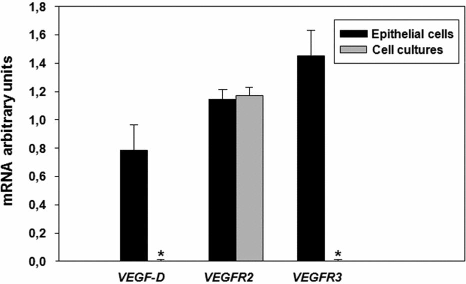
VEGF-D, VEGFR2 and VEGFR3 expression in ex vivo BOEC and in vitro cultivated cells were analyzed by RT-PCR. All the studied transcripts were detected in freshly obtained BOEC. In vitro cultured BOEC expressed VEGFR2 mRNA but not VEGF-D or VEGFR3 transcripts (Fig. 1).
VEGF-D, VEGFR2 and VEGFR3 expression in bovine oviductal epithelial cells before and after in vitro conditions. Arbitrary units were determined by relating band density to the corresponding β-ACTIN for each sample. Values are the mean ± standard error (SE) (n = 4). *Indicates significant differences (P < 0.05) before and after culture.

Expression of uPA, uPAR and PAI-1 genes in BOEC cultures stimulated with VEGF-D
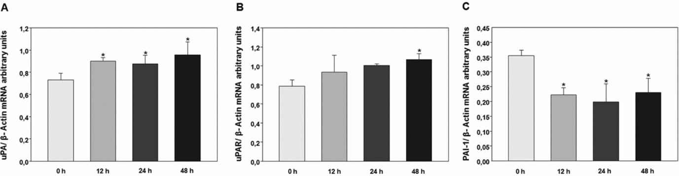
In order to test if VEGF-D affected mRNA abundance of the uPAS system, primary BOEC cultures were obtained and treated with VEGF-D (1 and 10 ng/ml) at 0, 12, 24 or 48 h. Then, uPA, uPAR, and PAI-1 mRNA abundance was analyzed by RT-PCR. The expression of uPA was significantly increased at 12, 24 and 48 h of induction with VEGF-D 10 ng/ml compared with the control at 0 h (Fig. 2A). uPAR expression increased at 48 h (Fig. 2B) and PAI-1 mRNA significantly decreased after 12 h of incubation (Fig. 2C). No significant differences were observed when cells were incubated with VEGF-D 1 ng/ml (data not shown).
Comparison of the relative density of (A) uPA/β-ACTIN, (B) uPAR/β- ACTIN and (C) PAI-1/β-ACTIN RT-PCR products in bovine oviductal epithelial cells cultures stimulated with VEGF-D. Values are the mean ± standard error (SE) (n = 4). *Indicates significant differences (P < 0.05) between treatments with the control at 0 h.

Wound healing assay
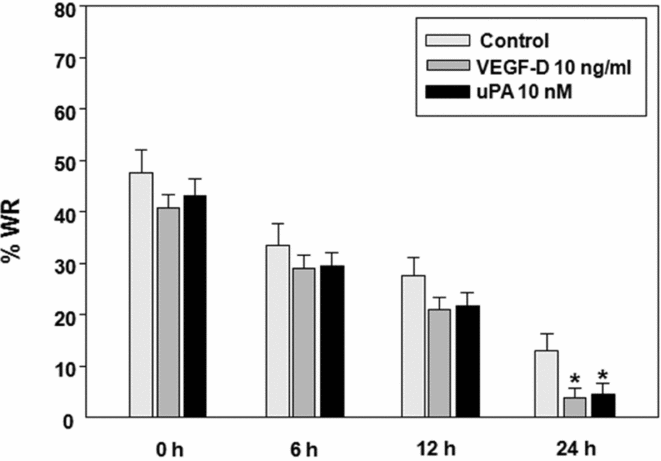
The capability of VEGF-D and uPA to induce BOEC migration was assayed in in vitro cultured BOEC monolayers that were cultured for 24 h in the presence of recombinant VEGF-D (10 ng/ml) or uPA (10 nM) after a scratch was made in the monolayers. Then, the scratched area was measured at 0, 6, 12 and 24 h after the addition of VEGF-D or uPA and compared with non-treated control groups. As shown in Fig. 3, both VEGF-D and uPA significantly increased the ability of the cells to close the scratched area at 24 h.
Quantification of the in vitro wound region (WR) process in bovine oviductal epithelial cell cultures. Bars represent % WR in cultures with VEGF-D or uPA at different times. Values are the mean ± standard error of the mean (SEM) (n = 3). Different letters represent significant differences between samples (P < 0.05). *Indicates differences between control and treatment samples at the same time.

Discussion
The oviduct plays a pivotal role in mammalian reproduction by providing an optimal environment for sperm capacitation, fertilization and transport of gametes and embryos (Maillo et al., Reference Maillo, Sánchez-Calabuig, Lopera-Vásquez, Hamdi, Gutiérrez-Adán, Lonergan and Rizos2016b). Previous works reported the localization of VEGF-A and its receptor transcripts in oviductal epithelial cells and fluids (Gabler et al., Reference Gabler, Einspanier, Schams and Einspanier1999; López-Albors et al., Reference López-Albors, Olsson, Llinares, Gutiérrez, Latorre, Candanosa, Guillen-Martinez and Izquierdo-Rico2017).
In the present work, RT-PCR analysis was used to evaluate VEGF-D gene expression and its receptors (VEGFR2 and VEGFR3) in BOEC. Bovine oviductal epithelial cells expressed VEGF-D, VEGFR2, and VEGFR3 mRNAs; when cultured in vitro, BOEC lose the VEGF-D and VEGFR3 transcripts but still express VEGFR2 mRNA.
It is also known that the VEGF-A presence in BOEC cultures could regulate the expression of genes that contribute to the contractile activity of the bovine oviduct (Wijayagunawardane et al., Reference Wijayagunawardane, Kodithuwakku, Yamamoto and Miyamoto2005). Also, others authors determined an increase in uPA and uPAR expression in vascular endothelial cells in vitro in a dose- and time-dependent manner in response to VEGF-A (Mandriota et al., Reference Mandriota, Seghezzi, Vassalli, Ferrara, Wasi, Mazzieri, Mignatti and Pepper1995). This increase could allow the appropriate regulation of the extracellular matrix remodelling (Behzadian et al., Reference Behzadian, Windsor, Ghaly, Liou, Tsai and Caldwell2003). However, VEGF-D action on the regulation of uPAS gene expression and its participation in the bovine oviduct has not been previously described.
In this study, we examined the effect of VEGF-D in the expression of uPA, uPAR and PAI-1 in BOEC cultures. Cells were treated with VEGF-D at 1 or 10 ng/ml at different times and the expression of these genes was analyzed by RT-PCR. Results showed that VEGF-D has a regulatory effect on uPAS gene expression in a time-dependent and dose-dependent manner, inducing an increase in uPA and uPAR mRNA abundance and a decrease in PAI-1 mRNA after 24 h of VEGF-D treatment only at a concentration of 10 ng/ml, while no significant differences were observed when VEGF-D at 1 ng/ml was employed. As primary cultured BOEC expresses VEGFR2, the action of this growth factor may be due to the interaction with its receptor and, through signal transduction, might lead to the activation or inhibition of the genes under study (Zhao et al., Reference Zhao, Zhao, Meng, Liu, Chen and Sun2014; Varricchi et al., Reference Varricchi, Granata, Loffredo, Genovese and Marone2015). The effect of VEGF-D would be similar to VEGF-A action as reported in previous studies (Mandriota et al., Reference Mandriota, Seghezzi, Vassalli, Ferrara, Wasi, Mazzieri, Mignatti and Pepper1995; Behzadian et al., Reference Behzadian, Windsor, Ghaly, Liou, Tsai and Caldwell2003). The present results suggested that VEGF-D would regulate uPAS activity and that it would be possible to find a similar effect with VEGF-A over uPAS gene expression as both factors act through VEGFR2 into the bovine oviduct.
The VEGF family is involved in cell proliferation and migration (Li et al., Reference Li, Bork, Krueger, Patsenker, Schulze-Krebs, Hahn and Schuppan2005). VEGF-D induces tyrosine phosphorylation pathways and multiple biological responses including cell survival, migration, tubulogenesis, and in vivo angiogenesis (Jia et al., Reference Jia, Bagherzadeh, Bicknell, Duchen, Liu and Zachary2004; Vlahakis et al., Reference Vlahakis, Young, Atakilit and Sheppard2005; Harris et al., Reference Harris, Davydova, Roufail, Paquet-Fifield, Paavonen, Karnezis, Zhang, Sato, Rothacker, Nice, Stacker and Achen2013; Costache et al., Reference Costache, Ioana, Iordache, Ene, Costache and Saftoiu2015).
In the present study, as an approach to the determination of the role of VEGF-D and uPA in BOEC, wound healing assays were performed on in vitro primary BOEC cultures. Results showed that both VEGF-D and uPA are capable of enhancing BOEC migration. Although no significant differences were observed between the in vitro VEGF-D and the uPA-induced cell migration, it is interesting to note that VEGF-D also induced an increase in uPA mRNA abundance in BOEC. It can be inferred that VEGF-D-induced uPA expression could have a synergistic effect on BOEC migration that might be important for maintaining epithelial lining integrity.
In conclusion, results showed that VEGF-D could trigger signals through its specific receptor VEGFR2 and upregulate uPA expression levels. Further studies of VEGF-D signalling pathways regulating the expression of uPAS components could contribute to the knowledge of the crosstalk between both systems in the bovine oviduct and their participation in the maintenance of the oviductal microenvironment necessary for the success of early reproductive events.
Acknowledgements
The authors wish to thank Frigorífico Calchaquí for allowing reproductive tract collection from their slaughtered cows.
Conflict of interest statement
The authors declare that there is not conflict of interest that could be perceived as prejudicing the impartiality of the research reported.
Financial support
This work was supported by Agencia Nacional de Promoción Científica y Tecnológica, ANPCYT, Argentina (M.R.O. grant BID PICT 2013 No. 0770), Consejo de Investigaciones de la Universidad Nacional de Tucumán, Argentina (D.C.M., PIUNT, grant 26/ D525, P.A.V., PIUNT, grant 26/ D551) and D.C.G. is grateful to CONICET for her postdoctoral fellowship.
Ethical standards
Not applicable.