INTRODUCTION
Nutrient fluxes among tissues follow the routes of blood and lymph, and the gastrointestinal tract plays an essential role in distribution of nutrients to peripheral tissues. In terms of whole-body metabolism, portal-drained viscera (PDV) have a disproportionate influence with respect to their masses and under certain circumstances their high metabolic rate may compromise nutrient availability to the periphery. In order to conduct gastrointestinal metabolism studies and/or quantify nutrient absorption, catheterization of the portal vein (PV) is indispensable. There are many combinations of catheter design and care, and surgical procedures, in the literature, although reports in pigs are limited. As van Leeuwen et al. (Reference Van Leeuwen, Leuvenink, Haasbroek, Priem, Bosch and Van Kleef1995) pointed out, methods of catheterization of the PV can be divided into two groups: indirect, where the catheter insertion is into a tributary mesenteric vein or a branch of the PV in the liver; and direct insertion in the PV. The same would apply for arterial catheterization, with respect to artery choice or the insertion technique. Also, the way of anchoring the catheter to the surrounding tissues, the tip design, dimensions and composition, and the catheter maintenance protocol vary among authors.
In pigs, most surgical procedures have been applied in modern breeds, following a direct catheterization in PV and the carotid artery (CA), with occlusion flow in the latter. However, differences in anatomical characteristics among breeds could be an added difficulty for the catheter implantation procedure.
The aim of the present paper was to improve the design of catheters, the surgical procedures and catheter care, to study nutrient metabolism of PDV in a native obese (Iberian) and a modern (Landrace) pig breed.
MATERIALS AND METHODS
Animals and diet
The experimental protocol was reviewed and approved by the Bioethical Committee of the Spanish Council for Scientific Research (CSIC), Spain.
The experiment was performed with three Landrace and three Iberian (Silvela strain) gilts (25±0·4 kg average body weight; BW) supplied by Granja El Arenal (Córdoba, Spain) and Sánchez Romero Carvajal, Jabugo S.A. (Sevilla, Spain), respectively. The pigs were allowed ad libitum access to a standard diet (160 g crude protein (CP)/kg; 14–14·5 MJ metabolizable energy (ME)/kg dry matter; DM) and allocated individual pens with free access to water in a controlled-environment room (21±1·5 °C). After surgery, the pigs were fed at 2·4×ME for maintenance (444 kJ/kg0·75 BW; NRC 1998). The daily ration was offered in two portions, at 09.00 h (0·25) and at 15.00 h (the remaining 0·75).
Catheter design
Spooled tubing was used to prepare three catheters, 600 mm long. The diameter of the PV catheter (Tygon, internal diameter 1·27 mm, outside diameter 2·29 mm; Cole-Parmer, Vernon Hills, IL, USA) was larger than those for the ileal vein (IV) and the CA (Tygon, internal diameter 1·02 mm, outside diameter 1·78 mm; Cole-Parmer).
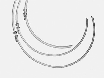
Each catheter was assembled with two rings, distal and proximal to the insertion point of the catheter tip. The rings were 2 and 5 mm wide (Tygon, internal diameter 1·6 mm, outside diameter 3·2 mm; Cole-Parmer), respectively, and glued with Loctite (Super Glue; Henkel Iberica, S.A., Barcelona, Spain), allowing a space of c. 2 mm between them. Before gluing, the proximal side of the first (proximal) catheter ring was cut at an angle of 30°, which allowed the catheter to be kept as tangential as possible to the vessel, while still avoiding blockage due to a possible bend in the tube, and the walls of both rings were longitudinally cut to facilitate installation around the catheter. The ring also stimulates tissue adherence to the vessels by increasing the surface contact, facilitating the anchoring of the catheter.
The proximal rings were 60, 100 and 120 mm from the insertion points of the catheter tips for PV, IV and CA, respectively. The insertion tips of the three catheters were cut to an angle of 45° rather than 90° to avoid catheter obstruction in the event of the tips locating against the vessel walls, and to enlarge the opening. As the tubing used for catheters is purchased in spools, the tendency is to coil. The 45° cut opposite the side of catheter twisting tendency circumvents the contingency that the orifice tip leans over the wall vessel. All these features can be observed in Fig. 1. The catheter insertion tip edge was smoothed with scissors to prevent vessel irritation and blood coagulation. Catheters were sterilized by steam autoclaving.
From top to down: PV, IV and CA catheters.

Surgery and catheterization
To avoid stressing the animals, gilts were adapted to close contact with humans prior to surgery. When pigs were around 25 kg BW, they were surgically fitted with the aforementioned chronic indwelling catheters in PV, IV and CA.
Pigs were fed 24 h prior to surgery and had free access to water. General anaesthesia was induced using an intramuscular (i.m.) combination of Ketamine (15 mg/kg BW; Imalgene 1000, Merial, Barcelona, Spain) and Azaperone (2 mg/kg BW; Stresnil, Steve, Barcelona, Spain), and maintained throughout the surgical procedure by administering halothane in the first two surgeries and isoflurane in the last four, and O2 (22–44 ml/kg BW/min) in a closed circuit, with soda lime for CO2 removal, through a face mask. The doses of halothane and isoflurane were settled at 5% during the first minute and 0·5–2% throughout the surgery. A dose of 5 ml (i.m.) of N-butyl hyoscine bromide+sodium metamizol (Buscapina Compositum; Boehringer Ingelheim Spain S.A., Barcelona, Spain) was administered as an analgesic and anti-spasm agent. The right side, back and neck of the gilt were clipped closely (Oster, USA; blade 50, 1/20 mm), washed and scrubbed three times using iodine soap before the gilt was moved to the surgery room and placed on a thermoregulated table. Incision area was sprayed with povidone iodine (7·5%) and alcohol (70%). Strict aseptic and sterile conditions were applied throughout the whole procedure.
Portal and ileal veins
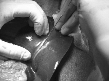
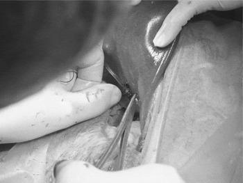
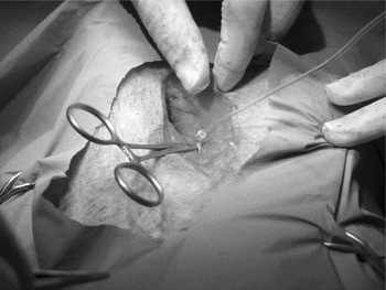
A paracostal incision (200–250 mm) was made through the skin, parallel to and as close as possible (20–30 mm) to the last rib on the right side of the gilt. Muscle layers and the peritoneum were cut with scissors. A sterile towel (400×400 mm) soaked in warm sterile saline (9 g/l) was used to hold the viscera away from the liver. The left lateral lobe of the liver was identified, grasped with gauze and lifted to locate the large PV branches in the visceral surface. To insert the catheter, a modification of the Seldinger technique (Seldinger Reference Seldinger1953) was followed. An 18G (1·2 mm×40 mm) needle was inserted through the liver tissue into the portal branch, towards the end of the visible part of the vein, and removed. Through the opening (Fig. 2), a wire guide (0·89 mm×700 mm; medCOMP, Harleysville, PA, USA) was used to insert the PV catheter, filled with physiological saline containing 250 international units (IU) heparin/ml (Fragmin, 5000 IU/0·2 ml; PHARMACIA Spain S.A., Barcelona, Spain), 60 mm upstream towards the origin of the PV ramification into the liver until the first ring of the catheter touched the liver wall. Subsequently, it was double sutured to the liver parenchyma (Fig. 3) using a non-absorbable suture (Silkam 0, 1/2 round needle; B. Braun VetCare S.A., Barcelona, Spain). Before the catheter insertion, the first suture was tied between the pre-placed catheter rings, which facilitate the first stitch and anchoring to the liver. The first stitch was c. 2 mm towards the catheter proximal tip with respect to the insertion point to keep the first ring against the liver wall, ensuring that the catheter is immobile with respect to the insertion point and its anchoring. A second suture was applied after the second ring. The suture puncture was always done avoiding the risk of damaging the portal branch underneath the catheter by making a wide loop in the liver parenchyma to go round the catheterized portal branch. Special care is needed to avoid parenchyma ripping when the suture knots are made; a good trick is to insert the tip of, for example, forceps through the suture loop before tightening the knot.
Wire guide inserted into a portal branch. It is inserted through the needle used for the puncture.

PV catheter gently double sutured to the liver parenchyma.

To verify proper catheter location, a sample of blood was collected simultaneously from the catheter and by puncturing the PV directly for determination of O2 saturation of haemoglobin (OSM 3 Hemoximeter; Radiometer Corporation, Copenhagen, Denmark). Portal branches are very close to the visceral surface of the liver, whereas branches of the hepatic vein are closer to the diaphragmatic side (Ortigues et al. Reference Ortigues, Durand and Lefaivre1994).
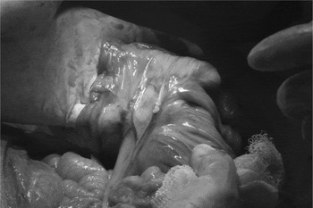
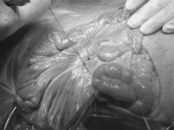
After the PV catheter was placed, the soaked towel remained within the abdominal cavity to ensure that the viscera remains covered. Through the same initial incision, the terminal ileum was located, a segment brought towards the incision and the IV catheter installed. After separating the surrounding connective tissue of a branch of the IV, a small puncture with an 18G needle was made and the catheter inserted 100 mm downstream into the vein (Fig. 4). The catheter was secured by two non-absorbable sutures (Silkam 1, 1/2 round needle; B. Braun VetCare S.A., Barcelona, Spain) around the vessel, one between the two rings and another behind the distal ring (Fig. 5).
Catheter inserted into a branch of the IV.

Ileal catheter secured by two non-absorbable sutures, one between the two rings and the other behind the distal ring.

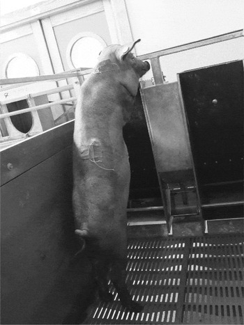
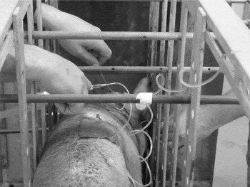
During the catheterization procedure, catheters were filled with sterile heparinized saline (250 IU/ml) and the distal ends were locked by two consecutive overhand knots using no more than 10 mm of the tips. Special care was always taken to clip the catheter with a haemostat when it was filled up and while heparinized saline was infused with the syringe to ensure that no blood entered the catheter, then the knot was fastened and the haemostat removed. The distal tips of the portal and ileal catheters were threaded through the eyelet of a regular needle (sack needle type, 150×2·5 mm) and exteriorized, by means of pliers, near the lateral process of the last thoracic vertebra situated between the caudal part of the diaphragm and the cranial side of the right kidney. Care must be taken to avoid occlusion of a loop of small intestine with the catheter tubing (van Leeuwen et al. Reference Van Leeuwen, Leuvenink, Haasbroek, Priem, Bosch and Van Kleef1995). The incision was closed in three separate layers and c. 1 ml of Penicillin G Procaine+Dihidrostreptomicine sulphate (Duphapen Strep; Fort Dodge Vet. S.A., Gerona, Spain) was poured onto each layer. The peritoneum was included with the first muscle layer. Before closing the first layer, viscera were rinsed with warm (39 °C) sterile saline until clean saline overflowed, and filled up again with sterile saline plus 2 ml of Duphapen Strep. A continuous absorbable suture (Polisorb 1, ½ round needle; Tyco Healthcare UK Ltd, Gosport, UK) was used for the first and second layers. A single interrupted suture, using non-absorbable suture (Silkam 0, 1/2 cutting needle; B. Braun VetCare S.A., Barcelona, Spain) was used to close the skin. A cohesive bandage (Askina Haft Color 0·08×20 m; B. Braun, Melsungen, Germany) patch (100×80 mm) was glued to the skin together with the catheters (Figs 6 and 7) c. 25 mm away from the externalization orifice using contact glue, in order to protect and keep the catheters coiled over the mid-line of the back. Special care was taken to ensure that the glued patch remained adhered to both the catheters and the skin throughout the experiments.
Pig following recovery from the surgery with the patches glued to the skin together with the catheters (carotid on the neck, portal and ileal on the back).

Pig in the cage during a sampling session, with the patches glued to the skin.

Carotid artery
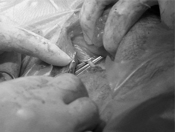
Pigs were moved to a dorsal recumbency position and an incision (c. 100 mm) was made along the jugular furrow. After incising the superficial cutaneous muscles, which is a relatively bloodless approach, the area between the sterno-cephalic and brachio-cephalic muscles was dissected. The CA was located within a sheath together with the vagus nerve and the internal jugular vein on the lateral aspects of the ventral surface of the cervical vertebrae. A blunt dissection over the dorsal surface of the sterno-cephalic muscle exposed the sheath containing the artery. A section of the artery wall (20–40 mm) was stripped free of associated connective tissue and then some droplets of lidocaine (Lincaina 2% S/N; B. Braun Medical, S.A., Barcelona, Spain) were dripped over the exposed carotid to relax it and avoid vasoconstriction. After 2 min, two small curved haemostats were situated underneath the artery, cranial and caudal to the catheter insertion site to lift the carotid towards the incision site, which stops the blood flow. A purse-string non-absorbable suture (Daclon Nylon 4/0, 3/8 cutting needle; smi, St. Vith, Belgium) was made on the CA surface (Fig. 8), making a small square (2 mm side) with four stitches going through the artery wall without perforating it. Then, the artery wall was cut with small iris scissors to create an opening into the artery in the middle of the purse-string. The catheter was inserted (and the cranial haemostat removed simultaneously) towards the aorta arch until the first catheter ring touched the carotid wall, and the purse-string suture was tied and knotted in its turn between the catheter rings (Fig. 9). A second suture was tied after the distal ring and sutured to surrounding tissues. The CA catheter was exteriorized through the neck wall down and caudal from the ear using a sack needle. Before continuously suturing the subcutaneous muscular layer (Polisorb 1, ½ round needle; Tyco Healthcare UK Ltd., Gosport, UK), c. 1 ml of Duphapen Strep was dripped into the wound and skin closed using a simple interrupted non-absorbable suture (Silkam 0, 1/2 cutting needle; B. Braun VetCare S.A., Barcelona, Spain).
Artery wall stripped free of associated connective tissue, lifted towards the incision site by two curved haemostats underneath the artery and making the first stitch of a purse-string non-absorbable suture on the artery surface.

Catheter inserted into the CA with the purse-string suture tied and knotted in its turn between the catheter rings.

A patch (100×80 mm) was glued to the skin together with the catheter (Figs 6 and 7) close to the exteriorization point, guided to the shoulder, fixed again with a second patch and kept coiled if necessary.
Post-surgery and catheter care
Following surgery, pigs were placed in metabolism stalls and heat lamps were used to provide additional warmth during recovery from the anaesthetic. The animals were fasted for 18 h with free access to water and given 0·25, 0·60 and 1·00 of their pre-surgery daily feed ration, respectively, on days 1, 2 and 3 after surgery. Feed and water intake and body temperature were monitored. The day after surgery, an analgesic and anti-spasm agent (Buscapina Compositum; Boehringer Ingelheim Spain S.A., Barcelona, Spain) was administered. The surgical wounds and catheter exteriorization sites were kept clean and sprayed with antibiotic (Veterin Tenicol; Lab. Intervet S.A., Salamanca, Spain) to prevent infection and aid healing. Animals were injected i.m. with a broad spectrum antibiotic (Duphapen Strep; Fort Dodge Vet. S.A., Gerona, Spain) the day of surgery and for 4 days (5–10 mg/kg BW per day). Stitches were removed 10 days after surgery.
Patency of catheters was checked weekly. Catheters were sprayed and cleaned with alcohol using sterile gauze, cut with scissors as close as possible to the proximal knot and a 5 ml syringe with a needle connected for flushing with sterile heparinized saline (250 IU/ml). Catheters were closed as described earlier by two knots. Each time the catheter was checked, c. 10 mm of catheter length was used.
Portal blood flow measurement
Portal blood flow was measured when the animals were fully recovered. Portal blood flow was calculated based on the Fick principle of arterio-venous concentration difference and the indicator infusion rate. It was determined by the indicator dilution method using para-aminohippuric acid (PAH) as described by Katz & Bergman (Reference Katz and Bergman1969). Forty-five minutes prior to blood sampling a 15 ml pulse dose of PAH (2% w/v; Sigma-Aldrich Quimica S.A., Madrid, Spain), prepared as described by Yen & Killefer (Reference Yen and Killefer1987), was infused into IV, followed by a continuous infusion of 16 mg/min using a syringe pump (Harvard Pump 33; Harvard Apparatus, Holliston, MA, USA). Apyrogenic filters (MILLEX GP, Syringe Driven Filter Unit, 0·22 μm; Millipore, Carrigtwohill, Ireland) were fitted to infusion syringes. Blood samples (4·5 ml) were taken simultaneously from CA and PV 5 min before and 0·5, 1, 1·5, 2, 2·5, 3, 3·5, 4, 5 and 6 h after feeding 0·25 of total daily ration, into a tube (Monovette VetMed LH; Sarstedt, Nümbrecht, Germany) with heparin-coated glass beads, centrifuged and plasma harvested and stored at −20 °C until analysis for PAH (Smith et al. Reference Smith, Finkelstein, Aliminosa, Crawford and Graber1945) using the gravimetric approach (Lobley et al. Reference Lobley, Connell, Lomax, Brown, Milne, Calder and Farningham1995). During serial sampling sessions (Fig. 7), catheters were set up with a needle with locker, and physiological saline containing 5 IU heparin/ml was used for flushing. After sampling, catheters were filled up with sterile heparinized saline (250 IU/ml) and closed as described earlier by two knots.
Statistical analysis
Repeated measures analyses were carried out using the mixed procedure of SAS (SAS Institute Inc., Cary, NC). The procedure included breed, time and breed×time. Blood sampling time was considered as the time variable. Animal was a random effect and was used as the experimental unit. Differences between means for the main effects were established using the HSD procedure of Tukey.
RESULTS
The diameter of PV catheter was increased (internal diameter from 1·02 to 1·27 and outer diameter from 1·78 to 2·29 mm) after initial surgeries when blood sampling was not successful 1 week after surgery, with no problems thereafter. The length (60 mm) of catheter inserted in the portal branch from the insertion point to the bifurcation of PV to the left and right lobes was selected after post-mortem observation of livers from Landrace and Iberian pigs of similar BW.
The time taken for the surgical procedure was variable, 3–4 h depending on anatomical individual variations or the amount of visceral fat.
Left lateral recumbency was utilized for the incision behind and parallel to the last rib on the right side of the pig and an indirect access to the PV through the left liver lobe, with good results.
Catheter blockage and a local infection surrounding the carotid insertion site was found 2 weeks after CA was ligated unilaterally, so a purse string suture allowing blood flow was used instead.
No specific differences were found between Landrace and Iberian pigs except that the Iberian pigs showed greater subcutaneous and intra-abdominal fat deposits. Special care was needed when abdominal catheters were exteriorized through the body wall in Iberian pigs, as it is necessary to get very close to the cranial side of the right kidney and care must be taken not to puncture the caudal part of diaphragm, since the space between both is very narrow.
After surgery, pigs did not show any symptom of discomfort, fever or appetite deprivation. Full ration was offered 3 days after surgery.
The catheters were manipulated and flushed once per week to avoid, as much as possible, contamination that could potentially block the catheters. As mentioned earlier, each day that a catheter was used, c. 10 mm was wasted. During the 8 weeks that the catheters’ patency was checked, there was no difficulty with blood withdrawal when catheters were flushed with heparinized saline solution before blood sampling, which indicated that no blockage caused by thrombi formation occurred.
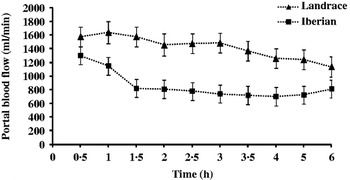
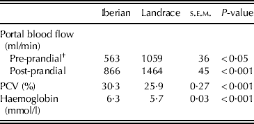
Both pre- and post-prandial portal blood flow determined by indicator dilution (PAH) was lower (41%; P<0·05) in Iberian compared to Landrace gilts (Table 1 and Fig. 10).
Post-prandial portal blood flow of Iberian and Landrace pigs fed 0·25 of their daily ration.

Pre- and post-prandial portal blood flow, packed cell volume (PCV) and haemoglobin concentration in Iberian and Landrace pigs (n=3; values are means±s.e.m.* for 10 post-prandial measurements)
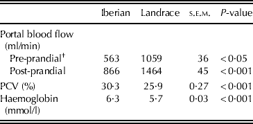
* s.e.m., standard error of the mean.
† Values are mean for one pre-prandial measurement.
DISCUSSION
The dimensions and designs of catheters used throughout the literature are variable. The same diameter as that in the present work was used for PV catheter by van der Meulen et al. (Reference Van Der Meulen, Bakker, Bakker, De Visser, Jongbloed and Everts1997) and Jørgensen et al. (Reference Jørgensen, Serena, Theil and Engberg2010), but this was broader than the size used by van Leeuwen et al. (Reference Yen and Killefer1995) and Hooda et al. (Reference Hooda, Matte, Wilkinson and Zijlstra2009). Catheter rings attached to catheters have been used previously in pigs. Van Leeuwen et al. (Reference Van Leeuwen, Leuvenink, Haasbroek, Priem, Bosch and Van Kleef1995) used a rosette sheeting, glued to the catheter at an angle of 45° to fix it to the vessel. Yen & Killefer (Reference Yen and Killefer1987) used a piece of nylon mesh attached to the catheter and sutured to the adjacent connective tissues of the vessel for anchoring. An intermediate design was described by Jørgensen et al. (Reference Jørgensen, Serena, Theil and Engberg2010), consisting of a cuff of silicone tubing glued to a piece of polyvinyl net which was placed around the PV catheter and polyvinyl net sutured to the hepatic tissue. Hooda et al. (Reference Hooda, Matte, Wilkinson and Zijlstra2009) used a PV catheter with two rings, but the first catheter ring was inserted into the PV and then a purse-string suture was closed; the catheter was also secured to the sheath using a suture around the second ring. Moreover, Hooda et al. (Reference Hooda, Matte, Wilkinson and Zijlstra2009) opened small holes in the sides of the catheter between the tip and first ring to have an alternate route of blood collection. Van Leeuwen et al. (Reference Van Leeuwen, Leuvenink, Haasbroek, Priem, Bosch and Van Kleef1995) observed that keeping the catheter as tangential as possible to the vessel avoided blockage due to bending of the tube. For this reason, in the present study the first (proximal) ring was cut with an angle of 30° to facilitate its tangential position to the vessel.
Blockage and loss of patency is always a problem in this kind of study. During post-mortem examinations of pigs, Yen & Killefer (Reference Yen and Killefer1987) revealed that lack of patency of arterial catheters was due to the formation of thrombi and fibrous tissue engulfing the tips, whereas complete enclosure of the tips by fibrous tissue caused the blockage of PV catheter. Jackson et al. (Reference Jackson, Cook and Gill1972) ascribed catheter blockage to the formation of small valve-like thrombi at the tip.
The duration of the surgical procedure was within the range of similar procedures in the literature (Hecker Reference Hecker1974; Hooda et al. Reference Hooda, Matte, Wilkinson and Zijlstra2009).
Regarding position of the animal for PDV surgery, dorsal recumbency for mid-line incision and left lateral recumbency for incision behind and parallel to the last rib on the right side of the pig have been utilized. Most published papers describe direct access to the PV (Rerat et al. Reference Rerat, Vaugelade, Villiers, Low and Partridge1980; Yen & Killefer Reference Yen and Killefer1987) and only a few report indirect access through the liver, by an incision in the left medial (Olesen et al. Reference Olesen, Sjøntoft and Tronier1989) or in the left liver lobe (Paschen & Müller Reference Paschen and Müller1986), as in the present work. Paschen & Müller (Reference Paschen and Müller1986) ligated, cut and stretched, by insertion of the tip of the scissors, a small tributary of the PV in the caudal angle of the left liver lobe and a Swan–Ganz catheter was passed into the PV, and secured to the surface of the liver by stitches parallel to the vessels. Jørgensen et al. (Reference Jørgensen, Serena, Theil and Engberg2010) described a similar approach where c. 30–40 mm from the top of a liver lobe was cut-off transversally, a portal vessel identified and catheter inserted until it appeared in the PV as verified by palpation.
It is well known that it is possible to ligate the CA on either side unilaterally without circulatory complications (Swindle Reference Swindle2009). It has been pointed out (Rerat et al. Reference Rerat, Vaugelade, Villiers, Low and Partridge1980; Yen & Killefer Reference Yen and Killefer1987) that occlusion of one of two CA should not result in any physiological detriment because of the anastomosis with the vertebral artery in the submaxillary zone. Nevertheless, lack of patency after 2 weeks has been found in previous studies, so in the current study the artery was prevented from occlusion using a purse-string suture which does not stop the blood flow, to lessen the risk of local infections with optimum results.
The criteria for success in the current paper were rapid restoration of normal feed intake, absence of infection at the incision site and patency of catheters. After surgery, pigs showed good condition and appetite. It is crucial to ensure that sterile and aseptic techniques are practised throughout all phases of the catheterization procedure for fast and satisfactory recovery. The use of a sterile warm towel to ‘pack’ the viscera away from the liver, viscera rinsing with warm (39 °C) sterile saline and filling the abdominal cavity up with sterile saline plus a broad spectrum antibiotic before closing it may have prevented cavity infection and accelerated the healing process. In addition, the indirect access using needle puncture through the pig liver parenchyma implied a minimal wound compared with incision or cutting of liver lobe, which produce temporary hepatic damage. For example, Jørgensen et al. (Reference Jørgensen, Serena, Theil and Engberg2010) reported 10–14 days to recover and regain full appetite after surgery accessing the PV by cutting the liver lobe.
The majority of the literature describes daily catheter flushing (e.g. Yen & Killefer Reference Yen and Killefer1987; Hooda et al. Reference Hooda, Matte, Wilkinson and Zijlstra2009) instead of once per week. Indeed, human studies (Smith et al. Reference Smith, Dawson, Hennessey and Andrew1991; Kelly et al. Reference Kelly, Dumenko, Mcgregor and Mchutchion1992) have demonstrated no significant difference in central venous catheters complications after switching from flushing daily or twice per day to a weekly flushing regimen. About 10 mm of the catheter was wasted each day when used, with the compensation that when no serial sampling was done there was no bleeding or clotting in the catheter because of locker loss.
Catheter patency is variable, ranging from 18 days (Rerat et al. Reference Rerat, Vaugelade, Villiers, Low and Partridge1980) to 6–12 weeks (Yen & Killefer Reference Yen and Killefer1987; van Leeuwen et al. Reference Van Leeuwen, Leuvenink, Haasbroek, Priem, Bosch and Van Kleef1995; Hooda et al. Reference Hooda, Matte, Wilkinson and Zijlstra2009). In the present study, the catheters were checked for 8 weeks, a period sufficient to perform nutrient kinetics studies of PDV, especially taking into account that the gilts were growing rapidly.
The indicator dilution (PAH; Katz & Bergman Reference Katz and Bergman1969) and electronic devices surrounding the vessels are the most frequent methods used to measure blood flow. Electronic devices are electromagnetic (Rerat et al. Reference Rerat, Vaugelade, Villiers, Low and Partridge1980) or ultrasonic (Ellis et al. Reference Ellis, Roberts, Low and Morgan1995) flow probes attached as a cuff around the blood vessel. They only allowed reliable measurements for 2–4 and 6 weeks after surgery, respectively, because the formation of fibrous tissues around the probe disturbed the signal and as a consequence the flow rate was underestimated. Nevertheless, Hooda et al. (Reference Hooda, Matte, Wilkinson and Zijlstra2009) reported similar portal blood flow measurements using an ultrasonic probe after 52 days compared with 10 days post-surgery. In the present work, the PAH dilution method was chosen. It has been widely and successfully utilized in pigs since Yen & Killefer (Reference Yen and Killefer1987) described their catheterization technique.
Organ and total body blood flow are very variable and depend on the cardiac output. As Ten Have et al. (Reference Ten Have, Bost, Suyk-Wierts, Van Den Bogaard and Deutz1996) pointed out, it is important that the experimental conditions are as constant as possible. Thus, in humans a standing position increases the cardiac output by 20% and Brown et al. (Reference Brown, Halliwell, Qamar, Read, Evans and Wells1989) showed that blood flow rate was significantly reduced in upright compared with supine position. Ellis et al. (Reference Ellis, Roberts, Low and Morgan1995) observed that the behaviour of the pig on the day of sampling affected blood flow. Therefore, care is needed when comparing literature data from different experiments and conditions. Overall, the portal blood flow of Landrace pigs (Table 1 and Fig. 10) was in the lower range of data found in the literature (Yen & Killefer Reference Yen and Killefer1987; Yen et al. Reference Yen, Varel and Nienaber2004; Hooda et al. Reference Hooda, Matte, Wilkinson and Zijlstra2009), probably as a result of the low feeding intake used (0·25 of the total daily ration for a 6 h sampling period, i.e. 0·25 of day length). The lower portal blood flow of Iberian compared with Landrace pigs repeatedly found in previous studies (Fernández-Fígares et al. Reference Fernández-Fígares, Rodríguez-Lopez, González-Valero, Lachica and Crovetto2010; Rodríguez-López et al. Reference Rodríguez-López, Lachica, González-Valero and Fernández-Fígares2010) may partly explain the lower ME requirement for maintenance of Iberian compared with modern pigs (Nieto et al. Reference Nieto, Miranda, García and Aguilera2002; Barea et al. Reference Barea, Nieto and Aguilera2007).
The present surgical procedure, together with catheter design and care, have been used for comparative studies of PDV energy expenditure (Fernández-Fígares et al. Reference Fernández-Fígares, Rodríguez-Lopez, González-Valero, Lachica and Crovetto2010; Rodríguez-López et al. Reference Rodríguez-López, Lachica, González-Valero and Fernández-Fígares2010) and portal appearance of lysine, threonine and methionine (González-Valero et al. Reference González-Valero, Rodríguez-López, Lachica and Fernández-Fígares2012) in Iberian and Landrace pigs fed diets with different CP content, and to measure glucose absorption and energy expenditure in Iberian pigs fed acorns (González-Valero et al. Reference González-Valero, Rodríguez-López, Lachica, Fernández-Fígares and de Pedro2010; Lachica et al. Reference Lachica, Rodríguez-Lopez, González-Valero, Fernández-Fígares and Crovetto2010).
CONCLUSIONS
Variations in surgical procedure, and catheter design and care to study PDV nutrient metabolism in pigs are described. No surgical problems or differences associated with breed were found during and after surgery. Post surgery recovery was very fast. Catheters could successfully be used for several weeks in both breeds.
The methodology followed is suitable for pigs, permitting evaluation of the kinetics of nutrients across the PDV. It could be used to explain differences in digestive and absorptive capacities between native and modern breeds.
J.M. Rodríguez-López and L. González-Valero are grateful to the Ministry of Science and Education of Spain for the award of a PhD student scholarship. This research was supported by grants no. AGL 2006-05937/GAN and AGL 2009-08916 from the Spanish Ministry of Science and Education.