Introduction
Multicomponent reactions (MCRs) include a combination of three or more constituents in a single vessel to fabricate an anticipant product with properties of all the reactant components, which avoided the intricacy separation and purification of complex to attain the purpose of high effect and low cost [Reference Nagarajaiah, Mukhopadhyay and Moorthy1, Reference Rotstein, Zaretsky, Rai and Yudin2, Reference Tao, Zhao, Yang, Wei and Wu3, Reference Wu, Yang and Tao4]. Moreover, MCRs have been a fascination as versatile tools for chemists to construct heterocyclic compounds, which would pave a straightforward way to fabricate novel libraries of polymers and might broad study of MCRs in interdisciplinary fields [Reference Xue, Zhao, Wu, Wang, Yang, Wei, Wang and Tao5]. Currently, owing to the atoms economy, excellent modular, efficient, and environmentally friendly properties [Reference Dong, Liu, Jiang, Huang, Wan, Wen, Tian, Dai, Zhang and Wei6, Reference Jiang, Liu, Liu, Tian, Huang, Huang, Wen, Cao, Zhang and Wei7, Reference Zhu, Yang, Zhao, Fu, Tao and Wei8], a variety of MCRs have been reported such as Mannich reaction, Passerini reaction, Hantzsch reaction, MALI reaction, Biginelli reaction, and Ugi reaction [Reference Mao, Liu, Wu, Wei, Gou, Wang and Tao9]. As end-products of Biginelli reaction, the 3,4-dihydropyrimidin-2(1H)-one (DHPM) derivatives were considered as a well-known class of cardiovascular drugs serving as calcium channel modulators and confirmed to be the central structure of many medicines such as the barrier reagent of Ca2+ ion avenue [Reference Wang, Fu, Wei and Tao10, Reference Kappe11]. Hence, they are very important in the pharmacological applications such as calcium channel modulators, mitotic kinesin inhibitors, antimalarial, antitubercular, and A2B adenosine receptor antagonists [Reference Crespo, Maatougui, Biagini, Azuaje, Coelho, Brea, Loza, Cadavid, Garcia-Mera, Gutierrez-de-Teran and Sotelo12, Reference Kaur, Machado, Kock, Smith, Chibale, Prudencio and Singh13, Reference Joshi, Barchha, Khedkar, Pissurlenkar, Sarkar, Sarkar, Joshi, Joshi, Shah and Coutinho14]. Therefore, the number of publications and patents about the synthesis of DHPMs derivatives reaction is increasing every year [Reference Simurova and Maiboroda15]. Many researches indicated that some heterocycles from Biginelli reaction could restrain the combination of HIV viruses and CD4 cells, which would possibly be a novel drug for curing AIDS [Reference Akhaja and Raval16]. Recently, due to the high efficiency and yield, tolerance to the extensive reactions conditions, and varied functional groups, Biginelli reaction has been a very powerful tool in polymer chemistry. A thiourea-contained polycondensate with highly reactive activity was easily synthesized via the Biginelli polycondensation, which might be a versatile platform to fabricate novel functional polymers [Reference Zhao, Wu, Zhang, Wang, Yang, Zhang, Ren, Fu, Wei, Wang, Wang and Tao17]. Inspiration from nature’s two-stage strategy for the efficient synthesis of numerous proteins using limited amino acids, a two-stage fabrication method was successfully developed via the combination of ultra-fast RAFT polymerization and Biginelli reaction, which opened a new way for the effective preparation and characterization of new series of polymers with abundant diversity and functions [Reference Ren, Yang, Zhao, Zhang, Wang, Wei and Tao18].
Living polymerization has been a favorite tool to fabricate functional copolymers with well-defined and predesigned structures [Reference He, Jiang, Zhang, Cheng and Zhu19]. Among the normal living polymerization methods of anionic polymerization, cation polymerization, reversible addition-fragmentation chain transfer (RAFT) polymerization, and atom transfer radical polymerization (ATRP), RAFT is versatile technique to fabricate functional polymers due to its excellent tolerance to many functional monomers and solvents [Reference Huang, Hou and Cao20]. Amphiphilic PPEGMA-b-PMMA block copolymer nanoparticles were successfully prepared by RAFT polymerization-induced self-assembly (PISA), the particle diameter of which gradually increased with the target DP of the PMMA increasing [Reference Peng, Tian, Zhang, Cheng and Zhu21]. Otherwise, linking multiblock copolymers to solid substrate’s surfaces was very important for both fundamental studies and applications. One combination of RAFT polymerization and click chemistry was employed to synthesize highly pure multiblock copolymers tethered to silica particles, and the one-pot strategy seems more attracting than the incremental route [Reference Huang, Hou and Cao20]. By combination of tandem RAFT polymerization, azide–alkyne cycloaddition reaction, and a subsequent de-grafting reaction, a series of silica-polymer hybrids with highly pure block copolymers was described in detail, which played an important role in material properties [Reference Zhao, Zhang, Zhang and Zhao22]. Moreover, RAFT was also actively applied for the construction of fluorescence-active functional polymers [Reference Jiang, Liu, Huang, Huang, Wan, Wen, Tian, Cao, Zhang and Wei23]. A self-healable hydrogel with fluorescence activity was prepared by ring opening polymerization (ROP) and RAFT via incorporation of ionic block copolymers (BCPs) with fluorescence response, which may be a potential for smart materials in the applications of tissue engineering and sensing fields in the near future [Reference Banerjee, Hoskins, Swift, Rimmer and Singha24]. By photoinitiating RAFT polymerization, TPE-poly(St-PEGMA) copolymers with aggregation-induced emission (AIE)-activity were successfully fabricated, which showed great potential for biomedical applications due to their optical and biocompatibility [Reference Zeng, Liu, Jiang, Huang, Huang, Wan, Dai, Wen, Zhang and Wei25]. As an attracting candidate for monitoring dynamic processes and bioimaging therapies, dual-functional copolymeric nanoparticles (NPs) with near-infrared (NIR) fluorescence imaging and prodrug were prepared in situ RAFT-mediated aqueous dispersion polymerization [Reference Tian, Niu, Wei, Xu, Zhang, Cheng and Zhu26].
The one-pot MCRs synthesis depended on the most economical step to obtain the products. The idea of constructing one-pot polymerization system was attracting wide attention in the area of polymer chemistry. The substrates of Biginelli reaction have cheap price, low toxicity, and smell with high safety, the DHPMs products of which have extensive biology and pharmacology activity. In this contribution, novel AIE-based copolymers were successfully prepared via one-pot smart combination of RAFT polymerization and in situ Biginelli reaction for the first time. First, the AEMA functional monomers participated in RAFT polymerization with a hydrophilic PEGMA monomer and resulted in functional copolymers, which would in situ occurred Biginelli reaction with other two components of TPB dye and urea, and produced a new fluorescent copolymer with DHPM ring. The as-prepared PEG-PTE would tend to self-assemble into fluorescent organic nanoparticles (FONs) in aqueous solution and exhibited promising biology imaging applications due to high water dispersibility, good fluorescence, and excellent biocompatibility. Furthermore, in order to fully evaluate the combination of RAFT polymerization and Biginelli reaction, the other effects of the reactants feeding ratio and two-step method on the copolymers structure were also investigated in detail.
Results and discussion
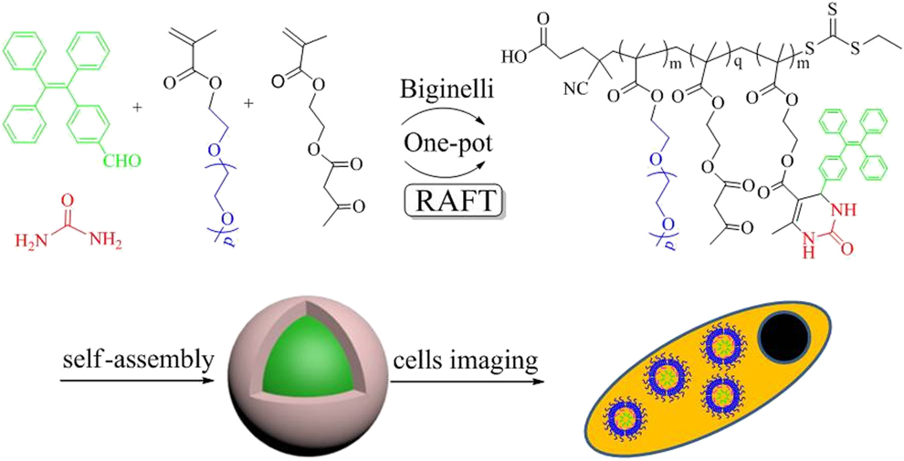
In this paper, the fabrication of amphiphilic fluorescent PEG-PTE copolymers via “one-pot” combination of RAFT polymerization and multicomponent Biginelli reaction is demonstrated in Scheme 1. Here, taking TPB dye and urea as the other two model components of Biginelli reaction, AEMA was used as the vinyl monomer for the 1,3-dione functional group source. The multicomponent Biginelli reaction would bring a pyrimidin ring, which was linked to AEMA monomer, producing another new monomer PTE-MA. The as-prepared hydrophobic PTE-MA monomer would participate in RAFT polymerization with hydrophilic PEGMA monomer to obtain amphiphilic fluorescent copolymers PEG-PTE with transformed DHPM rings. In aqueous solution, the amphiphilic PEG-PTE copolymers would tend to self-assemble into NPs and internalized by cells.
One-pot synthesis process of PEG-PTE copolymers based on RAFT polymerization and Biginelli reaction and their cell imaging application.
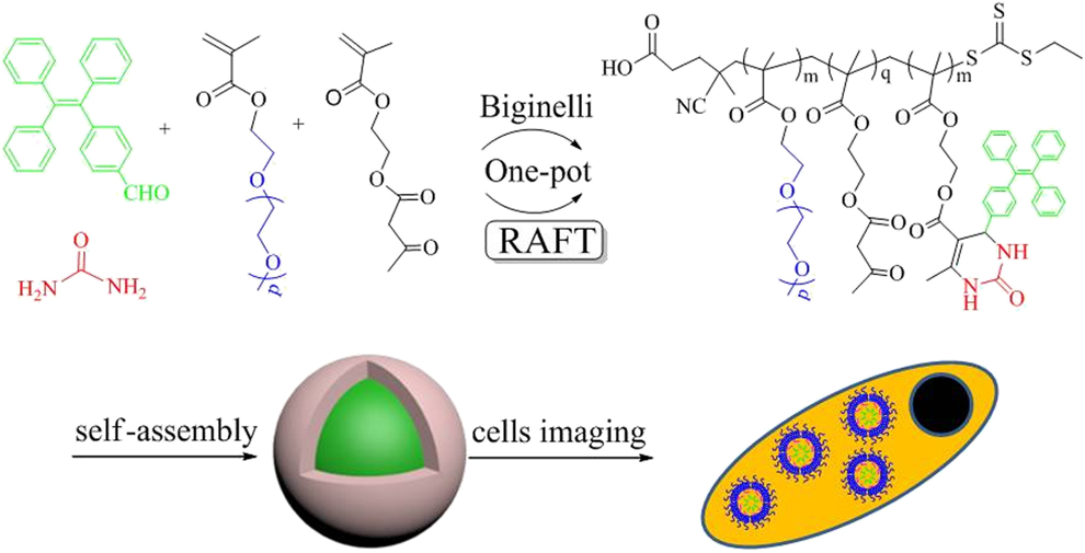
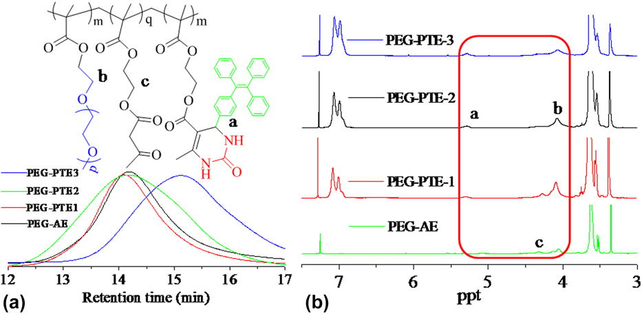
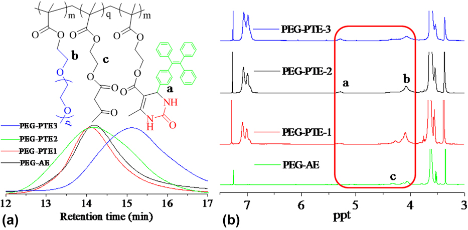
Figure 1 described the gel-permeation chromatograph (GPC) with PDIs and 1H NMR curves of the PEG-AE and PEG-PTE copolymers with the various molar fractions of AEMA and PEGMA. The GPC analysis showed that the RAFT procedure was well controlled, and PDIs of the copolymers were about 1.30, which indicated that the process had the characteristic of living polymerization. As compared with PEG-AE copolymers, the weight average molecular (Mn) of PEG-PTE1 copolymers increased to 59,300 from 52,800 of PEG-AE copolymers; moreover, the 1H NMR spectrum of PEG-PTE1 obviously presented the characteristic CH proton peak of the pyrimidin-2-one ring at 5.30 ppm with the aromatic hydrogen peaks at the range of 7.00–7.06 ppm [Reference Wang, Yu, Li, Yang, Zhao, Liu, Wei, Wang and Tao27], implying that the TPB dye was successfully incorporated into the PEG-AE intermediate chains via Biginelli reaction. The peaks at 4.05 ppm and 4.26 ppm were, respectively, assigned to the ester groups of poly-(PEG) and AEMA. Referring to the integral ratio of the peaks at 5.30, 4.05, and 4.26 ppm, the molar fraction of TPB and AEMA in PEG-PTE1 copolymers was about 14.2 and 19.0%. Instead of the two-step method by one-pot method, the Mn of PEG-PTE2 decreased to 52,300 from 59,300 of PEG-PTE1, implying that the Biginelli reaction possibly affected the chain propagation rate to a certain degree; however, the peak at 5.30 ppm in PEG-PTE2 copolymers was more obvious and the peak at 4.26 ppm in PEG-PTE2 copolymers obviously decreased, indicating that the Biginelli reaction of one-pot method was more complete than that of two-step method. On the basis of the above computational method of PEG-PTE1, the molar fraction of TPB dye and AEMA in the PEG-PTE2 was, respectively, about 22.5 and 10.5%. In order to further investigate the influence of various reaction conditions on the copolymers structure, when the feed ratio of TPB increased to 48.6% of PEG-PTE3 copolymers from 33.5% of PEG-PTE2 copolymers, the corresponding Mn of PEG-PTE3 decreased to 32,800 from 52,300 of PEG-PTE2 and the molar fraction of TPB dye increased to about 32.6% in PEG-PTE3 copolymers.
(a) The GPC traces (DMF) (PEG-AE, Mn = 52,800, PDI = 1.23; PEG-PTE1, Mn = 59,300, PDI = 1.24; PEG-PTE2, Mn = 52,300, PDI = 1.35; PEG-PTE3, Mn = 32,800, PDI = 1.24) and (b) 1H NMR spectrum (CDCl3) of the obtained PEG-AE and fluorescent copolymers PEG-PTE.
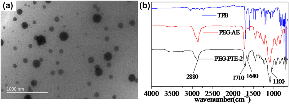
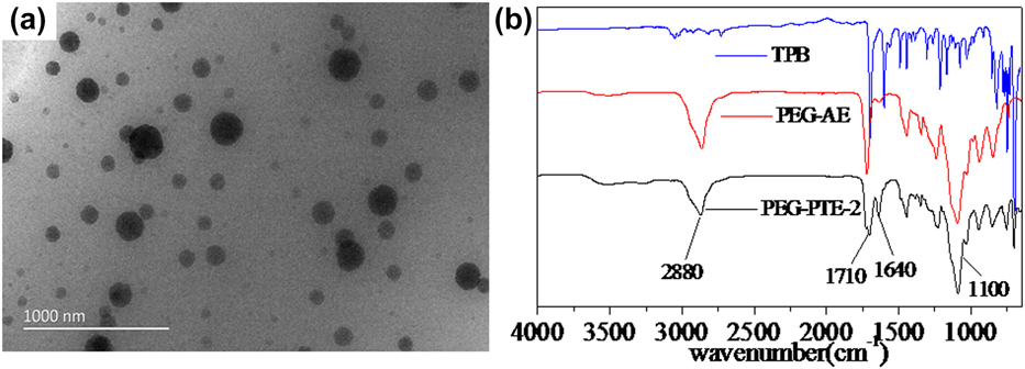
To investigate the amphiphilic property and effect of self-assembly of PEG-PTE copolymers in aqueous solution, Fig. 2(a) gave typical TEM images of microphase separated morphologies of PEG-PTE2 FONs. From the TEM images, the dark regions represented PEG-PTE2 subphase [Reference Wei, He and Wang28] and the size was 100–180 nm with the spherical morphology, indicating that the effective self-assembly owing to the successful incorporation of PEGMA monomers and TPB dye into the PEG-PTE2 copolymers via RAFT and Biginelli reaction. Moreover, successful fabrication of PEG-PTE2 copolymers is further confirmed by FT-IR spectra as shown in Fig. 2(b). For the spectrum of PEG-PTE2 copolymers, the characteristic peak at 1100 cm−1 was assigned to C–O stretching vibration in PEGMA, and the stretching vibration peaks C=O in PEGMA and AEMA obviously presented at 1710 cm−1 with C=C peaks of TPB at 1640 cm−1. Another peak at 2880 cm−1 should attribute to the stretching vibration of characteristic –CH2- and –CH3 in PEGMA and AEMA. From the above analysis, the anticipant copolymer structure was successfully prepared by the one-pot combination of RAFT polymerization and Biginelli reaction.
(a) TEM image of PEG-PTE2 FONs in water solution, scale bar = 1000 nm; (b) FT-IR spectra of TPB dye, PEG-AE, and PEG- PTE2 copolymers.
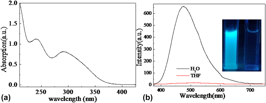
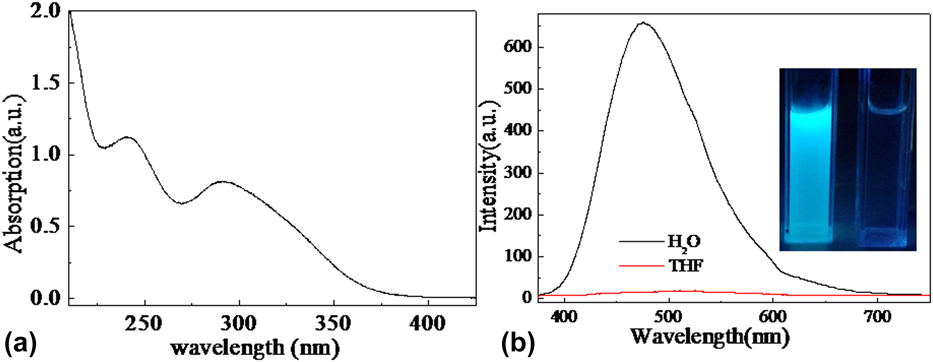
In order to continually evaluate dispersibility and optical characteristics of PEG-PTE2 in water solution, their UV and fluorescence curves were described in Figs. 3(a) and 3(b). The UV curve presented obvious absorption in the ultraviolet region with two major absorption peaks at 240 and 290 nm, which should assign to the π → π* and n → π* transition of the TPB polycyclic aromatic and pyrimidin rings [Reference He, He, Liu, Gu, Wei and Wang29, Reference Wei, Wang and He30, Reference Wei, Wang and He31]. Moreover, PEG-PTE2 copolymers had very little absorption when the incident wavelength was more than 400 nm, implying that absorbance of PEG-PTE2 copolymers was not nearly affected by the Mie effect or light scattering of the NP suspensions due to their excellent dispersibility in the aqueous solution [Reference Zhang, Chi, Zhang, Li, Xu, Li, Liu, Zhang and Xu32]. The PEG-PTE2 copolymers are expected to present excellent fluorescence owing to the incorporation of TPB dye. As shown in Fig. 3(b), the fluorescence spectrum of PEG-PTE2 FONs in aqueous solution had the evident fluorescence with the maximum emission peak at 480 nm; however, in the THF solution, the fluorescence was not almost be observed, indicating the clear AIE feature. When PEG-PTE2 molecules in the ground state S0 were excited by external ultraviolet light λ1, they would absorb the corresponding radiation energy λ1 and were excited to higher excitation states S1 and S2. The PEG-PTE2 molecules in the excitation states S1 and S2 were astable, and soon afterward, they would return to S0. When PEG-PTE2 were in THF solution, the relaxation energy from S1 and S2 to S0 would be consumed by molecules rotation or thermal energy owing to the active intramolecular rotation. While in water solution with the FONs aggregation forms, this rotation was restricted due to the intramolecular interactions such as C–H⋯π and C–H⋯H–C and the nonradiative path was blocked; thus, the radiation decay is activated with emitting radiation λ2 [Reference Hong, Lam and Tang33, Reference Tong, Dong, Hong, Häussler, Lam, Sung, Yu, Sun, Williams, Kwok and Tang34].
(a) UV-Vis curve of PEG-PTE2 copolymers in water solution; (b) fluorescence emission spectra of PEG-PTE2 FONs, inset is the fluorescent image of PEG-PTE2 FONs at 365 nm UV light [left bottle (in water), right bottle (in THF)].
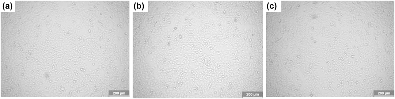
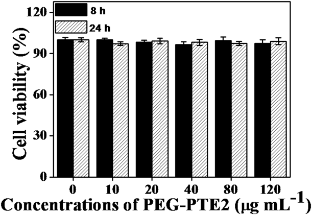
It was basic and necessary for FONs to have great biocompatibility for their promising biomedical application [Reference Zhang, Yin, Peng, Hu, Zhu, Li, Fan and Huang35, Reference Zhang, Wang, Zhu, Liu, Ji, Feng, Tao and Wei36, Reference Zhang, Zhang, Wang, Liu, Tao and Wei37, Reference Zhang, Hu, Li, Tao and Wei38]. As shown in Figs. 4(a)–4(c), optical microscopy images indicated that HepG2 cells could keep their normal morphology after they were incubated with PEG-PTE2 FONs. Even when the concentration of PEG-PTE2 FONs increased to 80 mg/mL, the morphology did not obviously change. In order to evaluate the biocompatibility of obtained PEG-PTE2 FONs, their cytocompatibility for HepG2 cells was investigated by the CCK-8 assay as shown in Fig. 5 through the 450 nm absorbance value of formazan dye taking 620 nm as the reference value [Reference Zhang, Qi, Wang, Feng, Ji, Tao, Li and Wei39, Reference Zhang, Hui, Yang, Yang, Fan, Liu, Tao and Wei40]. After incubating with 10–120 μg/mL PEG-PTE2 FONs for 8 and 24 h, no obvious decrease of cells viability could be observed; moreover, it was still more than 95% even when the concentration of PEG-PTE2 FONs increased to 120 μg/mL. From the above analysis, the as-prepared PEG-PTE2 FONs exhibit great cytocompatibility with HepG2 cells.
Biocompatibility evaluations of PEG-PTE2 FONs. (a–c) Optical microscopy images of HepG2 cells incubated with different concentrations of PEG-PTE2 FONs for 24 h, (a) control cells, (b) 20 μg/mL, and (c) 80 μg/mL.

Cell viability of PEG-PTE2 FONs with HepG2 cells.

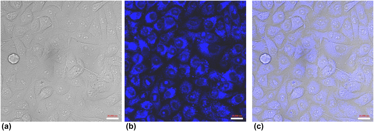
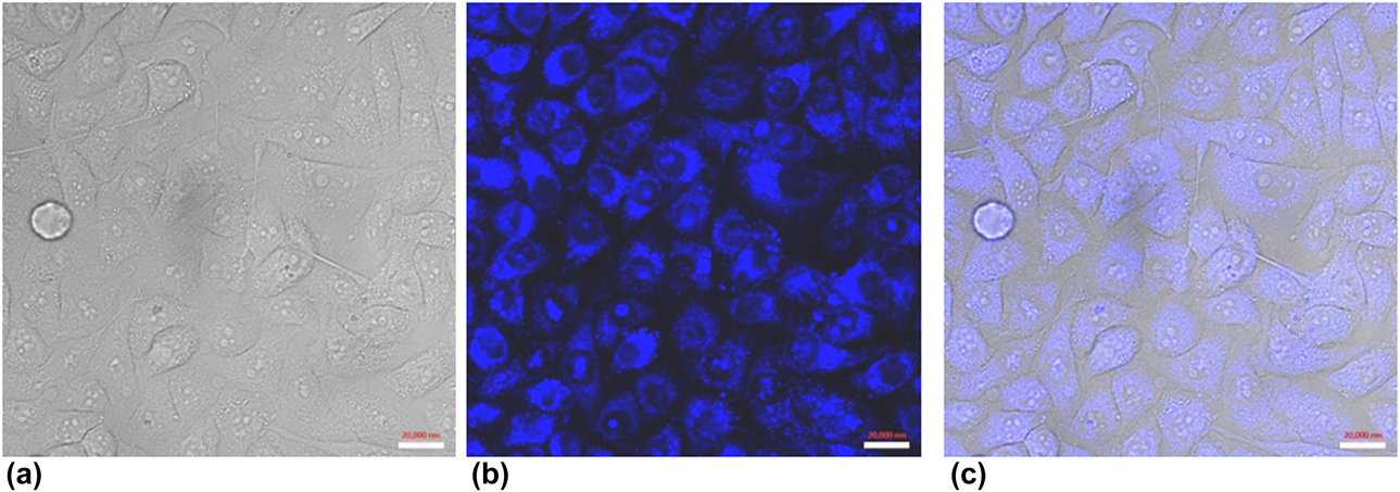
In consideration of the uniform spherical morphology, good fluorescence, great biocompatibility, and high dispersibility of PEG-PTE2 FONs, their effect on biological imaging and cell uptake behavior was further investigated by CLSM referred to the reported literature [Reference Wan, Liu, Xu, Huang, Mao, Zeng, Deng, Zhang and Wei41, Reference Wan, Wang, He, Liu, Zeng, Huang, Deng, Zhang and Wei42]. As shown in Figs. 6(a)–6(c), the black regions should be the location of cell nucleus, which were surrounded by the areas with strong fluorescence. These results indicated that PEG-PTE2 FONs could enter into cells by the endocytosis of the HepG2 cells and mainly distributed in the cytoplasma owing to the size difference of PEG-PTE2 FONs and cell nucleus [Reference Li, Zhang, Zhang, Yang, Yang and Wei43]. DHPMs derivatives from Biginelli reaction have the excellent pharmacology activity and biological reaction activity, which was the important intermediates of hypotensor, anticarcinogen, antibacterial drug, antagonist of calcium ion, and natural base [Reference Deshmukh, Salunkhe, Patil and Anbhule44, Reference Chitra, Devanathan and Pandiarajan45]. Moreover, due to the high efficiency, atom economy, and reliability under broad conditions [Reference Ren, Yang, Zhao, Zhang, Wang, Wei and Tao18], it was very convenient and effective for Biginelli reaction to incorporate some multicomponents such as imaging agents, drugs, prodrug monomers, and targeting agents into functional polymers with a combination of RAFT polymerization. Hence, one-pot combination of RAFT polymerization and Biginelli reaction provided an attractive avenue for multifunctional imaging and theranostic platforms.
CLSM images of HepG2 cells incubated with 40 μg/mL of PEG-PTE2 FONs: (a) bright field, (b) excited with a 405 nm laser, and (c) merged image of (a) and (b). Scale bar = 20 μm.
Conclusions
In summary, we have developed a novel strategy for fabrication of AIE-active PEG-PTE FONs via one-pot smart combination of RAFT polymerization and MCRs Biginelli reaction for the first time. The product structure from one-pot method was similar with that from two-step method but has obviously higher incorporation of TPB dye in PEG-PTE copolymers. Owing to high water dispersibility, good fluorescence, excellent cell dyeing behavior, remarkable photostability, and excellent biocompatibility, the as-prepared PEG-PTE copolymers were promising for biological imaging application. As compared with the strategy of AIE-active FONs on the basis of polymerizable or functional AIE monomer, this novel contribution has many advantages. First, it avoided the complex purification process of intermediates for the fabrication of AIE-active copolymers. Second, the chemical structure together with the physicochemical properties of the fluorescence copolymers can be easily adjusted via the RAFT polymerization and Biginelli reaction. More importantly, this work provides novel ideas to fabricate some clean and multifunctional FONs for cell imaging application through incorporating some various functional components such as drugs, imaging agents, and targeting agents by Biginelli reaction.
Experimental procedure
Materials and characterization
2-(Acetoacetoxy)ethyl methacrylate (AEMA, Aladdin, 95%), urea (Aladdin, AR), anhydrous magnesium chloride (Aladdin, 99%), and poly(ethylene glycol) monomethacrylate (PEGMA, Mn = 475, J & K Chemical, AR) were all used as purchased in China. Azodiisobutyronitrile (AIBN, Aladdin, 99%) was recrystallized twice from acetone before use. 4-(1,2,2-triphenylvinyl)benzaldehyde (TPB) dye and the chain transfer agent (CTA) of 4-cyano-4-(ethylthiocarbonothioylthio)pentanoic acid were fabricated according to the previous literature methods [Reference Zhang, Chi, Xu, Chen, Zhou, Zhang, Liu and Xu46, Reference Zhang, Liu, Yang, Zhang and Wei47, Reference Tao, Liu and Davis48, Reference Huang, Zhang, Zhang, Yang, Zhang, Wang, Yuan, Tao and Wei49].
The molecular weights and molecular weight distributions of PEG-AE and PEG-PTE copolymers were acquired from a Shimadzu LC-20AD (Japan) GPC system equipped with a Shimadzu RID-10A a refractive index (RI) detector, which was carried out at 35 °C taking N,N-dimethyl formamide (DMF) as the eluent with 1.0 mL/min flow rate and was calibrated by standard polystyrene. 1H NMR spectra of PEG-AE and PEG-PTE copolymers were recorded on a JEOL JNM-ECA 400 (400 MHz) spectrometer by using CDCl3 as the solvent and tetramethylsilane as an internal standard. Transmission electron microscopy (TEM) was acquired from a high-resolution electron microscope JEOL JEM-2100F (Japan), and the samples for TEM observation were fabricated by placing and then drying a drop of PEG-PTE2 suspension on a carbon-coated copper grid. UV-Vis spectrum of PEG-PTE2 in aqueous solution was recorded on a Shimadzu UV-2450 UV-Vis spectrophotometer. Fluorescence spectra of PEG-PTE2 in water and THF solution were recorded on a PE LS-55 spectrophotometer. Fourier transform infrared (FT-IR) measurements of TPB dye, PEG-AE, and EG-PTE2 copolymers were carried out on a PerkinElmer (Waltham, MA) Spectrum 100-IR spectrophotometer by a KBr method and a reflection mode.
Preparation of poly(poly(ethylene glycol) monomethacrylate-2-(acetoacetoxy)ethyl methacrylate) (PEG-AE)
AIBN (4.6 mg, 0.028 mmol) and CTA (7.75 mg, 0.029 mmol) were first added into a Schlenk tube with a magnetic stir bar. After the Schlenk tube was deoxidized through the vacuum-nitrogen cycle method for five times, AEMA (0.288 g, 1.35 mmol) and PEGMA (1.272 g, 2.68 mmol) in 5 mL toluene solvent were injected into under a N2 atmosphere. The above mixture was kept at 70 °C of oil bath for 36 h, and subsequently, the mixture was purified three times by precipitation from THF to petroleum ether after the solvent was removed by rotary evaporator under low pressure. Finally, the product was dried under vacuum at 45 °C, which was named as PEG-AE, yield: 1.15 g.
Preparation of poly(poly(ethylene glycol) monomethacrylate-(3,4-dihydropyrimidin-2(1H)-one-tetraphenylethylene)) (PEG-PTE1)
TPB (93.3 mg, 0.259 mmol), urea (21.6 mg, 0.36 mmol), magnesium chloride (2.3 mg, 0.024 mmol), and PEG-AE (300 mg) were first added into a Schlenk tube with a magnetic stir bar, which was then deoxidized five times through the vacuum-nitrogen cycle method. Subsequently, 2.0 mL of acetic acid was injected into the Schlenk tube under a N2 atmosphere, which was kept in an oil bath at 70 °C for 36 h. The reaction mixture was successively dialyzed in water, methanol, and acetone for 2 days. The copolymers was purified four times by precipitation from THF to petroleum ether after the solvent was removed by rotary evaporator under low pressure. Finally, the product was dried under vacuum at 50 °C, which was named as PEG-PTE1, yield: 0.32 g.
One-pot preparation of poly(poly(ethylene glycol) monomethacrylate-(3,4-dihydropyrimidin-2(1H)-tetraphenylethylene)) (PEG-PTE2 and PEG-PTE3)
TPB (93.2 mg, 0.259 mmol), urea (22.3 mg, 0.37 mmol), magnesium chloride (2.4 mg, 0.022 mmol), AIBN (1.0 mg, 0.0060 mmol), and CTA (1.54 mg, 5.8 × 10⁻³ mmol) were first added into a Schlenk tube with a magnetic stir bar, which was then deoxidized for five times through the vacuum-nitrogen cycle method. Then, a 2.0 mL mixed solvent of toluene and acetic acid (v/v = 3:1) with AEMA (55.4 mg, 0.259 mmol) and PEGMA (244 mg, 0.513 mmol) were injected into the Schlenk tube under a N2 atmosphere, which was kept in an oil bath at 70 °C for 36 h. Subsequently, the reaction mixture was successively dialyzed in water, methanol, and acetone for 2 days. The copolymers were purified four times by precipitation from THF to petroleum ether after the solvent was removed by rotary evaporator under low pressure. Finally, the product was dried under vacuum at 50 °C, which was named as PEG-PTE2, yield: 0.32 g. Reading the above procedure for reference, when the feed molar ratio of TPB increased to 48.6%, the corresponding copolymers were named as PEG-PTE3, yield: 0.29 g. Finally, the samples of 10 mg PEG-PTE1, PEG-PTE2, and PEG-PTE3 were, respectively, added to 10 mL H2O, then shaken until it had been dissolved completely, which was used to investigate its self-assembly in H2O solvent.
Acknowledgments
This research was supported by the Natural Science Foundation of Guangdong Province (2018A030313784), the National Science Foundation of China (Nos. 51673107 and 21574073), and the Science and Technology Project of Zhongshan City of China (2018B1112).