I. INTRODUCTION
Nanoporous metal foams represent the ultimate form factor of metal and have attracted much more attention for their unique mechanical properties and excellent functional properties. Reference Tappan, Steiner and Luther1–Reference Li, Chen, McCue, Snyder, Crozier, Erlebacher and Sierzdzki3 These materials, characterized by nanoporous structures and high specific surface area, have been considered in electrode, catalyst, sensors, actuators, and filtration applications. Reference Ghosh4,Reference Qi and Weissmueller5 Synthetic strategies for preparing nanoporous foams are dealloying, Reference Erlebacher, Aziz, Karma, Dimitrov and Sieradzk6 templating, Reference Shin, Leach, Goldbach, Kim, Jho, Tuominen, Hawker and Russell7 laser etching, Reference Naeth, Stephen, Roesler and Vollertsen8 and combustion synthesis. Reference Tappan, Huynh, Hiskey, Chavez, Luther, Mang and Son9 Most researches presently conducted using dealloying techniques in an aqueous solution. Reference Erlebacher, Aziz, Karma, Dimitrov and Sieradzk6,Reference Qi, Vainio, Kornowski, Ritter, Weller, Jin and Weissmueller10 In this method, one or more base constituents of an alloy are removed by selective corrosion in an acid or alkaline solution, resulting in the formation of noble nanoporous metals with fine and homogeneous structures. This method is most effective for preparing relatively noble nanoporous metals such as those of Au, Reference Qi, Vainio, Kornowski, Ritter, Weller, Jin and Weissmueller10 Pt, Reference Thorp, Sieradzki, Tang, Crozier, Misra, Nastasi, Mitlin and Picraux11 Pd, Reference Hakamada and Mabuchi12 Cu, Reference Tang, Liu, Lian, Zhou and He13 and Ni Reference Hakamada and Mabuchi14 and so on.
Titanium (Ti) is an attractive material with excellent mechanical properties, corrosion resistance, and biocompatibility. Reference Zhang, Weidmann, Nebe, Beck and Burkel15 Therefore, nanoporous Ti may provide new functional applications using as filters for gases or fluids, media for gas storage or adsorption, gas sensing, and biomaterials. Reference Wada, Yubuta, Inoue and Kato16,Reference Panagiotopoulos, Moreira Jorge, Rebai, Georgarakis, Botta and Yavari17 However, titanium and titanium alloys can’t generate nanoporous structures by the normal dealloying method because of their low standard electrode potential. The dealloying based on corrosion phenomenon lead to either the formation of titanium oxide or selective dissolution of Ti. Reference Tsuchiya, Berger and Macak18,Reference Abe, Sato, Nishikawa, Takemoto, Fukuhara and Inoue19 Recently, Wada et al. Reference Wada, Yubuta, Inoue and Kato16 developed a dealloying method using magnesium (Mg) melt instead of aqueous solution. They have reported the preparation of nanoporous α-Ti, Reference Wada, Yubuta, Inoue and Kato16,Reference Tsuda, Wada and Kato20 β-Ti Reference Wada, Setyawan, Yubuta and Kato21 and other metals Reference Kim, Tsuda, Wada, Yubuta, Kim and Kato22,Reference Wada and Kato23 using this Mg melt method. In this Mg melt method, the aim for Mg melt immersion of the master alloys is to form nanocomposites of Mg with Ti or other metals based on mixing enthalpy relationship among constituent elements. The elements with positive values of mixing enthalpy have repulsive atomic forces and negative values for attractive forces. Reference Tsuda, Wada and Kato20 The nanoporous structures were actually formed by subsequent etching of Ti/Mg nanocomposites in acidic solutions since the Mg has lower standard electrode potential (Mg2+, −2.375 V) than that of Ti (Ti2+, −1.628 V). As known, the Ti/Mg alloys or nanocomposites with high Mg amounts are hard to be fabricated directly by using casting or powder metallurgy methods due to the positive mixing enthalpy of Ti with Mg and large differences in their melting points. During immersion in the Mg melt, some constituent elements in the master alloys have been selectively removed and dissolved into the Mg. Therefore, the Mg melt method can be treated as a dealloying process to a certain extent. It is suitable for fabrication of nanoporous Ti and other related metals with lower standard electrode potentials.
However, one problem has to be pointed out that the Mg metal and its alloys are explosive hazards and highly flammable in their pure form when molten. The molten Mg metal reacts violently with water and input foreign matter. Thus, the smelting of Mg need special equipment in helium atmosphere or protection layers. Thus, it limits this Mg melt method as a general route for further applications. 3D morphology of the nanoporous Ti foams was characterized by using X-ray micro- and nano-tomography. Reference Chen-Wiegart, Wada, Butakov, Xiao, De Carlo, Kato, Wang, Dunand and Maire24 But the properties of the nanoporous Ti foams have not been well studied. As reported, Reference Wada, Yubuta, Inoue and Kato16,Reference Tsuda, Wada and Kato20,Reference Wada, Setyawan, Yubuta and Kato21 the molten Mg was conducted in a melting furnace with a carbon crucible in helium atmosphere, and the titanium master alloys were fabricated by arc-melting and melt-spinning methods. It is also known that Ti and its alloys are difficult to be produced from the liquid state due to the high melting point, the high reactivity at high temperature, and the contamination susceptibility. Reference Dunand25 The powder metallurgy method is the suitable source of Ti based, net shaped components, and systems without extensive machining requirement. Reference Dunand25,Reference Zhang, Otterstein and Burkel26 The spark plasma sintering (SPS) is a relative new sintering technique for powder metallurgy and ceramic processing. The SPS is an electric field assisted sintering process utilizing ON–OFF DC pulse energizing, and is a high efficient and energy saving powder consolidation and sintering technology capable of processing new and traditional materials. Reference Guillon, Julian, Dargatz, Kessel, Schierning, Rathel and Herrmann27,Reference Zhang, Reich, Kessler and Burkel28
In this study, a new method for dealloying using Mg powders instead of Mg melts is proposed for the first time. The SPS technique was used to fabricate the titanium copper (TiCu) master alloys. The dealloying was accomplished by a solid state heat-treatment of Mg powder compacts with TiCu immersed inside by a pressureless sintering, and then followed by a normal etching in acidic solution. The preparation and microstructures of the Ti nanoporous foams were analyzed by X-ray diffraction, scanning electron microscopy, transmission electron microscopy, and nitrogen adsorption techniques. The kinetic mechanism for the formation of the nanoporous structures during the solid state dealloying was investigated. The nanomechanical properties of the nanoporous foams with various ligament sizes were also studied.
II. EXPERIMENTAL DETAILS
A. Materials and powder processing
The Ti, Cu, and Mg powders with purity of 99.99% and particle size of 325 mesh were obtained from Xingye metals Co. (Xingtai City, China). The Ti and Cu powders with atomic ratio of 30:70 were mechanical alloyed by using a high energy planetary ball milling machine (QM-3P2). Chromium hardened steel vials and balls were used as grinding media, with ball to powder ratio of 15:1. Wet-milling in hexane was performed for different hours at 250 rpm. Hexane was used as process control agent to prevent oxidation and contamination from the grinding media.
B. Preparation procedures
SPS experiments were conducted in a spark plasma sintering system (FCT-HP-D5; FCT Systeme GmbH, Rauenstein, Germany) installed at Southeast University. The powder mixtures were pressed into a 20 mm diameter graphite die. A heating rate of 100 °C/min was adopted and the sintering process lasted for 5–30 min to get homogeneous master alloys. The sintering temperatures were measured by pyrometer and tested at temperatures ranging from 650 to 850 °C. The uniaxial pressure has been kept at 80 MPa. The sintering process was performed under vacuum condition (<6 Pa). The applied direct pulsed current for SPS was 1000–2000 A with pulse lengths of 12 ms on-pulses and 2 ms off-pulses. The final samples were disk-shaped samples with diameters of 20 mm and height of 6 mm. Subsequently, the disk shaped samples were cut into 0.2–0.3 mm thick slices by wire-electrode cutting machine for microstructure analysis and nanoindentation tests. The slices were cleaned, polished, and immersed in Mg powders, cold pressed at 50 MPa and heat treated in a pressureless sintering furnace. The weight ratio of Mg powders to TiCu alloy is 10 to 1. The heat treatment was conducted at temperatures of 500–600 °C for holding time 10–60 min with argon gas protection. After cooling down, the slices were taken out from the powders and etched by a 1 M nitric acid for less than 10 min at room temperature. Finally, the slices were washed in deionized water and ethanol, dried in a vacuum oven at 40 °C for 2 h. Disk-shaped TiCu samples with diameter of 10 mm and thickness of 1.2 mm were prepared directly by the SPS with 10 mm diameter’s graphite die. They were partly dealloyed using the above mentioned conditions. The thickness of Mg diffusion into the TiCu alloy was measured for analyzing the mechanism of the heat-treatment dealloying reaction.
C. Characterization methods
The phases of the samples were identified using X-ray diffraction with Cu kα radiation (D8-Discovery, Bruker). The morphologies of the foams were investigated by using field-emission scanning electron microscopes (SEM; Sirion-FEI and Quanta-FEG, FEI Company, Eindhoven, the Netherlands). The microstructure of the foams was analyzed by using transmission electron microscopy (TEM; Tecnai G2-20, FEI Company). Nitrogen adsorption on the foams were measured by the Autosorb Surface Area and Porosimetry System (Autosorb-IQ2) Analyzer. The titanium foams (with sample mass of at least 200 mg) were degassed within two stages. In the first stage, samples were degassed under an evacuation phase for 2 h and later in a heating phase for 2 h at 300 °C. After cleaning, the samples were plugged into the porosimetry Analyzer. Incrementally dosing of N2 into adsorbent tubes at different pressures was applied to analyze. The specific surface areas (SSA) were calculated by using the Brunauer Emmett and Teller (BET) equation through N2 adsorption isotherms collected at −196 °C. The pore size distributions in the materials were measured using Barrett–Joyner–Halenda (BJH) analysis following the adsorption of N2 in the foams. Nanoindentation tests with a nanomechanical test system (Micro Materials-NanoTest) were carried out on the surfaces of the specimens that had been polished before dealloying. A calibrated diamond Berkovich indenter tip was used for indentation. The maximum load was 1.0 mN, and the loading and unloading rates were 0.25 mN/s. Total 21 indents per samples were made at a separation of 10 μm to prevent the hardening effects. The average values of hardness and elastic modulus (E) were calculated.
III. RESULTS AND DISCUSSIONS
A. Preparation of master alloys
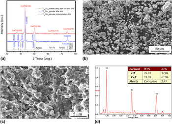
The Ti and Cu powders have been mechanical alloyed by a high energy ball milling machine. Figure 1(a) shows the X-ray diffraction patterns of the Ti30Cu70 powder mixtures before mechanical alloying (MA), the powders after MA for 20 h, and the spark plasma sintered Ti30Cu70 alloys. The Ti30Cu70 powder mixtures before MA consist of α-Ti and Cu phases. After MA, the Ti30Cu70 powders are still α-Ti and Cu phases, but some Ti peaks of Ti (102), (110), (103), (112), and (201) have disappeared. After SPS processing at 800 °C for 30 min, all the α-Ti and Cu phases are disappeared and Cu3Ti2 (ICDD No. 18–0459) and Cu4Ti3 (ICDD No. 18–0460) phases become dominate. Figures 1(b)–1(d) shows the SEM micrographs of the mechanical alloyed Ti30Cu70 powders and fractured surface of the spark plasma sintered alloys with EDX spectrum analysis. The powders are agglomerates with mean particle size of 10 μm in diameter [Fig. 1(b)]. The fractured surface of the spark plasma sintered Ti30Cu70 alloy shows a homogenous microstructure and grain size of about 1 μm with a fracture mode of intergranular cracking [Fig. 1(c)]. The relative density of the Ti30Cu70 alloy is measured to be 99.9%. The EDX spectrum of the area scanning in the sintered alloys shows elemental compositions of 32:68 (at.%), which corresponds to the initial designed 30:70 (at.%) in the powders.
X-ray diffraction patterns (a) and SEM morphologies of the mechanical alloyed Ti30Cu70 powders (b) and fractured surface of the spark plasma sintered Ti30Cu70 master alloy (c) with EDX spectrum exhibiting the elemental compositions in the master alloys (d).

B. Preparation and characterization of nanoporous α-Ti
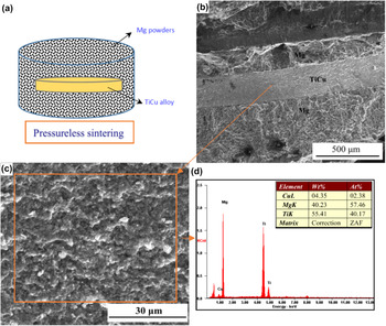
Figure 2(a) shows the schematic illustration of the solid state dealloying process. The slice of TiCu master alloy was immersed in magnesium powders, cold pressed, and finally heat treated in a pressureless sintering furnace. The temperatures are in the range of 500–600 °C for 10–60 min in argon atmosphere. Figure 2(b) shows the fractured cross section of the dealloyed TiCu alloys in the Mg powders after heat-treatment at 600 °C for 30 min. It can be seen that the TiCu slice is imbedded into the Mg matrix. A high magnification SEM micrograph on the central core of the TiCu slice is shown in Fig. 2(c). It shows a uniform and smooth fractured surface. The EDX spectrum in the red square area indicates that the elements are Ti, Cu, and Mg. It exhibits that the composition of Cu has reduced to 2.38 at.% but the content of Mg has increased to 57.46 at.% [Fig. 2(d)]. It means that the Cu atoms in the TiCu alloys have been selectively removed and dissolved into the Mg powder matrix during the solid state heat-treatment dealloying process.
Schematic illustration of the solid state dealloying process (a), SEM micrographs of fractured surface of the Ti30Cu70/Mg (b) and Ti30Cu70 alloy after heat-treatment in Mg powders (c) with EDX spectrum showing the elemental compositions in the TiCu sample (d).

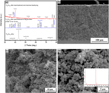
The phase compositions of the TiCu alloys during the solid state dealloying process are revealed in the XRD patterns of Fig. 3(a). After heat-treatment in Mg powders at 600 °C for 15 min, α-Ti, Ti2Cu (ICDD No. 15–0717), Cu2Mg (ICDD No. 65–5831), and Mg phases can be seen in the XRD pattern of TiCu alloys. It means that the Cu atoms have been diffused into the Mg powders and formed new phase of Cu2Mg. The Cu3Ti2 and Cu4Ti3 phases have transformed into Ti2Cu phase due to the release of Cu atoms. Finally, the TiCu slice was heat treated for 30 min at 600 °C for a complete reaction. After etching in the dilute nitric acid, it shows pure hcp structured α-Ti phase. But there is a weak and broad Ti2Cu peak at 2θ of 39.5°. It is because of the presence of residual Cu elements in the foams. Figures 3(b)–3(d) shows the SEM micrographs of the fractured surfaces of the heat-treatment and etched porous α-Ti with inserted EDX of the porous structure. The nano-pores are distributed in the cross section uniformly in the edge and center of the 300 μm wide foam. The open-cell nanoporous structure is uniformly spread throughout the entire cross section. As measured on the SEM image, the mean value of ligament size is around 650 nm. The EDX shows that it has just Ti element without Cu and Mg compositions in the Ti nanofoam. Therefore, relative pure nanoporous α-Ti foam was fabricated successfully.
X-ray diffraction patterns of the Ti30Cu70 alloy after heat-treatment and etching (a) and SEM micrographs of fractured surfaces of the nanoporous α-Ti foam after heat-treatment and etching with inserted EDX spectrum (b–d).

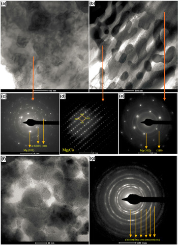
Figure 4 shows the TEM micrographs of the Ti30Cu70 alloys after solid state dealloying in Mg powders with corresponding selected area diffraction (SAD) patterns. Figure 4(a) exhibits the TEM morphology of the Ti/Mg nanocomposite. The SAD pattern in Fig. 4(c) confirms the presence of α-Ti and Mg phases. The Cu atoms have diffused into the Mg after the solid state heat-treatment dealloying, as shown in Fig. 4(b). The SAD pattern on the dark skeleton in Fig. 4(b) reveals that it is Mg2Cu single crystal phase [Fig. 4(d)]. The bright matrix in Fig. 4(b) is Mg phase [Fig. 4(e)]. However, the Mg2Cu phase has not been detected in the XRD result in Fig. 3(a), where just the Cu2Mg phase was found. It means that the diffusion of Cu atoms into the Mg formed Mg2Cu phase firstly. With increasing of Cu concentration, it was transformed into Cu2Mg phase. Figures 4(e) and 4(f) shows the TEM micrograph and SAD of the nanoporous α-Ti foam. It shows that the particle size of the Ti is about 20–50 nm and the pore size is in the range of 5–20 nm. The SAD pattern indicates that it is polycrystalline α-Ti phase. The particle sizes of the raw Ti and Cu powders are 325 mesh (45 μm). After MA, the TiCu powders show particles size about 10 μm [Fig. 1(b)]. After SPS processing, the TiCu alloys show grain size around 1.0 μm [Fig. 1(c)]. However, the Ti shows 20–50 nm particles size after the heat-treatment solid state dealloying. It is owing to the dealloying of Cu element from the Cu3Ti2 and Cu4Ti3 solid solutions where the Ti and Cu atoms have nanometer compounding after MA and SPS. It is also due to the formation of Ti/Mg nanocomposite after the solid state dealloying [Fig. 4(a)].
TEM micrographs of the Ti30Cu70 alloys after heat-treatment in Mg powders with corresponding SAD patterns showing the Ti/Mg nanocomposite (a, c), Mg2Cu phase (b, d) and Mg phase (b, e); the obtained nanoporous α-Ti foam (f) with SAD pattern (g).

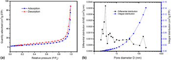
The porous structure and specific surface area of the Ti foams were characterized by N2 absorption–desorption isotherms. Figures 5(a) and 5(b) shows the nitrogen adsorption/desorption isothermal plot of the nanoporous Ti foam, BJH adsorption dV/dD pore volume and pore integral distribution in the Ti foams. The adsorption isotherm for the resulting foam has hysteresis loops along with a relative pressure (p/p 0) from 0.0 to 0.99. These isotherms are type IV, typical of mesoporous character materials. The BET surface area was calculated based on the isotherm results. The Ti nanofoam exhibits a BET surface area of (34.4 ± 0.8) m2/g. The total porosity is calculated to be about 60%. The porous properties of the Ti foams were further confirmed by pore size distribution analysis determined by the Barrett-Joyner-Hallender (BJH) method. The BJH desorption result showed that the smallest pores in the Ti foams are 2 nm. The values of pore width are from 2 nm up to 125 nm around. However, most of the pore sizes are distributed in the range of 2–50 nm, as shown in Fig. 5(b) of the BJH desorption dV/dD pore volume with a function of pore diameter. It is displayed in Fig. 5(b) that there is a sharp peak at pore size of 3.20 nm for sample of the nanoporous Ti foams. It means that the Ti foams are in nanoporous structures within mesoporous range (2–50 nm). Therefore, it is a nanoporous foam with a relative uniform pore size distribution.
Nitrogen adsorption/desorption isothermal plot of the nanoporous α-Ti foam (a), BJH desorption of pore differential and integral distributions with a function of pore diameter (b).

The dealloying method, present here, in the solid state powders is effective and powerful. The mechanism for the heat-treatment solid state dealloying is a solid state chemical reaction based on mixing enthalpy relationship of elements. The entropy between Mg and Cu is negative (−3 kJ/mol) while for the case of Ti and Mg is positive (16 kJ/mol). Reference Takeuchi and Inoue29 The selective removal of components takes place by the attractive (negative entropy) or repulsive force (positive entropy) among the constituent elements. It is independent of the nobilities of constituent elements. During the heat-treatment, the Cu elements have been diffused into the Mg powder compacts and selectively dissolved. Nanocomposite of Ti/Mg is formed because of the attractive force for Cu–Mg but the repulsive force for Ti–Mg. The solid state chemical reaction process proceeds as that the solid solutions of Cu3Ti2 and Cu4Ti3 are reacted with Mg during heat-treatment and formed to be Ti2Cu and Mg2Cu phases, finally to be Cu2Mg phase and Ti/Mg nanocomposite. In the end, the Ti/Mg nanocomposite was rapidly etched in dilute HNO3. Ti has higher value (Ti2+, −1.628 V) of standard electrode potential than that of Mg (Mg2+, −2.375 V). Therefore, the Ti has better corrosion resistance compared with Mg and Cu2Mg in the dilute HNO3. At last, the Mg and Cu2Mg phases have been selectively removed by the etching in the acid and the pure nanoporous α-Ti foams have been fabricated. It is noticed that the nanoporous structures were formed actually by the etching of the Ti/Mg nanocomposite in nitric acid. But during this step, no dynamic rearranging of the atoms occurs as in the dealloying, so it is merely an etching/dissolution process that chemically removes one of the two phases from the nanocomposite. During the solid state dealloying in Mg powders, there are dynamic rearranging of the atoms, in addition some constituent elements in the master alloy have been selectively removed and dissolved into the Mg powders, which is similar to an aqueous dealloying process. Therefore, the Mg powder method can be treated as a dealloying process to a certain extent.
C. Mechanisms of the solid state dealloying
The surface diffusion is the mechanism for dealloying in aqueous solution by the conventional electrochemical dealloying. Reference Jin, Wang, Parida, Wang, Seo and Weissmuller2–Reference Erlebacher, Aziz, Karma, Dimitrov and Sieradzk6 For example, Erlebacher et al. Reference Erlebacher, Aziz, Karma, Dimitrov and Sieradzk6,Reference Erlebacher and Seshadri30 revealed that the formation of nanoporous Au structures is caused by diffusion of Au atoms at the Au–Ag solid/electrolyte interface. The mechanism for the formation of the Ti nanoporous structures in Mg melt is surface and volume diffusion. Reference Tsuda, Wada and Kato20 The pore-forming mechanism in the solid state dealloying using powders here need investigations.
To study the pore-forming mechanism, disk-shaped Ti30Cu70 samples with diameter of 10 mm and thickness of 1.2 mm were used. One disk-shaped Ti30Cu70 sample was dealloyed by heat-treatment at 600 °C for holding time of 10 min, and then chemical dealloyed in dilute nitric acid. It can show an uncompleted dealloying state. There are three parts in the sample: dense Ti30Cu70 (inner core), porous TiCu (Ti2Cu, Ti3Cu) intermediate part, and nanoporous α-Ti part (out surface). The total thickness of the α-Ti and TiCu layers can be used to study the depth of Mg atoms diffusion into the Ti30Cu70 alloy. Therefore, the average thickness of the porous α-Ti (l α-Ti) and porous TiCu (l TiCu) can be calculated with the following equation, Reference Meguro, O and Kajihara31
 $${l_{\rm{x}}} = {{{A_{\rm{x}}}} \over {{W_{\rm{x}}}}}\quad ,$$
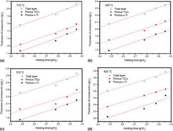
where, l x is the thickness of Mg diffusion into the TiCu alloy, A x means the total area of the compound x layer on the cross section, W x refers to the total length parallel to the compound x interface. Figure 6 shows the plots of logarithm of layer thickness [lg(l x)] with a function of logarithm of holding time t [lg(t/t 0)] at various heat-treatment temperatures of 510, 540, 570, and 600 °C. The total thickness, the thickness of porous TiCu and α-Ti are noted with triangle, circle, and square shape, respectively. As can be seen, the thickness lg(l x) increase linearly as a function of lg(t/t 0) at various temperatures. It indicates that all the logarithm of the Mg diffusion thickness increase linearly with the logarithm of holding time. The relationship between the thickness l x and the holding time t obeys the following equation,
 $${l_{\rm{x}}} = k{\left( {{t \over {{t_0}}}} \right)^n}$$
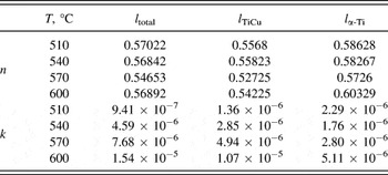
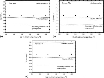
where, t 0 is the unit time, 1 s, that is adopted to make the argument t/t 0 of the powder function dimensionless, k x is the rate constant, n is the reaction exponent and it is the slope in the logarithmic formula. This formula is usually used for analyzing reaction diffusion interfaces. Reference Meguro, O and Kajihara31 The values of n and k x are determined by the least-square method using the data in Fig. 6 and summarized in Table I. The exponent n is affected by the rate-controlling process. When the layer growth is controlled by volume diffusion, n is equal to 0.5. The index n is equal to 0.25 when only grain boundary diffusion occurred. Reference Corcoran, King, Lanerolle and Kim32 The value of n is equal to 1 when growth of the phase is controlled by interface reaction. Figure 7 shows the relationship between reaction exponent n versus heat-treatment temperature of the disk-shaped sample for α-Ti phase, TiCu phase and total layer. The n value for growth of the total porous phase in Fig. 7(a) is determined by the α-Ti layer and TiCu layer. In Fig. 7(b), the value of n for the porous TiCu phase decreases with increasing of the dealloying temperature. The value of n is equal to 0.56 at dealloying temperatures of 510 and 540 °C, but around 0.5 at 570 and 600 °C. Thus, the growth of the porous TiCu phase is controlled by interface reaction at 510 and 540 °C, and by volume diffusion at relative higher temperatures of 570 and 600 °C. In Fig. 7(c), the n of the porous α-Ti phase is greater than 0.5, close to 0.6. So, the growth of the α-Ti layer is mainly controlled by the interface reaction. Consequently, the pore-forming process in the solid state dealloying using powders is controlled by both of interface reaction and volume diffusion. The dealloying within metallic powders is a water-free process. During the heat-treatment, diffusion is activated for mass transport. The constituent of Cu in the TiCu alloy was dissolved into the Mg powder compacts thanks to the mechanism of interface reaction and volume diffusion.
Plots of thicknesses of compound layers lg(l x) with a function of lg(t/t 0) at various heat-treatment temperatures of 510 °C (a), 540 °C (b), 570 °C (c) and 600 °C (d).

Values of rate constant k and exponent n of disk-shaped samples at 510, 540, 570, and 600 °C.

The relationship between exponent n versus heat-treatment temperature of the disk-shaped sample for total layer (a), porous TiCu (b) and porous α-Ti phase (c).

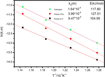
To better understand the mechanism for the formation of the porous layers, the activation energy for the growth of the porous layers was analyzed. Figure 8 shows the relationship between lnk of the total porous phase, the porous TiCu phase and porous α-Ti phase versus reciprocal heat-treatment temperature in Kelvin rating for the growth of each layers (1/T). As can be seen from Fig. 8, the plotted points are well located on straight lines. Thus the activation energy can be obtained by fitting an Arrhenius-type equation,
 $$k = {k_0}\exp \left( { - {E \over {RT}}} \right)\quad ,$$
where k 0 is the pre-exponential factor, R is the gas constant, and E is the activation energy. The activation energy of the total porous phase, the porous TiCu and the porous α-Ti are calculated to be 117.80, 127.01, and 104.99 kJ/mol, respectively. These values are higher than those in the Mg melts (35–45 kJ/mol). Reference Tsuda, Wada and Kato20 It indicates that the dealloying in solid state powders need much higher activation energies. Taguchi et al. Reference Taguchi, Iijima and Hirono33 reported that the activation energies are 79–125 kJ/mol for the growth of reaction layers in the reaction diffusion of a solid TiCu couple. Our values are very close to these in the solid TiCu couple. It means that the formation of α-Ti during the heat-treatment dealloying in Mg powders can be attributed to a solid-state interdiffusion phenomenon based on mixing enthalpy relationship among constituent elements. Finally, the nanoporous structured α-Ti are formed by subsequent etching in an acidic solution.
Plot of rate constant k versus reciprocal of heat-treatment temperature T in Kelvin rating.

D. Nanomechanical properties
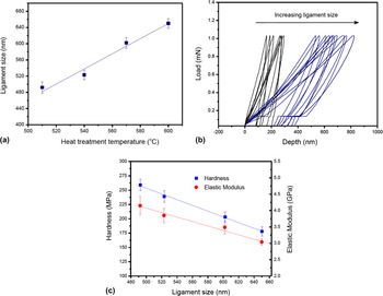
The ligament coarsening of the nanoporous α-Ti foams with a function of the heat-treatment temperatures was investigated and illustrated in Fig. 9(a). It can be clearly seen that the ligament sizes grow up with increasing of the heat-treatment temperature at a constant holding time of 30 min. It coarsely obeys a linear increment phenomena. The mean values of ligament size are 492, 523, 602, and 650 nm at temperatures of 510 °C, 540 °C, 570 °C, and 600 °C, respectively. Figure 9(b) shows the nanoindentation loading/unloading curves of the nanoporous Ti foams. The curves in blue color are for the 600 °C heat treated foams with ligament size of 650 nm. The curves in black color are for the 540 °C processed foams (523 nm). The overlapping loading sections of each data set demonstrate the excellent reproducibility of the measurements. The contact depth which represents plastic deformation intensity, increased with ligament size. Based on the nanoindentation results, the mean values of nanohardness and elastic modulus are calculated. Figure 9(c) demonstrates the relationship between hardness, elastic modulus and ligament size of the nanoporous α-Ti foams. As shown, the hardness and elastic modulus are both reduced with the ligament size increasing. The 492 nm ligament sized sample prepared at 510 °C shows hardness of 259 MPa and elastic modulus of 4.15 GPa. However, the 650 nm sample (600 °C processed) displays hardness of 178 MPa and elastic modulus of 3.04 GPa. The hardness and elastic modulus both follow linear decay fit as a function of ligament size increasing. These mechanical values are higher than those obtained by Mg melt method. Reference Wada, Setyawan, Yubuta and Kato21 Hakamada and Mabuchi reported that the hardness is equal to yield strength in the nanoporous foams. Reference Hakamada and Mabuchi34 In the current study, we found that the hardness and elastic modulus of nanoporous α-Ti decrease as the ligament size increases following linear decay. It means that the yield strengths of the nanoporous α-Ti foams follow the same trend.
The ligament size with a function of heat-treatment temperature (a). Nanoindentation loading/unloading curves (b) and the relationship between hardness, Elastic modulus with ligament size of the nanoporous α-Ti foams (c).

This study provides a new dealloying route by heat-treatment in solid state powders. The heat-treatment temperatures (500–600 °C) are lower than the melting point of Mg (650 °C) and much lower than the Mg melt method (670–870 °C). Reference Wada, Yubuta, Inoue and Kato16,Reference Tsuda, Wada and Kato20,Reference Wada, Setyawan, Yubuta and Kato21 The holding time in the solid state dealloying (10–60 min) is longer than the method of Mg melts (0.1–20 min). Reference Wada, Yubuta, Inoue and Kato16,Reference Tsuda, Wada and Kato20,Reference Wada, Setyawan, Yubuta and Kato21 Recently, Wada et al. reported the preparation of nanoporous FeCr alloy via a solid-state interfacial dealloying reaction. Reference Wada, Yubuta and Kato35 They used Mg plate (1 × 12 × 12 mm) as solid dealloying agent, where the plate was fixed tightly with precursor of (FeCr)Ni using stainless-steel bolts and heat treated at 440–480 °C for 6–72 h. Reference Wada, Yubuta and Kato35 In our study, Mg powders are used as solid dealloying agent, where the precursor was immersed in the powders and heated treated at 500–600 °C for 10–60 min. Both of them using Mg plate and powders belong to the solid state dealloying method. But the powder method is relatively simple and easy to implement since it’s no need for fixing with the precursor tightly. Its dealloying time is much shorter (10–60 min) than that of the Mg plate method (6–72 h). The method of solid state dealloying using powders need just a normal pressureless sintering furnace. The mean particle sizes of the Mg powders are not fine about 45 μm without hazard to flame. This method allows the shape of master alloy to be largely maintained during dealloying. Therefore, it is a simple and convenient method. It can be used alternatively besides the Mg melt and plate method. It could be used for the fabrication of nanoporous metals those have lower standard electrode potentials and are not suitable with electro chemical dealloying method, for example, nanoporous Ti, Mo, Nd, W, V, Ta, Zr, Hf, and so on. These metals all have very high melting point and are very suitable for the SPS processing and solid state dealloying. People can choose other elemental powders to instead of the Mg powders. It depends on the mixing entropy of elements in the alloy compounds and the entropy of the alloy constitution with the dealloying element. From an application perspective, Ti is valuable because it has the highest specific strength of any elemental metals and highly corrosion resistant, as well as is considered to be biomedical inert. The nanopores may be able to be loaded with stored bioactive compounds such as proteins or antibacterial drugs for slow release such that nanoporous networks could act as reservoirs for these bioactive compounds and prevent infections and inflammations that lead to septic loosening of implants during healing. Reference Panagiotopoulos, Moreira Jorge, Rebai, Georgarakis, Botta and Yavari17,Reference Necula, Apachitei, Fratila-Apachitei, van Langelaan and Duszczyk36 In addition, fabrication of nanoporous Ti foams would have implications in many fields. The Ti nanoporous metals can serve as precursors to other nanoporous ceramic materials, such as, titanium oxides, nitrides, and carbides. The nanoporous Ti foams may find very broad functional applications in the near future, especially in biomedical field as a new structured implant with drug release properties.
IV. Conclusions
-
(1) 3D open-cell nanoporous α-Ti foams are fabricated by a solid state dealloying using heat-treatment of Ti30Cu70 master alloy in Mg powders. Fully densified Ti30Cu70 (at.%) master alloys have been fabricated by SPS with nanometer compounded Cu3Ti2 and Cu4Ti3 phases. Pure nanoporous α-Ti foams are fabricated after pressureless sintering at 600 °C for 30 min in Mg powders and etching in nitric acid. The BET surface area of the foams is 34.4 ± 0.8 m2/g with pore size distribution mostly in the range of 2–50 nm.
-
(2) The dealloying in powders is a solid state chemical reaction process. It proceeds as that the solid solutions of Cu3Ti2 and Cu4Ti3 are reacted with Mg during heat treatment and then formed to be Ti2Cu and Mg2Cu phases, finally to Cu2Mg phase and Ti/Mg nanocomposites. The nanoporous structures are formed by subsequent etching in a nitric acid solution.
-
(3) The constituent of Cu in the Ti30Cu70 alloy was dissolved into the Mg powder compacts thanks to the mechanism of interface reaction and volume diffusion. The growth of porous α-Ti layer is mainly rate-controlled by interface reaction in addition to volume diffusion in the powder compacts. The activation energies for the transition layer growth are in the range of 104–128 kJ/mol. This suggests that mechanism for α-Ti formation in the Mg powders is a solid-state interdiffusion phenomenon.
-
(4) The ligament coarsening of the nanoporous α-Ti foams with a function of the heat-treatment temperatures was investigated. The ligament sizes grow up (492–650 nm) with increasing of the heat-treatment temperatures (510–600 °C). The nanoindention hardness and elastic modulus in nanoporous α-Ti follow linear decay fit with ligament size increasing. The hardness decreased from 259 to 178 MPa, and elastic modulus from 4.15 to 3.04 GPa.
ACKNOWLEDGMENTS
The authors gratefully acknowledge the financial supports from the State Key Laboratory for Powder Metallurgy at Central South University, Natural Science Foundation of Jiangsu Province (No. BK20161419), Scientific Research Foundation for the Returned Overseas Chinese Scholars at State Education Ministry (No. 2015-1098), National Natural Science Foundation of China (No. 11572087), Jiangsu Key Laboratory for Advanced Metallic Materials (No. BM2007204) at Southeast University, and the Fundamental Research Funds for the Central Universities (No. 2242016K40013).