Introduction
In the context of tissue engineering and regenerative medicine (TERM), bioprinting technologies emerged as an enabling tool for creating three-dimensional (3D) functional tissue constructs with tailored biological and mechanical properties (see the Introduction article in this issue). Reference Murphy and Atala1,Reference Feinberg and Miller2 Bioprinting has been defined as “the use of computer-aided transfer processes for patterning and assembling living and non-living materials with a prescribed two-dimensional or 3D organization in order to produce bioengineered structures serving in TERM, pharmacokinetic, and basic cell-biology studies.” Reference Guillemot, Mironov and Nakamura3 Together with bioassembly, bioprinting is one of the two main approaches of biofabrication in TERM. Reference Groll, Boland, Blunk, Burdick, Cho, Dalton, Derby, Forgacs, Li, Mironov, Moroni, Nakamura, Shu, Takeuchi, Vozzi, Woodfield, Xu, Yoo and Malda4
Different technologies have been proposed for patterning, depositing, and 3D shaping bioinks, which can be grouped into (micro)extrusion, Reference Ozbolat and Hospodiuk5 inkjet, Reference Saunders and Derby6 and photopolymerization. Reference Wang, Abdulla, Parker, Samanipour, Ghosh and Kim7 The simultaneous printing of biomaterials and cells allowed the achievement of several milestones, such as increased seeding efficiency and avoidance of nonhomogeneous cell distribution due to postfabrication seeding. Reference Melchels, Barradas, van Blitterswijk, de Boer, Feijen and Grijpma8 Extrusion-based technologies are currently targeted as promising for building clinically relevant constructs. Reference Malda, Visser, Melchels, Jüngst, Hennink, Dhert, Groll and Hutmacher9 Although they have limited resolution, several companies are investing in this growing market, Reference Arslan-Yildiz, El Assal, Chen, Guven, Inci and Demirci10 mainly proposing extrusion-based bioprinters with related bioinks. Reference Hölzl, Lin, Tytgat, Van Vlierberghe, Gu and Ovsianikov11
However, recapitulating in vitro the 3D multiscale microarchitecture with multiple cell types as well as the extracellular matrix (ECM) physicochemical cues of living tissues have remained unsolved challenges. Reference Atala, Kasper and Mikos12 The diffusion of nutrients and waste products above a distance of 150–200 μm is no longer efficient, compromising cellular viability and function in a short time. Reference Zhu, Qu, Zhu, Ma, Patel, Liu, Wang, Lai, Gou, Xu, Zhang and Chen13 Growth factors are specifically located in the ECM to guide tissue development, Reference Jang, Kim, Kim, Kim, Kwon and Cho14 and different types of cells are in close contact and in continuous cross-talking. Reference Boland, Mironov, Gutowska, Roth and Markwald15 Bioprinting a single cell type together with a single biomaterial cannot, therefore, bring further advancement.
This article describes recent advances in controlling spatial heterogeneity of chemical and physical properties of scaffolds using bioprinting technologies. We also discuss how such bioprinted artificial tissues have a prescribed cellular composition and spatial arrangement. As different bioprinting technologies have been reviewed elsewhere, Reference Malda, Visser, Melchels, Jüngst, Hennink, Dhert, Groll and Hutmacher9–Reference Hölzl, Lin, Tytgat, Van Vlierberghe, Gu and Ovsianikov11,Reference Derby16 we focus on the advantages and constraints for multimaterial processing, by analyzing attempts to merge different bioprinting technologies.
General considerations for multicellular and multimaterial bioprinting
Regardless of the chosen technology and the well-established issues regarding pores (i.e., pore size and shape, interconnectivity, and total porosity) and mechanical properties that each scaffold must present, common considerations can be highlighted.
Bioprinting technologies usually follow a layer-by-layer approach, where each layer is built and stacked over the previous one until completion of the 3D structure. Bottom layers have to provide mechanical support and sustain the weight of the upper layers without collapsing. The overhanging components need to resist without large, gravity-induced deformations.
The fabrication of a layer can be described as transfer of energy (mechanical, thermal, chemical, and electromagnetic) from the bioprinting machine to the cell-laden material. This process can affect cell phenotype and viability (e.g., by interacting with DNA), damaging cell membranes or altering osmotic equilibrium between cells and the external environment. Furthermore, sterility has to be assured in each step of the bioprinting procedure, by an accurate control of the material reservoirs and of the building chamber.
Nanotopography plays a fundamental role in cell adhesion. Reference Salmasi, Kalaskar, Yoon, Blunn and Seifalian17 Many studies demonstrate this feature as an intrinsic average property of the bioprinted material. However, bioprinting technologies should achieve submicrometer resolution to offer greater control over the entire bioprinted construct. Although millimetric biological constructs can be sufficient for in vitro organs (i.e., reverse-engineered “mature” functional tissues, comprising fundamental elements, architectures, and physical/chemical stimuli), macroscale constructs will be necessary for clinical applications. Maintaining a submicrometer resolution for long distances (>3 cm) can increase the cost by a factor of 10 (e.g., 10 μm to 1 μm accuracy).
Hydrogels are considered the elected materials for biofabrication due to their similarities with the ECM. Reference Malda, Visser, Melchels, Jüngst, Hennink, Dhert, Groll and Hutmacher9 Several hydrogels have been processed. However, further advances in biomaterials are required to enable printing low-viscosity materials with improved biological properties that will enhance the resulting tissue function. Reference Skardal and Atala18 Extrusion-based and inkjet technologies require low viscosity bioinks, which allow the use of a thinner nozzle and an increase in the dispensing speed. These factors will result in higher printing resolutions, shorter fabrication time, and reduced (shear) stress on cells. Reference Rutz, Hyland, Jakus, Burghardt and Shah19 This rheological behavior should be coupled with a fast gelation process to retain the shape impressed by the printing procedure. Reference Malda, Visser, Melchels, Jüngst, Hennink, Dhert, Groll and Hutmacher9 Control over the gel stiffness and swelling is necessary for structural support to provide the proper mechanical cues to cells. Reference Schuurman, Levett, Pot, van Weeren, Dhert, Hutmacher, Melchels, Klein and Malda20 These cell-responsive bioinks allow for control of the spreading and migration of cells. Reference Skardal and Atala18 The use of a dense polymer matrix (generally with a concentration higher than 5 wt%), necessary to have the right viscosity for printing definition, Reference Chang, Boland, Williams and Hoying21 can prevent spreading, migration, and proliferation. Therefore, these hydrogels are not ideal candidates for cell-laden constructs. Reference Brandl, Sommer and Goepferich22
From a rheological point of view, even if bioinks form a solid structure, they are normally described just by indicating the viscosity before the gel point. Reference Fedorovich, Schuurman, Wijnberg, Prins, van Weeren, Malda, Alblas and Dhert23 For hydrogels, gelation time and storage modulus have to be determined by oscillatory time sweep and frequency sweep. Reference Rutz, Hyland, Jakus, Burghardt and Shah19,Reference Hanson Shepherd, Parker, Shepherd, Gillette, Lewis and Nuzzo24 These analyses will confirm the viscoelasticity of the gel and will determine if there is a specified time frame for printing. Reference Hölzl, Lin, Tytgat, Van Vlierberghe, Gu and Ovsianikov11,Reference Rutz, Hyland, Jakus, Burghardt and Shah19 Gelation is necessary for the stabilization of the intralayer and interlayer structures and is generally achieved by physical, thermal, Reference Wang, Yan, Pan, Xiong, Liu, Cheng, Liu, Lin, Wu, Zhang and Lu25 ionical, Reference Pataky, Braschler, Negro, Renaud, Lutolf and Brugger26 or chemical and photochemical means. Reference Montemurro, De Maria, Orsi, Ghezzi, Tinè and Vozzi27–Reference Fedorovich, Swennen, Girones, Moroni, van Blitterswijk, Schacht, Alblas and Dhert29 Several studies have explored the combination of a first, rapid, physical stabilization followed by a second stronger chemical bond. Reference Jang, Kim, Kim, Kim, Kwon and Cho14,Reference Fedorovich, Swennen, Girones, Moroni, van Blitterswijk, Schacht, Alblas and Dhert29
Rutz and co-workers customized several bioink formulations in terms of composition, degree of cross-linking, and polymer concentration, by exploiting the functionality of light cross-linking with long length chemical cross-linkers, based on poly(ethylene glycol). Reference Rutz, Hyland, Jakus, Burghardt and Shah19 The Khademhosseini group, instead, combined the initial structural integrity given by ionic cross-linking alginate fibers with the UV photopolymerization of cell-laden gelatine-methacryloyl coaxially extruded (see the next section). Reference Colosi, Shin, Manoharan, Massa, Costantini, Barbetta, Dokmeci, Dentini and Khademhosseini30
A final consideration of the so-called “scaffold-free bioprinting” can be outlined. This process, pioneered by Forgacs and co-workers, foresees the use of tissue spheroids, an aggregate of a large number (approximately 107 cells/ml) Reference Itoh, Nakayama, Noguchi, Kamohara, Furukawa, Uchihashi, Toda, Oyama, Node and Morita31 of heterogeneous cells that will be remodeled after bioprinting. Such spheroids are important for recapitulating the minimal functions of a targeted tissue or organ. Spheroid remodeling is due to the tissue liquidity driving force Reference Jakab, Neagu, Mironov, Markwald and Forgacs32 sustained by temporarily inserting a support matrix usually made of agarose. The scaling up of these tissue constructs implies that a large number of cells must be extracted from the patient and expanded in vitro, which will result in cell senescence (i.e., cells becoming older), and consequently, may impinge on their functionality. Reference Tan, Tan, Yeong and Tor33
Multinozzle and microfluidic approaches for extrusion-based bioprinting
Extrusion-based bioprinting is characterized by a robotic micropositioner, which describes trajectories in 3D space, whereas hydrogel bioinks are extruded in cylindrical filaments by a fluid-dispensing system. Comprehensive reviews of fluid dispensing systems are available, and can be grouped into pneumatic (with or without a valve) or mechanical (piston or screw-driven). Reference Ozbolat and Hospodiuk5,Reference Arslan-Yildiz, El Assal, Chen, Guven, Inci and Demirci10 Recently, even lab pipettes have been used as extruders, although the system, as indicated by the authors, was completely manual. Reference Tan, Tan, Yeong and Tor33
Generally, volumetric pumps offer higher control over fluid dispensing and do not need continuous calibration of the system if the bioink viscosity changes. Reference Hölzl, Lin, Tytgat, Van Vlierberghe, Gu and Ovsianikov11 Because of the large driving forces that can be generated, very viscous solutions can be printed, and screw-driven extruders can dispense molten polymers, such as poly(ε-caprolactone) (PCL).
Extrusion-based bioprinters are extremely flexible, allowing the use of heterogeneous bioinks, both in terms of biomaterial and cell type composition, even if the bioprinters present a single extruder. Cell-laden hydrogels containing multiple cell types and high-cell-density tissue spheroids (which are intrinsically heterogeneous) have been successfully printed. Reference Jakab, Neagu, Mironov, Markwald and Forgacs32,Reference Norotte, Marga, Niklason and Forgacs34,Reference Jakab, Norotte, Marga, Murphy, Vunjak-Novakovic and Forgacs35 Due to the high driving force available for extrusion, it was possible to load hydrogel-based bioinks (1) with porous microspheres where cells can adhere and proliferate before printing, while hydrogels offer lubrication and glue after gelation; Reference Tan, Tan, Yeong and Tor33 and (2) with 3D printed microstructures, such as the so-called lockyballs, two-photon polymerized cages that trap tissue spheroids and guide their spatial organization. Reference Rezende, Pereira, Kasyanov, Ovsianikov, Torgensen, Gruber, Stampfl, Brakke, Nogueira, Mironov and da Silva36
An extrusion-based bioprinter was successfully used to print decellularized ECM, which offers fibrous proteins, glycosaminoglycans (GAGs), and remnant growth factors mimicking the native tissue microenvironment, thereby enabling cells to preserve their original functionalities. The final structure needs to be cross-linked after printing to avoid dissolution in aqueous environment after physical (thermal) gelation. The use of vitamin B2-induced UVA radiation cross-linking was demonstrated to reinforce the structure and to be cell-friendly toward cardiac progenitor cells. Reference Jang, Kim, Kim, Kim, Kwon and Cho14
Self-assembling hydrogels, whose properties are driven by noncovalent interaction of their moieties, Reference Gao, Tang, Elsawy, Smith, Miller and Saiani37 have been recently investigated due to their shear thinning behavior, which allows easy extrusion. These materials are used as substrate/support materials in the so-called gel-in-gel printing because of their self-healing properties. Reference Highley, Rodell and Burdick38
Multimaterial and multicellular 3D scaffolds can also be fabricated by upscaling the complexity of the extrusion-based system. A first choice is the swapping of dispensing systems, which allows fabricating heterogeneous materials, such as the extrusion of PCL and alginate. Reference Chang, Nam and Sun39,Reference Tirella, De Maria, Criscenti, Vozzi and Ahluwalia40 This was also explored by building a bioprinter with multiple arms with independent motion paths and independent extruders. Reference Ozbolat, Chen and Yu41
The multiplication of print heads introduces the problem of alignment of multiple nozzles. Alternatives include coaxial extrusion, mainly used for dispensing noncross-linked gel and the cross-linking solution (ionic + covalent cross-linking), Reference Colosi, Shin, Manoharan, Massa, Costantini, Barbetta, Dokmeci, Dentini and Khademhosseini30 or printers with a single print head and independent syringe pumps, utilizing a single nozzle and a selector valve. Reference Campbell, McGuinnes, Wirz, Sharon and Sauer-Budge42 Another solution is the use of a single print head and a microfluidic system that mixes the various bioinks, coming from different reservoirs, before extruding a single multimaterial filament, characterized by a stable concentration gradient of the mixed bioinks across the filament section. Reference Liu, Zhang, Heinrich, De Ferrari, Jang, Bakht, Moisés Alvarez, Yang, Li, Trujillo-de Santiago, Miri, Zhu, Khoshakhlagh, Prakash, Cheng, Guan, Zhong, Ju, Zhu, Jin, Shin, Dokmeci and Khademhosseini43,Reference Snyder, Son, Hamid, Wu and Sun44 Microfluidic techniques assemble multiple material inlets in a single outlet channel without mixing, due to low inertial forces in microscale cross-sectional channels. Even if the microfluidic approach is limited by the mixing of the same fluid solution with different solutes (or different concentrations of the same solute), sub-needle (filament) resolution can be achieved. Currently proposed devices yield graded outlets by modulating the inlets velocities, but greater variety can be obtained by adding more complex microfluidic circuits. Reference Orsi, Fagnano, De Maria, Montemurro and Vozzi45
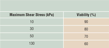
The main limitation in extrusion-based bioprinting is related to shear stresses in the flowing solution, which induce deformation in the cell membrane, affecting viability and phenotype stability during postprinting. Nair et al. Reference Nair, Gandhi, Khalil, Yan, Marcolongo, Barbee and Sun46 have discussed the effects of shear stress (see also Table I ).
Influence of shear stress on cell viability during extrusion-based 3D printing, using a pneumatic system with a microvalve. Reference Nair, Gandhi, Khalil, Yan, Marcolongo, Barbee and Sun46

Extrusion-based bioprinters have been successfully used to indirectly create thick vascularized tissues (up to 1 cm in thickness) by 3D printing a fugitive vascular template on which a cell-laden matrix is cast, avoiding shear stress on cells. In an approach proposed by Miller, Reference Miller, Stevens, Yang, Baker, Nguyen, Cohen, Toro, Chen, Galie, Yu, Chaturvedi, Bhatia and Chen47 a sacrificial carbohydrate glass was printed at elevated temperature (>100°C), protectively coated, and then removed before introducing a homogeneous cell-laden matrix. Lewis et al. Reference Kolesky, Homan, Skylar-Scott and Lewis48 instead proposed a more complex procedure that consisted of (1) a fugitive ink composed of pluronic (poloxamer or copolymers) and thrombin; (2) a cell-laden ink (deposited through extrusion) that contains gelatin, fibrinogen, and human mesenchymal stromal cells; and (3) a castable ECM material that contains gelatin, fibrinogen, human neonatal dermal fibroblasts, thrombin, and transglutaminase (TG). This matrix is cast over the printed inks. After casting, thrombin induces fibrinogen cleavage and rapid polymerization into fibrin in both the cast matrix, and through diffusion, in the printed cell ink. Similarly, TG diffuses from the molten casting matrix and slowly cross-links the gelatin and fibrin. The fugitive ink is dissolved by cooling, forming a perfusable vascular network, which is endothelialized with human umbilical vein endothelial cells and perfused via an external pump ( Figure 1 ). Interestingly, this complex tissue construct was observed over several weeks (>6 weeks), allowing a relatively long-term study of emergent biological phenomena in a complex microenvironment. Reference Kolesky, Homan, Skylar-Scott and Lewis48
Example of multicellular and vascularized scaffold, fabricated by combining a fugitive ink, a cell-laden ink, and a casting of a biomatrix. (a) Fabrication steps (i) Three-dimensional bioprinting of fugitive ink and cell-laden ink, (ii) casting of extracellular matrix material, (iii) removal of fugitive ink for creating the vascular network channels, and (iv) seeding of endothelial cells and perfusion of the vascular network. (b) Fabricated construct (fugitive [red] and cell-laden inks [green]; scale bar = 5 mm) and (c) confocal microscope image through a cross section of 1-cm-thick vascularized bone tissue construct after 30 days of active perfusion and in situ differentiation. Red: RFP-HUVECs, primary red fluorescent protein-expressing human umbilical vein endothelial cells; green, actin; violet, osteocalcin; blue, DAPI, 4′,6-diamidino-2-phenylindole. Scale bar = 1.5 mm. Adapted with permission from Reference Reference Kolesky, Homan, Skylar-Scott and Lewis48. © 2016 National Academy of Sciences.

Inkjet-based bioprinting
Inkjet-based bioprinters allow noncontact delivery of small droplets of bioinks at controlled positions on a substrate. Piezoelectric and thermal are the two most common mechanisms for drop-on-demand (DOD) formation. The piezoelectric inkjet printer uses piezoelectric crystals to produce acoustic waves to force the liquid through the nozzle. The thermal inkjet system produces pulses of pressure by vaporizing the bioink around the heating element, expelling the droplets out from the print head. Reference Saunders and Derby6 Heterogeneous patterns of structural proteins, Reference Kodadek49 enzymes, Reference Arrabito, Musumeci, Aiello, Libertino, Compagnini and Pignataro50 antibodies, Reference Cahill51 DNA/RNA, Reference Allain, Stratis-Cullum and Vo-Dinh52 and living cells Reference Yanez, Rincon, Dones, De Maria, Gonzales and Boland53 have been successfully printed, demonstrating the inherent multimaterial nature of this technology. However, intrinsic limitations and issues should be highlighted, such as those involving the printability of inks, described by the Ohnesorge number (Oh). This dimensionless number expresses the ratio of the viscous force to the surface tension during drop formation, and thus depends on the surface tension, density, viscosity, and nozzle size. Reference Derby54 High viscous forces (Oh > 1) dissipate the energy for expelling the drop, while high surface tension forces (Oh < 0.1) lead to many satellite droplets, limiting the printing quality. For biological fluids, the surface tension upper limit is that of pure water, because the density of electrolytes and buffers does not deviate more than a few percent from that of water. Hence, the only physical parameter in Oh that shows significant variation between biological fluids is the viscosity. Considering a typical nozzle diameter of 20–100 µm, a fluid viscosity of approximately 30 mPa × s is a practical upper limit for printability. Reference Saunders and Derby6
Even if the drop generation rate was increased up to 30 kHz, the bioprinting of clinically relevant constructs would be difficult to achieve, Reference Malda, Visser, Melchels, Jüngst, Hennink, Dhert, Groll and Hutmacher9 due to the small volume of each droplet. Currently, commercially available DOD printers are characterized by a drop volume of 1–100 pl, easily subjected to evaporation problems and related “coffee ring” effects.
Bioink damage has been indicated as the main weakness of inkjet-based bioprinters. Several studies have shown that biological materials (including cells) are not affected by the local high temperatures (300°C) of the heating element in the thermal DOD process, due to the short period of exposure (2 μs). Reference Derby54 The main concern about piezoelectric printing is possible sonication due to the frequency (8–20 kHz) used during printing, Reference Saunders and Derby6 although parametric studies on piezo-waveform parameters revealed a viability higher than 95%. Reference Saunders, Gough and Derby55 However, both thermal and piezo printers can induce temporary damage in cell membranes by shear stresses, due to the passage of the bioink through narrow nozzles. Reference Saunders and Derby6 Finally, cell viability can be influenced by the drop impact on the target substrate, where the drop velocity can range from 1–10 m/s. In this case, the stiffness of the substrate is fundamental to absorb the excess energy and dampen the impact, thereby protecting the cells. Reference Tirella, Vozzi, De Maria, Vozzi, Sandri, Sassano, Cognolato and Ahluwalia56
Despite these limitations, complex 3D structures have been created Reference Yanez, Rincon, Dones, De Maria, Gonzales and Boland53,Reference Cui, Dean, Ruggeri and Boland57 by printing cells and a gelation agent onto a prepolymer substrate, such as calcium ions onto alginate Reference Xu, Baicu, Aho, Zile and Boland58 or calcium ions and thrombin over fibrinogen. Reference Cui and Boland59 Orsi et al. demonstrated inkjet printing of a hydrogel and cross-linker contained in the same solution by printing gelatin and (3-glycidyloxypropyl) trimethoxysilane, a cross-linker that reacts once the solvent evaporates. Because of the versatility of inkjet printing, the researchers were also able to include pH-sensitive nanoparticles in the printing solution, creating a scaffold with in situ or built-in sensors. Reference Orsi, De Maria, Montemurro, Chauhan, Aylott and Vozzi60
Three-dimensional inkjet bioprinting of photopolymerizable gels is a well-defined approach that is currently under investigation. In particular, thermoreversible gelation can be useful for the initial fabrication of a scaffold, followed by a photopolymerization reaction to maintain the scaffold structure. Reference Di Biase, Saunders, Tirelli and Derby61 Cui et al., who printed chondrocytes (cartilage cells) directly into an explanted (ex vivo) articular cartilage defect, reported that being a noncontact technology, inkjet printing is well suited for decorating scaffolds fabricated with other technologies Reference Xu, Binder, Albanna, Dice, Zhao, Yoo and Atala62 or directly printing in situ. Reference Cui, Breitenkamp, Finn, Lotz and D’Lima63
Hybrid technologies
Electrospinning is the most versatile technique to produce nanofibers, as it allows large-scale production, combining different materials at the same time. Reference Teo, Inai and Ramakrishna64 The classical setup of an electrospinning system consists of a high voltage supply, a syringe with a metal needle of small diameter, and a metal collecting screen (collector). The presence of a high voltage between the needle and the collector allows the development of an electrically charged jet of polymer solution (or melted polymer) out from the needle. Before reaching the collector, the solvent evaporates from the solution, and the polymer solidifies as nanofibers, which impact the collector and develop a web of small fibers. The topology of the final structure can be modified by tuning the feed solution (solvent, concentration, viscosity, and molecular weight), processing conditions (electric field, distance, and shape of the collector), and environmental parameters (temperature and humidity). Reference Teo, Inai and Ramakrishna64
Combinations of extrusion-based bioprinting technologies and electrospinning have been investigated. Moroni et al. Reference Moroni, de Wijn and van Blitterswijk65,Reference Moroni, Schotel, Hamann, De Wijn and van Blitterswijk66 integrated 3D fiber deposition (extrusion-based additive manufacturing of molten material) with electrospinning techniques to fabricate a new set of multiscale scaffolds for cartilage regeneration. They fabricated poly(ethylene oxide terephthalate)/poly(butylene terephthalate) scaffolds that were characterized by periodic alternation between an additive manufactured structure, to obtain an architecture with appropriate mechanical properties, and electrospun fibers, to provide additional surface area and improve the cell-adhesion phenomena.
Park et al. Reference Park, Kim, Kim, Yang and Park67 followed a similar process, using PCL both for the additive manufactured structure and the electrospun mesh. Criscenti et al. fabricated a triphasic scaffold for the regeneration of the bone–ligament interface by combining a 3D fiber-deposited PCL structure and a poly(lactic-co-glycolic acid) electrospun mesh ( Figure 2 ). This scaffold presented a gradient of physical and mechanical properties, which elicited different biological responses from human mesenchymal stromal cells. Reference Criscenti, Longoni, Di Luca, De Maria, van Blitterswijk, Vozzi and Moroni68
Example of multiscale and multimaterial scaffold fabricated using hybrid technologies. The 3DF region was built by extruding poly(ε-caprolactone) strands, while PLGA was electrospun on a specific location to fabricate a triphasic scaffold. (a) 3DF region (scale bar = 500 μm); (b) mixed region with electrospun PLGA over 3DF strands (scale bar = 200 μm); and (c) ESP region (scale bar = 20 μm). Reference Criscenti, Longoni, Di Luca, De Maria, van Blitterswijk, Vozzi and Moroni68 Note: 3DF, three-dimensional fiber deposition; ESP, electrospinning; PLGA, poly(lactic-co-glycolic acid).

Multiscale structures combining synthetic and natural polymers have been demonstrated by the Vozzi group, where a PCL additive manufactured structure was functionalized with a gelatin spun mesh. The nanotexture produced by electrospinning enhanced the revascularization of ischemic tissues (i.e., tissues with a restriction in the blood supply, causing a local shortage of oxygen and nutrients) in a mouse model. Reference Carrabba, De Maria, Oikawa, Reni, Rodriguez-Arabaolaza, Spencer, Slater, Avolio, Dang, Spinetti, Madeddu and Vozzi69 The combination of melt electrospinning (i.e., the electrospinning of a molten polymer) with a three-axis micropositioner and the layer-by-layer deposition approach led to “melt electrohydrodynamic 3D printing.” PCL filaments of approximately 10 μm in diameter were precisely stacked into 3D straight walls with fine surface quality. Reference He, Xia and Li70 A hybrid inkjet printing/electrospinning system was used to fabricate viable tissues for cartilage tissue-engineering applications. PCL spun fibers alternated with inkjet-printed chondrocytes suspended in a fibrin–collagen hydrogel were fabricated up to a tissue construct thickness of 1 mm. The fabricated constructs formed cartilage-like tissues both in vitro and in vivo, as evidenced by the deposition of Type II collagen and GAGs. Reference Xu, Binder, Albanna, Dice, Zhao, Yoo and Atala62
Future trends
Fabrication of functional tissue is a complex process, comprising a hierarchical arrangement of multiple cell types, including a multiscale network of vascular, neural, and lymphatic networks. Bioprinting technologies offer the opportunity of hierarchical arrangement of cells and biomaterials in a 3D microenvironment. Hybrid processes, multiple materials, and heterogeneous cell types are necessary to recapitulate the geometry, complexity, and longevity of human tissues. This article gave an overview of current approaches for selecting the best features of the various bioprinting approaches available today. Technology development is helping in accuracy and reproducibility of 3D structures with micrometric features, but the bioink is the fundamental component of this framework—it provides the biochemical and physical cues for cells and its rheological properties, and its stability after printing determines the working windows for processability. A variety of synthetic and natural bioinks have been proposed, which are expected to drive the market.
The ambition to mimic the highly dynamic and constantly changing morphologies of native tissues in response to surrounding stimulants is pushing biofabrication research toward the so-called four-dimensional bioprinting, which is able to produce structures with the embedded ability of shape transformation in time. In this strategy, stimuli-responsive biomaterials are processed with 3D bioprinting technology to fabricate biologically active constructs that can alter shapes upon desired stimulation to achieve prescribed functionality. Reference Li, Zhang, Akpek, Shin and Khademhosseini71 Mathematical modeling for predicting these structural evolutions, and the related cellular response, will be an invaluable tool for a deeper understanding of phenomena and consequently the development of a better design of bioconstructs.
Advancements in bioprinting also can come from developments in other fields of 3D printing (e.g., the continuous liquid interface production technology), Reference Tumbleston, Shirvanyants, Ermoshkin, Janusziewicz, Johnson, Kelly, Chen, Pinschmidt, Rolland, Ermoshkin, Samulski and De Simone72 a stereolithography-derived approach, which allows an extreme reduction in the printing time of a 3D object. This technological innovation relies on the introduction of an oxygen-containing “dead zone” between the solid part and the liquid precursor where solidification cannot occur, thanks to an oxygen-permeable optical window. The precursor liquid is then renewed by the upward movement of the growing solid part, instead of by the movement of the machine, thus reducing the printing time. If applied to bioprinting, we could reduce the time the cells spend in the bioprinter and thus limit their possible suffering.
Once fabricated, the post-bioprinting maturation phase is as important as the bioprinting process itself. Bioreactors’ technologies can induce a more rapid maturation of tissues, multiscale vascularization, innervation, and lymphogenesis for survivability of tissues, and mechanical integrity. Multimaterial processing has already demonstrated the potential to completely fabricate not only 3D cell-laden constructs, but also the complete framework, which can support tissue perfusion and is essential for a 3D model of in vitro tissue or organ maturation.

Carmelo De Maria is an assistant professor of bioengineering in the Department of Ingegneria dell’Informazione and Research Center E. Piaggio at the University of Pisa, Italy, and is affiliated with the Research Center “E. Piaggio.” He is a guest professor in bioengineering at Addis Ababa University, Ethiopia, and a member of the African Biomedical Engineering Consortium secretariat. His research interests focus on additive manufacturing/rapid prototyping technologies, with a particular focus on biofabrication. He has published more than 30 papers. He received the 2016 Young Investigator Award from the International Society for Biofabrication. De Maria can be reached by email at carmelo.demaria@centropiaggio.unipi.it.

Giovanni Vozzi is an associate professor in bioengineering and vice director of the Degree Program in Biomedical Engineering at the University of Pisa, Italy. His research interests include biofabrication and mechanical characterization of biological materials to systems biology. He has published more than 90 papers, 11 book chapters, and holds 21 patents. He is a director’s board member of the International Society for Biofabrication and a member of IEEE and of the National Group of Bioengineering. Vozzi can be reached by email at g.vozzi@ing.unipi.it.

Lorenzo Moroni is a professor in biofabrication for regenerative medicine at the MERLN Institute for Technology-Inspired Regenerative Medicine at Maastricht University, The Netherlands. He is the author of more than 100 scientific publications and five pending patents. He received the 2014 Jean Leray Award from the European Society for Biomaterials, a European Research Council starting grant, and the 2016 Young Scientist Award from the tissue engineering and regenerative medicine international society. He is treasurer of the International Society for Biofabrication and was elected in 2016 as a member of the Young Academia of Europe. Moroni can be reached by email at l.moroni@maastrichtuniversity.nl.