


1. Introduction
Morphogenesis is the biological process through which the form of a cell, a tissue or an organism is established. In general, shape change occurs through a set of stereotypical, fundamental deformations driven by cell and organismal mechanics, for example, in animal tissues, bending, tissue flow or growth, which are organized in space and time. Tremendous progress in genetics and cellular biochemistry initially led to the doctrine that the morphogenetic information underlying this organization is deterministically encoded by genes, which encapsulate the developmental program. This view was reinforced by the discovery of so-called master genes (Halder et al., Reference Halder, Callaerts and Gehring1995), which mediate organ formation. However, this all-in-genes perspective does not fully capture the emergent nature of morphogenesis, nor the multiphysical, multiscale and nonlinear feedbacks that shape form, including those that regulate gene expression itself. Thus, the idea of genes forming the ‘blueprint of life’ somewhat has fallen out of favour (Noble, Reference Noble2024). Instead, a large body of work has examined morphogenesis as a self-organized physical phenomenon, for example, through Turing-type chemical instabilities in morphogen fields (Murray, Reference Murray2003); elastic instabilities in growing soft matter (Ben Amar, Reference Ben Amar2025; Goriely, Reference Goriely2017) or chemo-mechanical couplings in the cytoplasm leading to spontaneous phase separation, for example, in cell division (Mietke et al., Reference Mietke, Jemseena, Kumar, Sbalzarini and Jülicher2019).
Active matter refers generically to any matter that takes energy (e.g., chemical energy and heat) from its environment to perform work (e.g., to move or deform). Its study has generated a vast field of biophysics which has traditionally focused on contractile and fluid-like, motile systems, such as flocks (Toner, Reference Toner2024) or active gels, such as actomyosin networks, with a wealth of applications to animals or bacteria (Jülicher et al., Reference Jülicher, Grill and Salbreux2018; Marchetti et al., Reference Marchetti, Joanny, Ramaswamy, Liverpool, Prost, Rao and Simha2013; Saw et al., Reference Saw, Xi, Ladoux and Lim2018). This description has permitted considerable advances in the study of animal morphogenesis and helped develop physically grounded theories of living matter. This approach has helped show that not only patterns of gene expression, but also mechanics, physics and geometry itself may convey morphogenetic information and define the characteristic time and length scales controlling morphogenesis (Collinet & Lecuit, Reference Collinet and Lecuit2021).
Plants differ fundamentally from animals in that their cells are mostly non-contractile and embedded in a rigid matrix of cellulose – the cell wall – which prevents their migration. Hence, plant morphogenesis relies essentially on the accretion of material through growth and cell division. The kinematics of growth has been extensively measured, at the organ (Silk & Erickson, Reference Silk and Erickson1979) and cellular scales, for example, in the shoot apical meristem (Kwiatkowska & Dumais, Reference Kwiatkowska and Dumais2003). To explain the origin of observed growth kinematics, Coen et al. (Reference Coen, Rolland-Lagan, Matthews, Bangham and Prusinkiewicz2004) posited a direct causal link between gene-expression patterns and specific descriptors of the expansion rate. Using this paradigm in simulations, they demonstrated how complex forms can emerge by prescribing the spatial and temporal distributions of these parameters, thereby highlighting the essential role of genetically regulated growth and patterning in plant morphogenesis. Accordingly, molecular regulatory networks and gene-expression patterns have long been treated as the principal determinants of morphogenesis.
But more fundamentally, plant growth is an active, self-organized process governed by tightly coupled hydraulic, mechanical and biochemical interactions. Cells grow by absorbing water from their surroundings, a well-described hydromechanical phenomenon powered by high osmolarity, actively maintained within the cells. Water entry generates hydrostatic (turgor) pressure in the cell, balanced by mechanical tension within the cell walls. Simultaneously, high tensions cause the wall to yield and expand irreversibly (Cosgrove, Reference Cosgrove2005; Mirabet et al., Reference Ali, Mirabet, Godin and Traas2014). From a thermodynamic standpoint, continuous growth is sustained by metabolic activity, which maintains the chemical potential gradients that drive osmotic fluxes. This energy is then dissipated through mainly two processes: (i) wall yield, remodelling and synthesis mediated by cell wall tension and (ii) water transport towards growing regions through various pathways – typically via cell-to-cell membrane connections (symplastic pathway) or in spaces surrounding cells (apoplastic pathway).
Historically, the cell wall has attracted considerable interest in the plant biology community, and its structure and molecular mechanics are now relatively well characterized. The cell wall is composed of a complex composite material consisting of cellulose fibres – microfibrils and hemicellulose – embedded within a pectin matrix (Cosgrove, Reference Cosgrove2001). Directionally-biased microfibril alignment confers anisotropic mechanical properties to the cell wall, limiting growth in the direction of these fibres. In turn, cellulose microfibrils are deposited by cellulose synthases whose trajectories are guided by cortical microtubules at the inner face of the plasma membrane (Paredez et al., Reference Paredez, Somerville and Ehrhardt2006). Through this coupling, cells regulate growth by modulating the mechanical properties of their walls through microtubule alignment. A mechanism for this regulation was hypothesized by Hamant et al. (Reference Hamant, Heisler, Jönsson, Krupinski, Uyttewaal, Bokov, Corson, Sahlin, Boudaoud, Meyerowitz, Couder and Traas2008), who proposed a feedback loop between mechanical stresses within the cell wall and the organization of cortical microtubules. Although the underlying molecular basis for this coupling remains unclear, the hypothesis successfully accounts for the coordinated microtubule arrangements observed in epidermal cells and for characteristic patterns of apical morphogenesis.
Although more marginally studied in the context of morphogenesis, water plays a central role in plant development, and plant–water relations have been an important field of research as well (Kramer & Boyer, Reference Kramer and Boyer1995). Yet, in most growth studies, water is considered to be a non-limiting source of mechanical work available to deform the cell walls and trigger their expansion through pressure forces. Consequently, most mechanical models of plant growth have focused on the cell wall mechanical properties (see, e.g., Boudon et al., Reference Boudon, Chopard, Ali, Gilles, Hamant, Boudaoud, Traas and Godin2015), and turgor pressure has been treated as a constant parameter. However, the relation between turgor and growth is not straightforward in general (Ali et al., Reference Ali, Cheddadi, Landrein and Long2023). For instance, recent experimental and theoretical works have shown that cell pressure can be spatially heterogeneous across the shoot apex (Long et al., Reference Long, Cheddadi, Mosca, Mirabet, Dumond, Kiss, Traas, Godin and Boudaoud2020), with either negative or positive correlation with cell growth rate. This dependence could be described parsimoniously with a cellular model coupling water fluxes, wall mechanics and growth (Cheddadi et al., Reference Cheddadi, Génard, Bertin and Godin2019). More generally, the simple fact that water has to be transported to account for the gain in volume corresponding to growth reveals that a growing region, in essence, acts as a water sink (Cheddadi et al., Reference Cheddadi, Génard, Bertin and Godin2019; Oliveri & Cheddadi, Reference Oliveri and Cheddadi2025). Such a sink could inhibit growth in its neighbourhood, providing then a lateral inhibition mechanism, akin to molecular inhibition fields identified in phyllotaxis (Douady & Couder, Reference Douady and Couder1992). Such hydraulic inhibition was recently supported by the experimental observation of shrinking cells in the shoot apex at the boundary of growing primordia (Alonso-Serra et al., Reference Alonso-Serra, Cheddadi, Kiss, Cerutti, Lang, Dieudonné, Lionnet, Godin and Hamant2024), which was also shown to contribute to defining the cellular identity of the boundary domain.
Physics-based modelling of plant living matter is essential to understanding how these couplings generate and constrain plant form. As in animals, the time and length scales of morphogenetic patterns are defined by physical interactions. Here, we develop the idea that a theoretical description of both tissue mechanics and water transport is essential to achieving sound physics-based modelling of plant growth. We review the history and development of growth studies and their applications to plant development. By re-examining key concepts such as turgor pressure through biophysical principles, we argue for an active-matter view of plant tissue as a complex system regulated by coupled hydraulic, mechanical and chemical interactions.
2. Morphometrics: Measuring form
What is growth? In the introduction of his magnum opus On Growth and Form, D’Arcy Thompson already grappled with this question:
While growth is a somewhat vague word for a complex matter, which may depend on various things, from simple imbibition of water to the complicated results of the chemistry of nutrition, it deserves to be studied in relation to form. (Thompson, Reference Thompson1917, Chap. 1)
Thus, while growth is the result of complex mechanisms, early authors approached it primarily as a change in form, highlighting a description in terms of specific mathematical functions (Ambrosi et al., Reference Ambrosi, Ateshian, Arruda, Cowin, Dumais, Goriely, Holzapfel, Humphrey, Kemkemer, Kuhl, Olberding, Taber and Garikipati2011). Thompson explores various mathematical concepts of morphology (a term which he attributes to Goethe; p. 719). In Chapter 3, he discusses the notion of rate of growth. In Chapters 11–13, he reflects on the occurrence of spiral and helicoidal geometries in animals and plants. In Chapter 17, he presents his theory of transformation which maps the forms of related species onto one another through smooth deformations of a Cartesian grid.
Morphometrics, the measurement of living forms and their change, is an old problem – How quickly does a human grow from a baby to an adult? How tall and wide does a tree grow? What defines a normal body shape? How do the relative proportions of body parts change during development? Which part of a leaf or a hand grows the fastest? Or how can one define a rate of growth?
These questions led to the development of allometry – “the changes in relative dimensions of parts of an organism that are correlated with changes in overall size” (Gayon, Reference Gayon2000) – a reflection having origins in the work of Galileo and the anatomists Cuvier and Dubois, and synthesized in the 1920–1930s by (Huxley (Reference Huxley1924, Reference Huxley1932) and Huxley and Teissier (Reference Huxley and Teissier1936). In the context of morphogenesis, these ideas gave rise to ontogenetic allometry, which follows from the understanding that complex form requires different body parts of an organism to grow at different rates – a property termed heterogonic growth by Pézard (Reference Pézard1918). The idea then is to characterize the scaling relationship between the size of a body (x), and that of a subregion (y), in the form of a power law
$y\sim x^\beta $
 – an approach employed by Avery (Reference Avery1933) in a classic study on tobacco leaves.
This type of early characterization of growth nevertheless presents a number of evident mathematical shortcomings that hinder its physical interpretation (Goriely, Reference Goriely2017; Kavanagh & Richards, Reference Kavanagh and Richards1942; Needham, Reference Needham1934). Instead, authors have turned to more robust concepts of continuum kinematics, tracing their origins to the development of modern elasticity theory in the 19th century. These concepts resurfaced in the plant biology community, for example, as the relative elemental growth rate in the context of roots (Erickson & Sax, Reference Erickson and Sax1956), or the elemental growth rate in volume per unit volume to measure growth of leaves (Richards & Kavanagh, Reference Richards and Kavanagh1943). A generalized view was further developed by Hejnowicz and Romberger (Reference Hejnowicz and Romberger1984) and Skalak et al. (Reference Skalak, Dasgupta, Moss, Otten, Dullemeijer and Vilmann1982) with the introduction of various growth tensors (see also Silk, Reference Silk1984), synthesizing the earlier intuition of Thompson:
The form of an animalFootnote 1 is determined by its specific rate of growth in various directions; accordingly, the phenomenon of rate of growth deserves to be studied as a necessary preliminary to the theoretical study of form. (Thompson, Reference Thompson1917, Chap. 3)
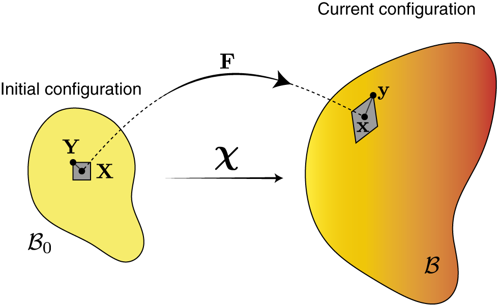
The deformation gradient tensor is a central concept in the mathematical study of body shape changes, characterizing the linear transformation undergone by infinitesimal volumes within the solid. More precisely, let
$\mathbf {X}$
 and
$\mathbf {Y} = \mathbf {X}+\Delta \mathbf {X} $
 be a pair of material points located in the growing body at an initial time
$t=0$
 . After growth, at a given time
$t\geq 0$
 , these two material points have moved to two new positions in space,
$\mathbf {x}$
 and
$\mathbf {y} = \mathbf {x} + \Delta \mathbf {x}$
 . The tracking of material points in time is represented by a mapping
$\chi $
 , such that
$\mathbf {x} = \chi \left ({\mathbf {X},\ t}\right )$
 and
$\mathbf {y} = \chi \left ({\mathbf {Y},\ t}\right )$
 . If
$\mathbf {X}$
 and
$\mathbf {Y}$
 are close to each other (in the sense that
$\Delta \mathbf {X} \rightarrow \mathbf {0}$
 ), the two vectors
$\Delta \mathbf {X}$
 and
$\Delta \mathbf {x}$
 are linked through the deformation gradient
$\mathbf {F} (\mathbf {X},\ t)$
 at
$\mathbf {X}$
 via the relation

(Figure 1). Here,
$\mathbf {F} (\mathbf {X},\ t) = {\partial {\chi }}/{\partial {\mathbf {X}}}$
 is the differential of
$\chi $
 with respect to the material position
$\mathbf {X}$
 , which describes the integrated local expansion and rotation of material lines around the point
$\mathbf {X}$
 of interest, between times zero and t (Goriely, Reference Goriely2017; Holzapfel, Reference Holzapfel2000). In three-dimensional space,
$\mathbf {F}$
 is a
$3\times 3$
 tensor. In one dimension, it reduces to the derivative
$ F(X,\ t) = {\partial {\chi }}/{\partial {X}} \approx \Delta x / \Delta X$
 .
Deformation of the initial configuration
$\mathcal B_0$
 by the smooth map
$\chi $
 to the current configuration
$\mathcal B$
 . The tensor
$\mathbf {F}$
 is the gradient of deformation tensor.

By differentiating
$\mathbf {F}$
 with respect to time, one obtains a rate of deformation tensor, defined as
$\mathbf {L} := \mathbf {\dot F}\mathbf {F}^{-1}$
 (Holzapfel, Reference Holzapfel2000), with the overdot denoting differentiation with respect to time t. Indeed, this definition can be identified with the gradient of the spatial velocity field
$\mathbf {v} (\mathbf {x},\ t)$
 (i.e., the instantaneous velocity of the material at a location
$\mathbf {x}$
 ) as
$\mathbf {L} = {\partial {\mathbf {v}}}/{\partial {\mathbf {x}}}$
 , providing a measure of how nearby points move relative to one another in physical space.
The success of continuum kinematics in the context of growth are not surprising:
“Like a flame or the wake of a boat, the form of a plant changes slowly, but the components are in continual flux. The motions of the components can therefore be analysed in terms of fluid flow.” (Erickson & Silk, Reference Erickson and Silk1980)
Following this analogy, empirical maps of strain rate and vorticity (rate of local rotation) have been constructed, for example, in leaves (Alim et al., Reference Alim, Armon, Shraiman and Boudaoud2016; Derr et al., Reference Derr, Bastien, Couturier and Douady2018; Richards & Kavanagh, Reference Richards and Kavanagh1943; Rolland-Lagan et al., Reference Rolland-Lagan, Coen, Impey and Bangham2005; Silk & Erickson, Reference Silk and Erickson1979; Wolf et al., Reference Wolf, Silk and Plant1986). Progress in microscopy and image analysis further enabled quantitative kinematic analysis at the cell scale, now a primary means of investigation in modern plant developmental biology (Barbier de Reuille et al., Reference Barbier de Reuille, Routier-Kierzkowska, Kierzkowski, Bassel, Schüpbach, Tauriello, Bajpai, Strauss, Weber, Kiss, Burian, Hofhuis, Sapala, Lipowczan, Heimlicher, Robinson, Bayer, Basler, Koumoutsakos and Smith2015; Dumais & Kwiatkowska, Reference Dumais and Kwiatkowska2002; Kwiatkowska & Dumais, Reference Kwiatkowska and Dumais2003). These various works have established the importance of quantifying form to understand its origin. However, the sole description of geometry and kinematics lacks the explanatory power of scientific models, which seek to capture phenomena in light of their causes, aiming not only to describe observed changes in form but also to account for their origin.
The origin of form is a longstanding problem in biology. Against the current of the Darwinian revolution, On Growth and Form revisits the old concept of form through the lens of material sciences, interpreting growing bodies as ‘diagrams of forces’. Richards and Kavanagh (Reference Richards and Kavanagh1943) later articulated this view, noting that:
Geometrical change alone [
$\ldots $
 ] may not give a completely satisfactory picture of the underlying growth activity. The change in size at a given point is due to both the functional activity of the cells located there and to the forces of stretch or compression exerted by the adjacent material.
This perspective has motivated the study of morphogenesis through physical and mechanical principles. In parallel, another tradition in developmental biology has approached morphogenesis from the standpoint of genes, their evolution and their expression. In the next few sections, we outline how these two views combine into a theory of form.
3. Morphogenetics: The chemical basis of morphogenesis
A chief approach to elucidating the mechanisms of morphogenesis in plants has centred on the study of chemical fields, for example, genes and hormones, viewed as determinants of growth. Prior to modern genetics and cell biochemistry, such a perspective appeared in botany in classical studies of tropism. In the 1870s, Charles Darwin and his son Francis explored phototropism, the bending of shoots towards light (Darwin, Reference Darwin1880). They proposed that a ‘substance’ propagated from the photosensitive apex of the shoot down to the growing regions, where bending occurs. This research culminated with the hormonal theory of tropism established based on work by Boysen Jensen, Cholodny, Thimann and Went at the beginning of the 20th century, and the isolation of auxin by Kögl and Haagen-Smit (Arteca, Reference Arteca1996; Whippo & Hangarter, Reference Whippo and Hangarter2006). Auxin is now associated with virtually all morphogenetic processes. In tropism, shoot curvature arises from differential growth of the tissue stimulated by heterogeneous auxin distribution (Moulton et al., Reference Moulton, Oliveri and Goriely2020; Muday, Reference Muday2001). The general mechanisms of spatial organization of auxin have become a crucial area of study in plant development, for example, in leaves (Bilsborough et al., Reference Bilsborough, Runions, Barkoulas, Jenkins, Hasson, Galinha, Laufs, Hay, Prusinkiewicz and Tsiantis2011; Scarpella et al., Reference Scarpella, Barkoulas and Tsiantis2010) and roots (Band et al., Reference Band, Wells, Fozard, Ghetiu, French, Pound, Wilson, Yu, Li, Hijazi, Oh, Pearce, Perez-Amador, Yun, Kramer, Alonso, Godin, Vernoux, Hodgman and Bennett2014; Grieneisen et al., Reference Grieneisen, Xu, Marée, Hogeweg and Scheres2007), or in phyllotaxis (Traas, Reference Traas2013; Vernoux et al., Reference Vernoux, Besnard and Traas2010, Reference Vernoux, Besnard and Godin2021).
Generally, a defining property of life lies in the ability of chemical determinants of development to self-organize in space to generate complex forms. How chemical patterning emerges is a key problem in development: How do cheetahs get their spots? How does the Drosophila embryo segment itself? How do Fibonacci spirals form at the surface of a fir cone? How do we get five fingers on each hand and thirty-two teeth in our mouths?
In his celebrated paper The chemical basis of morphogenesis, Alan Turing proposed a theoretical framework for studying such questions mathematically (Turing, Reference Turing1952). Turing posited a generic system of partial differential equations describing the concentrations of
$n\geq 2$
 chemicals interacting locally, and diffusing in the domain of the tissue. Remarkably, under certain theoretical conditions, these species may behave in such a way that the uniform state (which in the absence of diffusion would be stable) becomes unstable and gives way to a spontaneous, spatially heterogeneous pattern (cf. Murray, Reference Murray2003, Chap. 2). Turing then postulated that a family of growth determinants (broadly speaking, morphogens) capable of self-organizing heterogeneously could serve to generate complex forms by stimulating growth in a non-uniform manner.
Turing’s equations, and reaction–diffusion systems more generally, show how diffusion – a process usually associated with homogenization – can instead generate spatial heterogeneity. In doing so, they provide a classic example of emergence in biophysics.
Phyllotaxis provides a paradigm example of such dynamical self-organization in plants (Godin et al., Reference Godin, Golé and Douady2020). Turing developed an early interest in the topic, which he recognized as a promising application for a morphogen-based model (a work cut short by his death in 1954 and published posthumously as fragmented notes in 1992, albeit with relatively marginal impact; cf. Rueda-Contreras & Aragón, Reference Rueda-Contreras and Aragón2014; Swinton, Reference Swinton and Teuscher2004). Several authors have since extended his approach through more or less detailed continuum models (Meinhardt et al., Reference Meinhardt, Koch, Bernasconi, Barabe and Jean1998; Newell et al., Reference Newell, Shipman and Sun2008; Rueda-Contreras et al., Reference Rueda-Contreras, Romero-Arias, Aragon and Barrio2018; Smith et al., Reference Smith, Kuhlemeier and Prusinkiewicz2006b) or through discrete cell-scale descriptions (Barbier de Reuille et al., Reference Barbier de Reuille, Bohn-Courseau, Ljung, Morin, Carraro, Godin and Traas2006; Cieslak et al., Reference Cieslak, Runions and Prusinkiewicz2015; Hartmann et al., Reference Hartmann, Barbier de Reuille and Kuhlemeier2019; Jönsson et al., Reference Jönsson, Heisler, Shapiro, Meyerowitz and Mjolsness2006; Smith et al., Reference Smith, Guyomarc’h, Mandel, Reinhardt, Kuhlemeier and Prusinkiewicz2006a) integrating polar auxin transport by PIN proteins. The broad paradigm of spontaneous morphogen organization through inhibitory fields has become increasingly influential in phyllotaxis, with auxin–PIN interactions now a central focus of research (Traas, Reference Traas2013; Vernoux et al., Reference Vernoux, Besnard and Godin2021).
Yet within the context of morphogenesis, the knowledge of the spatial organization of morphogens does not directly reveal the form of the organism that arises from this organization. Turing already had a good grasp of this issue, which he described as “a problem of formidable mathematical complexity”:
In determining the changes of state one should take into account: (i) the changes of position [
$\ldots $
 ] as given by Newton’s laws of motion; (ii) the stresses as given by the elasticities [
$\ldots $
 ] taking into account the osmotic pressures as given from the chemical data; (iii) the chemical reactions; (iv) the diffusion of the chemical substances [
$\ldots $
 ] (Turing, Reference Turing1952)
This problem is inherently mechanical and especially hard; thus, as he recommended, it often necessitates the use of digital computers, a path followed with great flair by his successors.
In their paper The genetics of geometry, Coen et al. (Reference Coen, Rolland-Lagan, Matthews, Bangham and Prusinkiewicz2004) proposed a simple computational paradigm for linking genes to form. In this model, the action of genes is to control the local kinematic properties of growth directly. For example, a given gene with a high expression level in a tissue region may result in faster or slower growth. Similarly, the anisotropic expansion of a given region may be prescribed through the action of a growth polarizer defining a preferential growth direction locally.
At the root of this notion is the observation that the development of a general form can be locally broken down into a finite fundamental developmental ‘vocabulary’ given in terms of kinematic descriptors (Section 2): growth rate, anisotropy and direction, plus a rotation. The idea is then to link gene activity to these kinematic variables through explicit constitutive laws. This so-called paradigm of specified growth has since become conceptually influential and has been applied to many case studies, informed by experimental genetics and imaging.
Yet, the link between genes and growth, that is, the causative chain of events connecting cell chemistry to growth mechanics across multiple scales, is not captured in this approach. Thus, such morphogenetic models may be regarded as phenomenological, in the sense that their focus is on the morphological consequences of a given specified growth field, though without express consideration for its physical causes or feasibility.
Mathematically, there exists a fundamental difficulty in specifying growth. Indeed, it is well-known that an arbitrary specification of a strain field does not yield a compatible deformation in general (loosely speaking, the patches do not fit together when rejoined after growth), unless so-called geometric compatibility conditions are met (cf. Barber, Reference Barber2002, Chap. 2). To alleviate this issue, Coen and coworkers introduced the notion of resultant growth, referring to the deformation obtained by computationally correcting the specified growth field at each time step to satisfy these constraints, typically via an additional elastic energy minimization step (Kennaway et al., Reference Kennaway, Coen, Green and Bangham2011). Due to the discrepancy between the (reference) specified and resultant growth, this intermediate step leads to the build-up of internal stress, that is, internal cohesion forces arising from growth incompatibility. However, in most instances considered by authors, this stress is discarded from the computation, so that the resulting configuration at each time step is ultimately stress-free, preventing the accumulation of stress in the tissue as growth simulation proceeds. Then a new specified growth step can be performed, and the process continues iteratively. In practice, this growth procedure is usually formulated algorithmically rather than as a closed mathematical structure, such as a system of partial differential equations, that would be amenable to analysis (in particular, the existence of a continuous-time limit for the stress-release procedure is unclear).
In the study of morphogenesis, a tradition in continuum mechanics gave rise to the theory of morphoelasticity (a term coined by Alain Goriely in his 2005 lecture at the Rencontre du Non-Linéaire in Paris; cf. Goriely & Ben Amar, Reference Goriely, Ben Amar, Chazottes, Joets, Letellier and Ribotta2005). Morphoelasticity is a mechanical theory of growth built upon nonlinear elasticity and plasticity, seeking to formalize the geometry, mechanics and thermodynamics of a growing body mathematically. This approach offers a natural pathway towards a field theory of plant growth.
4. Morphoelasticity: The mechanics of growth
The understanding that form arises from forces has led authors to approach growth as a problem of solid mechanics, an effort that entails formulating appropriate balance laws and examining the physical origin of growth. In this context, the emphasis shifts from defining growth as a change in form, towards defining it more fundamentally as a change in mass, a perspective which offers a more direct link between physiology and form, and a stronger connection with the open-system thermodynamics of a growing body (Ambrosi et al., Reference Ambrosi, Ateshian, Arruda, Cowin, Dumais, Goriely, Holzapfel, Humphrey, Kemkemer, Kuhl, Olberding, Taber and Garikipati2011, Reference Ambrosi, Ben Amar, Cyron, DeSimone, Goriely, Humphrey and Kuhl2019).
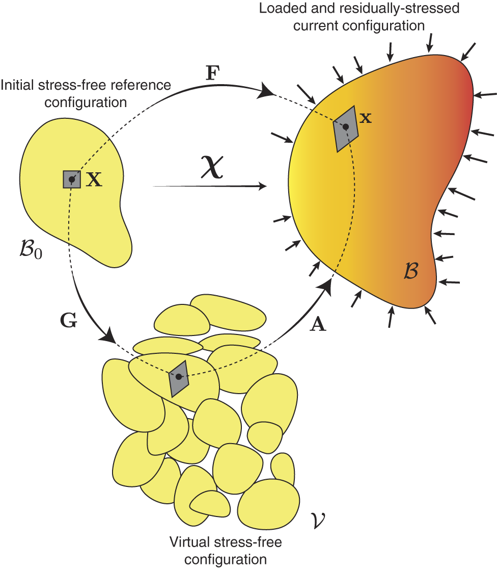
Translating a local gain in mass into a global deformation has been an important problem in mechanics. This has commonly been achieved through the introduction of the growth tensor (Rodriguez et al., Reference Rodriguez, Hoger and McCulloch1994) which quantifies the accumulated change in the resting configuration of a given infinitesimal volume upon local mass accumulation and reorganization. This growth tensor field (henceforth denoted
$\mathbf {G}$
 ) is not compatible in general (i.e., it does not derive from a deformation map; Section 2). Therefore, it is combined with an additional elastic deformation tensor (
$\mathbf {A}$
 ). Constitutively, the deformation gradient
$\mathbf {F}$
 at a given point is thus taken to reflect the two composed contributions given by the product

This instantaneous relation expresses the conceptual hypothesis of morphoelasticity known as multiplicative decomposition (Figure 2). Physically, the growth tensor
$\mathbf {G}$
 captures the slow anelastic expansion of the tissue through mass addition and remodelling. This component is associated with an intermediate, stress-free configuration, often loosely interpreted as a collection of disjoint and stress-free volume elements (cf. Goriely, Reference Goriely2017, Chap. 12). In contrast, the elastic deformation tensor
$\mathbf {A}$
 reflects the rapid elastic deformation of the growing constituents necessary to maintain the integrity of the body. This deformation may be associated with residual mechanical stresses (details on compatibility are given in Jones & Chapman, Reference Jones and Chapman2012 and Goriely, Reference Goriely2017).Footnote
2
The multiplicative decomposition of morphoelasticity. Starting from a stress-free initial configuration, a local growth deformation
$\mathbf {G}$
 is applied on volume elements, resulting in an incompatible intermediate configuration. A second deformation
$\mathbf {A}$
 ensures compatibility and results in a stressed configuration that includes residual growth stresses and external loads.

Figure 2. Long description
The diagram consists of three main shapes representing different states of a body.
* Top-Left: A yellow, bean-shaped body labeled B sub 0, described as the Initial stress-free reference configuration. It contains a small square element labeled X.
* Bottom-Center: A cluster of disconnected yellow oval elements labeled V, described as the Virtual stress-free configuration. A curved arrow labeled G points from the initial configuration to this state.
* Top-Right: A larger, orange-to-red gradient shape labeled B, described as the Loaded and residually-stressed current configuration. It contains a distorted element labeled x. A curved arrow labeled A points from the virtual configuration to this final state. Numerous small black arrows point inward around the perimeter of this shape, representing external loads.
* Direct Mapping: A large curved arrow labeled F spans from the top-left configuration to the top-right configuration. Below it, a straight horizontal arrow labeled chi also connects the two states.
* Internal Correspondence: Dotted lines trace the path of the specific volume element from X in the initial state, through the virtual state, to x in the final stressed state.
The existence of internal mechanical stress in plants has long been acknowledged (Kutschera, Reference Kutschera1989; Kutschera & Niklas, Reference Kutschera and Niklas2007; Lapointe et al., Reference Lapointe, Kaur, Routier-Kierzkowska and Burian2025; Peters & Tomos, Reference Peters and Tomos1996). The general understanding that tissues may be mechanically stressed by growth itself has generated a wealth of problems, particularly around the exploration of growth-induced instabilities – the loss of stability a body experiences at a critical growth threshold leading to a qualitative change in shape, such as buckling (Dervaux & Ben Amar, Reference Dervaux and Ben Amar2008; Guo et al., Reference Guo, Zhang, Paradiso, Long, Hsia and Liu2026; Huang et al., Reference Huang, Wang, Quinn, Suresh and Hsia2018; Liang & Mahadevan, Reference Liang and Mahadevan2009; Sharon & Efrati, Reference Sharon and Efrati2010), wrinkling, creasing (Ben Amar, Reference Ben Amar2025), cusping (Zhang et al., Reference Zhang, Cohen, Moshe and Sharon2025) or tendril perversion (Goriely & Tabor, Reference Goriely and Tabor1998). The mathematical study of growth-induced instabilities has provided crucial tools to understand the solid mechanics of morphogenesis, and the role of incompatibilities in the emergence of complex forms.
In their simplest instance, morphoelastic models have focused on a growth field prescribed as a bifurcation parameter governing the onset of instability. In other words, their focus is on the elasticity problems generated by the presence of a growth field, rather than the origin or dynamics of this growth field. In contrast, the problem of morphodynamics is to model morphogenesis as a dynamical system in which growth combines with other coupled state variables, such as mechanical stresses and morphogen fields, to form a coupled theory of growth.
5. Morphodynamics: Towards a coupled theory
The general paradigm of morphodynamics is to describe the emergence of form as a coupled dynamical system, with growth coupled with other variables. This perspective builds upon a self-organizing view of developing life where growth emerges as the manifestation of more fundamental physical processes operating within the body. Mathematically, the general problem is then (i) to integrate the physical mechanisms affecting the growth dynamics and (ii) to formulate growth laws which couple the growth kinetics to other evolving variables of the system.
For instance, in plant tropism, a shoot detects an external stimulus (e.g., gravity or light) through specialized cells, which, in response, affect the distribution of auxin in the tissue. Then, auxin stimulates differential growth in the tissue, eliciting curvature and altering the plant’s overall posture with respect to the stimulus. This loop can be captured through multiscale modelling and its emergent dynamics can be studied (Chauvet et al., Reference Chauvet, Moulia, Legué, Forterre and Pouliquen2019; Moulton et al., Reference Moulton, Oliveri and Goriely2020; Oliveri et al., Reference Oliveri, Moulton, Harrington and Goriely2024). In growing tissues, various feedback mechanisms have been postulated between stresses and cell mechanical (Hamant et al., Reference Hamant, Heisler, Jönsson, Krupinski, Uyttewaal, Bokov, Corson, Sahlin, Boudaoud, Meyerowitz, Couder and Traas2008) and chemical (Heisler et al., Reference Heisler, Hamant, Krupinski, Uyttewaal, Ohno, Jönsson, Traas and Meyerowitz2010; Nakayama et al., Reference Nakayama, Smith, Mandel, Robinson, Kimura, Boudaoud and Kuhlemeier2012) polarities, and models have been designed to study the behaviour of these feedbacks (Alim et al., Reference Alim, Hamant and Boudaoud2012; Bozorg et al., Reference Bozorg, Krupinski and Jönsson2014; Fruleux & Boudaoud, Reference Fruleux and Boudaoud2019; Hamant et al., Reference Hamant, Heisler, Jönsson, Krupinski, Uyttewaal, Bokov, Corson, Sahlin, Boudaoud, Meyerowitz, Couder and Traas2008; Hervieux et al., Reference Hervieux, Dumond, Sapala, Routier-Kierzkowska, Kierzkowski, Roeder, Smith, Boudaoud and Hamant2016; Khadka et al., Reference Khadka, Julien and Alim2019; Oliveri et al., Reference Oliveri, Traas, Godin and Ali2018; Ramos et al., Reference Ramos, Maizel and Alim2021; Zhao et al., Reference Zhao, Du, Oliveri, Zhou, Ali, Chen, Feng, Wang, Lü, Long, Schneider, Sampathkumar, Godin, Traas and Jiao2020). In these scenarios, growth dynamics results from the integration of multiple factors, with their combined effect giving rise to non-trivial emergent properties.
More fundamentally, growth itself is a mechanical phenomenon, and a crucial problem is to couple the rate of tissue expansion to more fundamental physical and mechanical fields (Vandiver & Goriely, Reference Vandiver and Goriely2009). This is the problem of the growth law. A common perspective has been to view cell wall expansion as a plastic-like yield to pressure forces, combined with remodelling through secretion of new wall material. While the molecular details of this expansion are relatively well described (Cosgrove, Reference Cosgrove2005, Reference Cosgrove2018), how exactly this anisotropic yield occurs, and how to model it in multiple dimensions and at the scale of the entire cell or tissue, is rather unclear. Detailed homogenized models for wall expansion in single cells offer a valuable step towards a physical theory, by integrating explicitly the rheology of wall microstructural components and their yield dynamics (Dyson et al., Reference Dyson, Band and Jensen2012; Smithers et al., Reference Smithers, Luo and Dyson2024), however typically in one dimension. Anisotropy, a distinctive feature of plants that makes their mechanics especially challenging, has yet to be modelled satisfactorily, and tissue-scale computational models have therefore typically represented wall expansion in multiple dimensions using more phenomenological approaches. In particular, linear strain-based growth laws easily account for the role of cellulose microfibrils in modulating anisotropic growth (Boudon et al., Reference Boudon, Chopard, Ali, Gilles, Hamant, Boudaoud, Traas and Godin2015; Bozorg et al., Reference Bozorg, Krupinski and Jönsson2016; Silveira et al., Reference Silveira, Collet, Haque, Lapierre, Bagniewska-Zadworna, Smith, Gosselin, Routier-Kierzkowska and Kierzkowski2025). However, biophysically, such phenomenological laws are insufficiently connected to cell wall micromechanics (Cosgrove & Coen, Reference Cosgrove and Coen2025), and, further, are not thermodynamically equivalent to a dissipative plastic yield because they may necessitate an entropy sink to operate (Oliveri & Cheddadi, Reference Oliveri and Cheddadi2025). Alternatively, authors have adapted the theory of linear plasticity (Dumais et al., Reference Dumais, Shaw, Steele, Long and Ray2006) or viscous fluids (Chakraborty et al., Reference Chakraborty, Luo and Dyson2021; Dyson & Jensen, Reference Dyson and Jensen2010) directly to plant walls in the context of single cells. Overall, determining an appropriate and broadly accepted growth law for cell walls remains an open problem.
Building growth models in a continuum where cells are not explicitly represented is especially difficult. To circumvent this challenge, multicellular computational models have been developed to represent the effect of pressure on individual cells explicitly (e.g., Boudon et al., Reference Boudon, Chopard, Ali, Gilles, Hamant, Boudaoud, Traas and Godin2015; Cheddadi et al., Reference Cheddadi, Génard, Bertin and Godin2019; Dupuy et al., Reference Dupuy, Mackenzie, Rudge and Haseloff2008; Fozard et al., Reference Fozard, Lucas, King and Jensen2013; Merks et al., Reference Merks, Guravage, Inzé and Beemster2011; Rudge & Haseloff, Reference Rudge and Haseloff2005). These models provide a refined mechanistic view of tissue expansion, enabling a more explicit integration of the wall mechanical properties and cellular topology, and avoiding the need to prescribe growth directly. However, while providing detailed insight, they are inherently computational and lack the generality, minimalism, scalability and analytic tractability afforded by a continuum mathematical framework.
Further, turgor pressure in most of these models is treated as a non-dynamical component, either constant in every cell, or enforced as an explicit function of time. These models may therefore be viewed as specified-turgor models. While this simplification largely facilitates the treatment of these systems, as resolving for pressure can turn difficult numerically, it nonetheless runs counter to general mechanical sense. In mechanics, hydrostatic pressure is a variable associated with a volume conservation relation; thus, it is typically not directly prescribable. A critical reevaluation of the role of water in growth and the nature of turgor is emerging in the community at both experimental and theoretical levels (Ali et al., Reference Ali, Cheddadi, Landrein and Long2023; Alonso-Serra, Reference Alonso-Serra2026; Alonso-Serra et al., Reference Alonso-Serra, Cheddadi, Kiss, Cerutti, Lang, Dieudonné, Lionnet, Godin and Hamant2024; Cheddadi et al., Reference Cheddadi, Génard, Bertin and Godin2019; Dumais, Reference Dumais2021; Laplaud et al., Reference Laplaud, Muller, Demidova, Drevensek and Boudaoud2024; Long et al., Reference Long, Cheddadi, Mosca, Mirabet, Dumond, Kiss, Traas, Godin and Boudaoud2020; Oliveri & Cheddadi, Reference Oliveri and Cheddadi2025; Zhang et al., Reference Zhang, Ramakanth and Long2024). In the next section, we discuss the challenge of modelling turgor pressure and how this discussion can serve as a basis to advance plant modelling towards a hydromechanical theory of morphogenesis.
6. Multiphysics: Towards a hydro-chemo-mechanical theory
6.1. The hydromechanical basis of cell expansion
The development of a physically grounded theory of morphogenesis, whether discrete at the cellular scale or continuum-based, requires a focus on the basic cellular physiology of growth.
The understanding that mechanical forces associated with turgor pressure power the expansion of cells traces back to the work of Schwendener (Reference Schwendener1878) and Sachs (Reference Sachs1882) in the 19th century (cf. Hamant & Traas, Reference Hamant and Traas2010). This notion, referred to as turgor-driven growth, is now relatively well-accepted; yet, its details and interpretation have sometimes generated confusion, and ignited various debates, for example, around the notion of the driving force of growth, between physiologists Hans Burström, and Peter Ray, Paul Green and Robert Cleland, as reflected in correspondences published in Nature (Burström, Reference Burström1971; Ray et al., Reference Ray, Green and Cleland1972). In his 1971 letter, Burström expresses scepticism regarding the prevailing notion of turgor-driven growth, observing that
The rigidity of the walls preventing the entry of water is the cause of the turgor pressure [
$\ldots $
 ] The driving force of any expansion is a difference in water potentials. Expansion is due to water uptake. (Burström, Reference Burström1971)
As a provocative conclusion, he recommends that
“The literature on plant cell growth would certainly improve if the notion of turgor expanding the cell was abandoned and replaced by accepted equations for water balance of fluxes.”
Burström’s perspective is partly correct: fundamentally, cells grow by absorbing water, which both causes and controls the build-up of turgor pressure, a process governed by mass balance. Indeed, hydrostatic pressure builds up as an effect of cell walls mechanically resisting water uptake. But without an explicit constitutive assumption for the cell wall – which must expand to allow for water intake – this principle alone is insufficient to complete the picture. In their response, Ray et al. write:
Burström fails to come to grips with the principle that irreversible increase in plant cell volume involves simultaneous water uptake (driven by a water potential difference) and cell wall yielding that depends on turgor stress, and in this sense is ‘driven by’ turgor pressure. (Ray et al., Reference Ray, Green and Cleland1972)
They conclude – this time advancing a more explicit hierarchy – that “clearly stress relaxation is the primary event in cell enlargement, whereas water uptake, volume increase and extension (strain) of the cell wall are secondary.” Here, Ray and colleagues emphasize the rheological nature of growth, noting that for cells to expand, their walls must yield to make room for water, a phenomenon indeed caused by turgor.
At the root of these controversies lies the model proposed a few years earlier by Lockhart (Reference Lockhart1965), which provides a systematic bridge between wall mechanics, water uptake and turgor. Lockhart’s seminal approach has seen numerous extensions, notably by Cosgrove (Reference Cosgrove1981) and Ortega (Reference Ortega1985), somewhat in a similar manner. The extended model can be summarized as follows. We consider the elongation of a long cylindrical cell of length
$\ell $
 , wall thickness
$\delta $
 , cross-sectional perimeter
$\mathcal P$
 and cross-sectional area
$\mathcal A$
 (such that
$\delta \ll \mathcal A/\mathcal P \ll \ell $
 ). We introduce the cell volume
$\mathcal V$
 and cell outer surface area
$\mathcal S$
 .
The expansion rate
$\dot \ell $
 of the cell can be expressed in terms of the volumetric flux of water across the thin wall, through the balance of mass equation (Dainty, Reference Dainty and Preston1963)

with
$k^*$
 the hydraulic conductivity of the cell wall;
$\pi $
 and p, respectively, the excess osmotic and hydrostatic pressures relative to the outside. The r.h.s. in (3) corresponds to the osmotic influx of water; with the quantity
$\psi = p-\pi $
 denoting the water potential of the cell relative to the outside, measuring the free energy of water (Forterre, Reference Forterre, Jensen and Forterre2022; Niklas & Spatz, Reference Niklas and Spatz2012; Nobel, Reference Nobel2020). Using
$\mathcal V=\mathcal A\ell $
 and
$ \mathcal S \approx \mathcal P \ell $
 and rearranging the terms, we obtain

with
$k:= k^* \mathcal P / \mathcal A \delta $
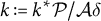 the effective hydraulic conductivity of the cell.
Note that (4) does not define a closed system, as the pressure p is related to the mechanics of the cell wall via the balance of forces between the wall and the water content. To allow for water influx, the cell wall must expand, which involves loosening, yielding under tension and remodelling of the cell walls (Cosgrove, Reference Cosgrove2005). By differentiating the multiplicative decomposition (2) with respect to time in this one-dimensional scenario, we obtain the kinematic relation

involving the (small) elastic strain
$\varepsilon _{\mathrm {e}} \ll 1$
 and the growth multiplier
$\gamma $
 measuring the accumulated elongation due only to irreversible processes. Postulating linear growth above a strain threshold
$\varepsilon _{\mathrm {y}}$
 , we introduce the constitutive growth law

with
$\left ({x}\right )_+:=\max \left ({x,0}\right )$
 the ramp function; and
$\tau $
 a chemo-mechanical characteristic time associated with wall yield, synthesis and remodelling process. From (5) and (6), we obtain an effective visco-elasto-plastic rheological law for the cell wall,

In this cylindrical cell considered in quasi-static equilibrium, the pressure p and longitudinal wall stress
$\sigma $
 are linked through

On combining this relation with Hooke’s law,

which links tension to elastic strain via the cell-wall Young’s modulus
$E^*$
 , we obtain the pressure–strain relation
$p = E\varepsilon _{\mathrm {e}}$
 , with
$E:= E^* \mathcal P\delta /\mathcal A$
 measuring the cell’s effective elastic stiffness under pressure loads. Thus, (7) can be recast as

with
$y := E\varepsilon _{\mathrm {y}}$
 a yield threshold pressure; and
$\phi := (E\tau )^{-1}$
 the so-called extensibility of the cell. Equations (4) and (10) now form a closed system for
$\ell $
 and p, for which a solution is straightforward to derive.
In the steady growth regime with constant pressure (
$\dot p = 0$
 ), p is fully determined by the three parameters y,
$\pi $
 and k as

and we obtain Lockhart’s equation from (10) and (11):

This single equation captures the cell elongation under quasi-static pressure conditions, expressed in terms of physiological and rheological parameters. Importantly, (12) applies only to a single elongating cell. This is, for instance, the case of hair cells of cotton, which can increase their volume by up to 1,000-fold compared to their initial meristematic size (Cosgrove, Reference Cosgrove2005; Hernández-Hernández et al., Reference Hernández-Hernández, Marchand, Kiss and Boudaoud2024; Ruan et al., Reference Ruan, Llewellyn and Furbank2001).
Turgor pressure here results from an equilibrium between mechanical (elastic) and osmotic forces controlled by
$\phi $
 and k through (11). By comparing the magnitude of these parameters, we identify two distinct regimes: (i) a wall-limited regime where
$\phi \ll k$
 and
$p \approx \pi $
 and (ii) a flux-limited regime with
$\phi \gg k$
 and
$p \approx y$
 (Ali et al., Reference Ali, Cheddadi, Landrein and Long2023; Cheddadi et al., Reference Cheddadi, Génard, Bertin and Godin2019; Dumais, Reference Dumais2021). In intermediate situations, we have
$y \leq p \leq \pi $
 (if
$y>\pi $
 , the cell cannot grow). A common, albeit debated assumption is that, in most scenarios relevant to morphogenesis, the cell operates in the wall-limited regime, so that the approximation

holds. In this situation, the cell is close to hydraulic equilibrium and turgor pressure is fully prescribed by the cell chemistry via
$\pi $
 . Yet, the relative contributions of water fluxes and wall synthesis to growth control remain unclear, and recent works tend to build a more nuanced picture where hydraulic resistance cannot be neglected (Laplaud et al., Reference Laplaud, Muller, Demidova, Drevensek and Boudaoud2024). In this scenario, growth, sustained through continuous osmolyte supply, maintains cells in a state of hydraulic imbalance with
$p < \pi $
 . In other words, growing cells are out-of-equilibrium systems, in which the chemical energy from the osmolytes is dissipated through water transport and wall extension. Growth and turgor both arise as emergent properties of this process.
We can now revisit Burström’s objection: Since turgor is a dynamical variable and lacks a straightforward correlation with growth rate, the notion of turgor-driven growth can contribute to an inaccurate picture of pressure viewed as an external force decoupled from the mechanics of the cell and from water transport.
6.2. From cell physiology towards a multiphysics theory of tissue morphogenesis
To advance our understanding of tissue growth, we need multiphysics theories that integrate the physical and mechanical processes that control morphogenesis within a single closed mathematical framework. Specifically, the challenge in plants is to integrate morphogens, growth, mechanics and hydraulics on the basis of physical principles. To that end, we must first revisit the specified-growth paradigm, which has proved highly valuable for linking pattern to form, but does not explicitly include detailed mechanisms of growth, which limits its predictive power. Many authors (Section 5) have sought to address this limitation by proposing mechanical frameworks where growth arises explicitly from wall yield by the action of turgor forces. However, as mentioned, these models have relied on prescribing turgor, thus, they do not account for potential hydromechanical effects in morphogenesis, which may potentially lead to misleading interpretations when such effects are relevant. The phenomenology of the Lockhart–Cosgrove–Ortega model is a good first step towards a non-prescribed theory of turgor and growth in tissues.
Lockhart’s model provides a physiology-based paradigm for plant growth that is now widely accepted in the plant community and has been directly extended to various applications, including growth models at the organismal scale, for example, modelling a fruit as a single Lockhart-type compartment (Fishman & Génard, Reference Fishman and Génard1998). Such compartment-based models seek to describe water and sugar relations between sinks (growing organs and fruits) and sources (roots and leaves) in complex organs, yet without any explicit dependency on the geometry. Other authors have attempted to extend Lockhart’s model directly to a phenomenological tensorial growth law for a continuum; however, this approach relies on an ad hoc tensorial notion of pressure which does not have a clear mechanical basis (Lewicka & Pietruszka, Reference Lewicka and Pietruszka2007; Pietruszka & Lewicka, Reference Pietruszka and Lewicka2007).
We stress that Lockhart’s model describes a cylindrical cell expanding longitudinally, for which the stress–pressure relation is independent of the length of the cell, yielding a description of growth kinetics directly in terms of pressure. In particular, the behaviour of this cell is described in terms of effective parameters k,
$\phi $
 , y and E, which are not intensive properties, as they depend on the cross-sectional geometry of the cell. Such a description does not directly apply to other cell shapes where the surface-to-volume ratio varies with growth and subtle geometric effects may influence the stress–pressure relationship as well (this is familiar to anyone who has blown into a spherical rubber balloon, where pressure first increases up to a threshold in radius, and then decreases). Thus, any effort to extend the hydromechanical phenomenology of Lockhart’s model should start by separately describing (i) water transport and gain (balance of mass), as in (3); (ii) mechanical equilibrium (balance of momentum) between cell pressure and wall stress, as in (8) and (iii) cell wall growth and elasticity constitutive relations, as in (6) and (9).
This principle is crucial when moving to multicellular tissues, where collective hydromechanical effects can emerge over finite spatial scales. Lockhart predicted that, in a tissue, “[t]he resistance of the plant to the flow of water from the source of water to the growing tissue will, in general, exert a marked influence on cell elongation” (Lockhart, Reference Lockhart1965). In contrast to a single cell growing on a hydrated medium, the growth of a region within a tissue may be hindered by hydraulic effects depending on its location within the tissue. In such a poro-morpho-elastic material, dimensional analysis indicates that pressure should vary over a characteristic hydromechanical length of order
$\sim \sqrt {K G \tau }$
 setting the lengthscale of hydraulic interactions (Oliveri & Cheddadi, Reference Oliveri and Cheddadi2025). Here, K and G are, respectively, the bulk water permeability and elastic shear modulus at the tissue scale, and
$\tau $
 the chemo-mechanical characteristic time of cell wall yield, synthesis and remodelling of Section 6.1.
Many works – ours included – using Lockhart’s rheological equation (4) as a premise, typically proceed with a multicellular model in which a fixed pressure is prescribed in every cell, implemented as a constant akin to other constitutive parameters such as the cell extensibility or Young’s modulus, or as an explicit function of time informed by data (Creff et al., Reference Creff, Ali, Bied, Bayle, Ingram and Landrein2023). By construction, such a simplification precludes hydraulic effects and, in this sense, actually departs from Lockhart’s general view. This modelling choice assumes that all cells in the tissue are in the wall-limited regime (13), that is, assuming that the cells grow slowly and close to hydraulic equilibrium. While this simplification likely offers a reasonable approximation in many cases, it immediately excludes a broader spectrum of richer regimes.
The hydrostatic pressure of a cell is linked to the mechanics of cell walls, their geometry, their location within the mechanical context of the tissue and water fluxes governed by balance relations. Any change in these properties, for example, through modifications of cell osmolarity, wall extensibility, permeability or elastic moduli, may, in principle, generate a change in cell pressure. These consequences of Lockhartian hydrostatics have the potential to influence the interpretation of experiments and simulations significantly, underscoring the need for a thorough rethinking and rigorous treatment of pressure in both theoretical and experimental works.
The extent to which water transport shapes morphogenesis remains poorly characterized and is an active area of research. Thus, to deepen our understanding of tissues, a prudent approach is to dispense with the costly assumption of prescribed pressure, with the idea that a sound, parsimonious physical theory should remain agnostic about the ongoing tensions between concurrent biological hypotheses. In recent years, we, with other colleagues, have sought to revive the debate and revisit the dynamical properties of plant matter through the lens of hydromechanical principles.
Cheddadi et al. (Reference Cheddadi, Génard, Bertin and Godin2019) proposed a vertex-based cellular-level model, including hydraulic fluxes between cells, growth and elasticity, thereby extending Lockhart’s approach directly. In this model, wall tensions and pressure are coupled through mechanical equilibrium, and the growth of the cell walls results from wall tension. In a tissue, this tension reflects both the cell hydrostatic pressure and the mechanical influences of neighbouring cells (Boudon et al., Reference Boudon, Chopard, Ali, Gilles, Hamant, Boudaoud, Traas and Godin2015). In all cases, any change of volume has to be accommodated by water flux, as reflected by (4), where pressure appears as a contribution to the cell water potential. Hence, pressure adapts to both mechanical and hydraulic constraints, and it is indeed a dynamical variable. As in Lockhart’s model, turgor pressure is lower than the osmotic pressure for a growing cell: growth brings cells out of equilibrium, so that turgor pressure can become heterogeneous between cells with finite water conductivity.
A striking example of such heterogeneous turgor distribution appears in tissues with heterogeneous cell topology, that is, in irregular tissues where growing cells have a variable number of neighbours (Long et al., Reference Long, Cheddadi, Mosca, Mirabet, Dumond, Kiss, Traas, Godin and Boudaoud2020). In principle, all other things being equal, cell geometry and mechanical balance are expected to result in slower growth of cells with fewer neighbours (Ali et al., Reference Ali, Cheddadi, Landrein and Long2023). Furthermore, these cells are expected to have higher turgor pressure, even though they are hydraulically connected to their neighbours. This phenomenon is well known in foams (Cantat et al., Reference Cantat, Cohen-Addad, Elias, Graner, Höhler, Pitois, Rouyer and Saint-Jalmes2013) and was also indirectly observed in oryzalin-treated meristems where cells with fewer neighbours have a convex shape, bulging outward into their neighbours, potentially indicative of higher turgor (Corson et al., Reference Corson, Hamant, Bohn, Traas, Boudaoud and Couder2009). Long et al. (Reference Long, Cheddadi, Mosca, Mirabet, Dumond, Kiss, Traas, Godin and Boudaoud2020) confirmed this interpretation with atomic-force-microscopy-based measurements of untreated and oryzalin-treated meristems, giving estimates for individual cell pressures. In foams, the pressure difference between bubbles is associated with gas exchanges, and smaller bubbles (with generally fewer neighbours and higher pressure) tend to shrink and eventually disappear, contributing to the growth of their larger neighbours, through the so-called von Neumann–Mullins coarsening (Cantat et al., Reference Cantat, Cohen-Addad, Elias, Graner, Höhler, Pitois, Rouyer and Saint-Jalmes2013). Plant cells differ from bubbles in that they possess an osmotic potential that prevents them from shrinking in general. Yet, one may anticipate that smaller cells would be less able to grow than their larger neighbours. This was observed in oryzalin-treated meristems (Long et al., Reference Long, Cheddadi, Mosca, Mirabet, Dumond, Kiss, Traas, Godin and Boudaoud2020), but surprisingly not in untreated ones. Indeed, smaller cells also benefit from a higher surface-to-volume ratio. In the untreated case, this hydraulic advantage may suffice to overcome the mechanical disadvantage arising from topology. Oryzalin could alter this balance. Interestingly, such a shift between these two regimes was also observed by Tsugawa et al. (Reference Tsugawa, Hervieux, Kierzkowski, Routier-Kierzkowska, Sapala, Hamant, Smith, Roeder, Boudaoud and Li2017) in growing Arabidopsis thaliana sepals, with smaller cells growing faster at early stages and the opposite later on. Whether this shift is regulated by a global change in the hydraulic and/or mechanical properties of the sepal remains unknown. However, these observations combined with modelling, provide a clear example of how moving beyond the prescribed-turgor paradigm (
$p = \pi $
 ) opens a window on new emergent properties and enriches the theoretical vocabulary available to model the regulation and patterning of growth, and thus deepens our understanding of these processes, potentially opening new avenues of experimental investigations.
As previously discussed, pressure adjusts to mechanical constraints, and cells with lower wall tension generally have lower turgor pressure. This effect can appear in primordia at the apical meristem: cells in this region have a much higher growth rate, resulting in the bulging of the primordia (Kwiatkowska & Dumais, Reference Kwiatkowska and Dumais2003), likely caused by cell wall loosening in these cells (Kierzkowski et al., Reference Kierzkowski, Nakayama, Routier-Kierzkowska, Weber, Bayer, Schorderet, Reinhardt, Kuhlemeier and Smith2012; Sassi et al., Reference Sassi, Ali, Boudon, Cloarec, Abad, Cellier, Chen, Gilles, Milani, Friml, Vernoux, Godin, Hamant and Traas2014). Then, lower turgor in these cells results in lower water potential, giving them an additional hydraulic advantage. This effect was analysed by Cheddadi et al. (Reference Cheddadi, Génard, Bertin and Godin2019) who showed that growing primordia act as water sinks that pump water from their neighbourhood in virtue of their lower turgor, in a way reminiscent of the growth-induced water potential concept developed by John Boyer and coworkers (Molz & Boyer, Reference Molz and Boyer1978). Cheddadi et al. (Reference Cheddadi, Génard, Bertin and Godin2019) showed that a situation of scarce water resources further amplifies competition between neighbouring cells. When cell-cell hydraulic connectivity is important, this flux-based lateral inhibition can create a growth rate heterogeneity large enough to generate the sharp change of tissue curvature at the primordium boundary seen in meristems. Strikingly, this effect can even result in boundary cells shrinking. Conversely, if the cells have unlimited access to water or if water exchanges between cells are weak, no hydraulic competition occurs, and the simulated curvature change at the boundary appears shallower.
This consequence of hydraulics was recently confirmed by experimental observations of shrinking cells in the apical meristem at the boundary of growing primordia, which was also shown to contribute to defining the cellular identity of the boundary domain (Alonso-Serra et al., Reference Alonso-Serra, Cheddadi, Kiss, Cerutti, Lang, Dieudonné, Lionnet, Godin and Hamant2024). Here again, the coupling between wall growth and mechanics and water transport affects the growth dynamics and the resulting form deeply. Taken together, these results point to hydraulic coupling as a plausible regulatory mechanism in morphogenetic patterning.
Recently, this modelling work was extended to a continuum theory (Oliveri & Cheddadi, Reference Oliveri and Cheddadi2025), combining morphoelasticity with poroelasticity, a theory of fluid-saturated solids (Forterre, Reference Forterre, Jensen and Forterre2022). This theory is summarized in Figure 3. This work synthesizes previous exploration by Molz and Boyer (Reference Molz and Boyer1978), Molz and Ikenberry (Reference Molz and Ikenberry1974), Molz et al. (Reference Molz, Truelove and Peterson1975), Passioura and Boyer (Reference Passioura and Boyer2003), Philip (Reference Philip1958), Plant (Reference Plant1982), Silk and Wagner (Reference Silk and Wagner1980) and Wiegers et al. (Reference Wiegers, Cheer and Silk2009) and establishes a field theory of growth in plants. In this model, the fluid and solid phases of the tissue are both modelled via explicit balance relations and constitutive assumptions; thus, the movement of water, in relation to the expansion of the tissue, can be included, enabling unbalanced water potentials and heterogeneous pressures. Integrating the phenomenology of Lockhart’s model within a continuum framework enables a mechanistic description of tissue growth dynamics. This approach relies on explicit, coupled laws that link cell expansion to stresses or strains.
This continuum formulation provides an analytic view on the macroscopic laws governing tissue expansion, which feature explicit biophysical parameters, ultimately controlled by genes. For instance, the model yields a complete description of the phenomenon of water competition generated by a heterogeneous stiffness, for which a closed-form asymptotic solution for the pressure and growth profiles can be derived. In contrast to a cell-based computational description, this approach relies on well-established tools of nonlinear solid mechanics in three dimensions, allowing us to describe the finite deformations of entire organs rigorously while freeing us from the constraints of cell-scale modelling. Based on these concepts, Oliveri and Cheddadi (Reference Oliveri and Cheddadi2025) revisited the classic problem of tissue tension in shoots (Peters & Tomos, Reference Peters and Tomos1996) through the lens of hydraulic principles (following an earlier idea by Passioura & Boyer, Reference Passioura and Boyer2003). In contrast to specified-growth models, which are essentially timescale-free, this framework portrays shoot expansion as a dynamic process, in which geometry and kinematics are constrained by hydraulics, defining the shoot’s attainable space of forms.
Poromorphoelastic theory of plant morphogenesis (Oliveri & Cheddadi, Reference Oliveri and Cheddadi2025). The tissue is modelled as a morphoelastic porous medium, capturing the seepage of water across the domain and the mechanics of the solid cell wall matrix. Both components are coupled via the hydrostatic pressure.

Figure 3. Long description
The diagram consists of three main shapes representing tissue states and one square inset for microscopic detail.
1. Top-Left: A yellow amorphous shape labeled Initial stress-free reference configuration and script B sub 0. A small square inside contains a point labeled X.
2. Top-Right: A larger, orange-to-red gradient shape labeled Loaded and residually-stressed current configuration and script B. It is surrounded by inward-pointing black arrows representing external pressure. Inside, blue curved arrows indicate Fluid seepage. A small tilted square contains a point labeled x.
3. Bottom-Right: A square inset labeled Cellular scale, connected by dashed lines to a region in the current configuration. It shows a network of light blue cells with brown walls and small blue arrows indicating local water movement.
4. Bottom-Left: A cluster of individual yellow oval shapes labeled Virtual stress-free configuration and script V.
Mathematical mappings are shown by arrows between the configurations:
- A curved arrow labeled F points from the initial to the current configuration.
- A straight arrow labeled chi points from the initial to the current configuration.
- A curved arrow labeled G points from the initial to the virtual configuration.
- A curved arrow labeled A points from the virtual to the current configuration.
7. Concluding remarks and perspectives
In retrospect, the tensions surrounding the concept of the driving force reflect a definitional – rather than phenomenological – debate: “the difference of opinion hinged on the definition of ‘driving force’ rather than any disagreement about the events that occur during expansion” (Money, Reference Money1997). In plant active matter, the driving force of growth – if we are to hold on to that notion – lies in the metabolic activity of the cells, which work to maintain the chemical potential needed to power their osmotic potential and, consequently, their growth. This energy comes from the environment through photosynthesis. Turgor and growth reflect a complex equilibrium between various chemical and physical processes, as their values emerge from water and solute fluxes, mechanical equilibrium and constitutive laws for cell wall elasticity and anelasticity. From this perspective, plant living matter may be viewed macroscopically as a poro-morpho-elastic material, introducing a characteristic length scale
$\sqrt {K G \tau }$
 , reflecting the coupling of hydraulics (permeability K), mechanics (elastic modulus G) and wall synthesis (characteristic time
$\tau $
 ) defining the typical length of hydraulic interactions. This length may, in principle, govern the water-based morphogenetic patterns emerging through amplification of growth heterogeneities by water fluxes, as proposed in the shoot apex (Alonso-Serra et al., Reference Alonso-Serra, Cheddadi, Kiss, Cerutti, Lang, Dieudonné, Lionnet, Godin and Hamant2024).
Plants display key attributes of active matter: they are open and out-of-equilibrium systems capable of generating non-local emergent behaviours. They respond and adapt their growth to their mechanical environment on their own timescale, as exemplified by the secondary growth of trees, which creep around obstacles in an almost fluid-like manner (a process that calls for a non-prescriptive physical theory). Yet, in contrast to animals and their swirling embryos, the active properties of plants remain subtle and reserved, quietly concealed behind their cellulose curtains. To engage with these properties, we must move beyond linear and reductive conceptions of growth mechanics. The general paradigm and concepts of active matter physics enable a conceptual leap towards a more systematic understanding of plant growth. In particular, the theoretical approaches emphasized in Section 6.2 highlight the inherently out-of-equilibrium nature of growth in plants, where fluxes of mass and energy remain unbalanced. This imbalance is the condition of plants: in a perpetual state of growth, a plant continually evades balance. By incorporating such active properties explicitly, these approaches reveal emergent spatiotemporal couplings that arise directly from hydromechanical principles, for example, water-competition phenomena between different regions of the tissue. Taken together, they offer a more nuanced picture of the physical nature of plants.
The main goals of this perspective paper were (i) to critically reassess key assumptions of plant modelling, especially the specified-growth and prescribed-turgor paradigms, in light of physical principles and recent experiments and (ii) to propose conceptual tools for developing a theory of emergence in plant forms. Important challenges and perspectives towards such a theory include:
-
1. Establishing a growth law based on well-accepted physical processes and first principles, and reproducing a range of experiments. This law should also account for the cellular properties of the tissues systematically; here, multiscale approaches (e.g., Boudaoud et al., Reference Boudaoud, Kiss and Ptashnyk2023; Ghysels et al., Reference Ghysels, Samaey, Van Liedekerke, Tijskens, Ramon and Roose2010) will be valuable.
-
2. Revisiting the relationship between water and growth, experimentally and theoretically. Understanding the role of water transport in morphogenesis, its pathways and regulations.
-
3. Developing a complete description of the thermodynamics of growth and water dissipation within a tissue. A promising avenue is to constrain growth laws thermodynamically by explicitly examining the dissipation resulting from various biological processes, as done, for example, in tumour growth via the so-called Coleman–Noll procedure (Ambrosi and Guana, Reference Ambrosi and Guana2007; Ambrosi et al., Reference Ambrosi, Preziosi and Vitale2012; DiCarlo & Quiligotti, Reference DiCarlo and Quiligotti2002; Dunlop et al., Reference Dunlop, Fischer, Gamsjäger and Fratzl2010; Erlich & Recho, Reference Erlich and Recho2023; Olaranont et al., Reference Olaranont, Wei, Lowengrub and Wu2025; Xue et al., Reference Xue, Li, Feng and Gao2016). Furthermore, other thermodynamics-based analyses at the cell scale may be of interest to achieve such global description (Ali & Traas, Reference Ali and Traas2016; Barbacci et al., Reference Barbacci, Lahaye and Magnenet2013).
-
4. Extending the description of ‘naked’ plant matter to a theory of smart active matter (Levine & Goldman, Reference Levine and Goldman2023), including self-regulation, such as mechanical feedbacks, complex hormonal interactions, ion transport and gene regulation. One question in particular is how water pathways integrate in a broader context of regulation by auxin and PINs, and stress-dependent cell mechanical properties.
One approach emphasized here involves viewing the tissue as a continuum, which yields simplified and mathematically tractable representations focusing on bulk properties rather than cytohistological details. This approach participates in an organismal view of plant multicellularity, in which the organism develops independently of cellular structure, and, conversely, cell proliferation is relatively separate from – or even subordinate to – overall growth (Kaplan & Hagemann, Reference Kaplan and Hagemann1991). This perspective supports a cell-free, solid-mechanics view of plant tissues and argues for shifting attention from individual cells seen as independent agents of form – a view long central to gene-focused analyses of morphogenesis – to the global mechanical properties of tissues:
The development of an organ proceeds with little relation to the manner in which it is cut up into organized cellular units [
$\ldots $
 ] Organization at one level seems independent of that at another. (Sinnott, Reference Sinnott1939)
The effort to formalize the mechanisms of form mathematically through a unified theory of morphogenesis that moves beyond ad hoc simulations is crucial if plant developmental biology is to make a quantitative leap. Overall, our goal is to clarify the physical constraints acting on plant growth and, in doing so, to define more systematically the space of forms achievable under genetic control.
Open peer review
To view the open peer review materials for this article, please visit http://doi.org/10.1017/qpb.2026.10048.
Author contributions
All authors contributed equally.
Funding statement
The authors acknowledge support from the Institut rhônalpin des systèmes complexes (IXXI). I.C. and C.G. acknowledge support from the French National Research Agency (ANR) under Research Grant ANR-20-CE13-0022-03 HydroField.
Non-financial support
This research was supported by a visiting research fellowship for H.O. at the Mathematical Institute, University of Oxford; and by the CNRS Groupement de Recherche Biophysique et biomécanique des plantes (GDR PhyP, No. 2007), in particular through the biennial GDR PhyP, No. 2007 meeting, whose organizers are warmly thanked for fostering discussions that were instrumental to this work.
Competing interests
The authors declare that no competing interests exist.





 Open access


Comments
To the Editor,
Please find enclosed our invited article, “Towards an active matter theory of plant morphogenesis,” submitted for the special issue on “Plant Hydraulics”.
In this contribution, we argue that plant morphogenesis can be reframed within a rigorous active-matter perspective, in which tissues are treated as open, out-of-equilibrium systems powered by metabolism and governed by coupled hydraulic, mechanical, and chemical processes. Building on the history of growth geometry and morphoelasticity, we critically reassess standard “specified growth” and “prescribed turgor” paradigms, and instead outline a hydromechanical field theory that integrates cell imbibition, tissue mechanics, and water fluxes with genetic regulation. Our aim is to clarify the physical basis of plant growth, to formalize growth laws consistent with continuum mechanics and thermodynamics, and to delineate open problems in plant active matter theory.
We believe this perspective article is well aligned with the goals of Quantitative Plant Biology and the theme of the special issue, as it connects classical plant physiology, modern continuum theory, and active matter physics into a unified conceptual framework for future quantitative work. This article should appeal to the broad readership of the journal, especially readers interested in morphogenesis, plant mechanics and modelling.
Thank you very much for your consideration.
Sincerely,
Ibrahim Cheddadi, on behalf of the authors