




No CrossRef data available.
Pulmonary hypertension in infancy and childhood
Published online by Cambridge University Press: 24 May 2005
Abstract
In this review, we discuss current concepts in the pathogenesis and management of pulmonary hypertension affecting infants and children, with special focus on left-to-right shunting, bronchopulmonary dysplasia, and primary pulmonary hypertension.
In patients of these ages, functional aspects, such as an imbalance between vasoconstricting and vasodilating mechanisms, and morphological alterations of the vessel wall, contribute to the pulmonary hypertension. In the past decades, strategies have emerged for treatment that are targeted at the pathophysiological basis. Thus, in patients with left-to-right shunting and pulmonary hypertension after intra-cardiac repair, treatment with nitric oxide has been introduced effectively, while treatment with prostanoids, such as iloprost, is under investigation. In patients with pulmonary hypertension and bronchopulmonary dysplasia, therapeutic strategies focus on the underlying chronic lung disease and use of vasodilators. The pathogenesis of primary pulmonary hypertension in children remains as yet unclear, although treatment with prostanoids has proven effectively to improve the long-term prognosis.
Information
- Type
- Review
- Information
- Copyright
- © 2003 Cambridge University Press
Pulmonary hypertension, when encountered in children, accompanies several conditions that can affect the lung or the pulmonary circulation proper. Pulmonary hypertension itself has been defined as an increase in mean pulmonary arterial pressure greater than 25 mmHg at rest, or greater than 30 mmHg with exercise at cardiac catheterisation.1 The definition was established in adults, but has been adopted for children and infants.2
The hypertension is considered to be primary whenever the elevation in pulmonary arterial pressure cannot be explained by any known cause, or as secondary whenever there is a coexisting disease that can account for the increase in pressure. In 1998, the World Health Organisation proposed a new classification: This has five main categories based on common clinical features:3
- Pulmonary arterial hypertension.
- Pulmonary venous hypertension.
- Pulmonary hypertension associated with disorders of the respiratory system and/or hypoxemia.
- Pulmonary hypertension resulting from chronic thrombotic and/or embolic disease.
- Pulmonary hypertension resulting from disorders directly affecting the pulmonary vasculature.
Most children with pulmonary hypertension usually present with pulmonary arterial hypertension, as in persistent pulmonary hypertension of the neonate, left-to-right shunting, or primary pulmonary hypertension, or they can be assigned to the group of patients with pulmonary hypertension resulting from respiratory disorders, as in bronchopulmonary dysplasia.
In pulmonary hypertension associated with left-to-right shunting or neonatal lung disease, the pulmonary circulation is affected early in life: the physiological process of postnatal structural remodelling of the pulmonary vasculature4 is then distorted. The resistance to pulmonary perfusion, which usually falls to one-tenth of the resistance that can be found in the systemic circulation,5 may remain at a high level. Besides structural remodelling of the vessel wall, functional aspects play an important role in regulating the pulmonary vascular tone. Both active and passive factors influence the balance between vasoconstriction and vasodilation under physiological conditions, as well as in diseases associated with pulmonary hypertension (Fig. 1).6

Figure 1. Important factors influencing the pulmonary vascular tone. Gaseous, humoral and neural factors influence the balance between vasoconstriction and vasodilation.
Oxygen is the most important vasodilating substance for the regulation of pulmonary vascular tone. Its precise mechanism of action remains to be elucidated, but purinergic receptors may participate in the mechanism of oxygen-induced pulmonary vasodilation.7 How the pulmonary vasculature “senses” the decreased tension of oxygen, and how this signal is coupled to initiate the contractile response of the smooth muscle cell, is presently undetermined. Recent studies suggest that the electron transport chain could serve as a sensor of oxygen in acute hypoxic pulmonary vasoconstriction.8, 9 Hypoxia induces the expression of several transcription factors, such as hypoxia-inducible factor-1, which in turn activates the expression of endothelin-1 in vascular endothelial cells.10 Endothelin-1 is a peptide hormone with dual effects on the pulmonary smooth muscle cell. When coupling to the receptor subtype B on the endothelial cell, it leads to production of nitric oxide, followed by smooth muscle relaxation. When binding to the receptor subtype A on the smooth muscle cell, it causes long lasting vasoconstriction. Levels of endothelin-1 are increased in the plasma from patients with primary pulmonary hypertension.11
Atrial natriuretic peptide is a pulmonary vasodilator.6 Levels of this peptide have been correlated with the right ventricular afterload found in patients with pulmonary hypertension.12 Nitric oxide is produced by vascular endothelial cells in response to shear stress produced by the flow of blood. It is an important pulmonary vasodilator, and its role in pulmonary hypertension has been recently reviewed.13 Prostacyclin is synthesised in the endothelial cell from arachidonic acid through the cyclooxygenase pathway.14 The very short half-life of prostacyclin suggests that it acts as a local hormone with vasodilating capacity. The main pharmacological properties of prostacyclin include inhibition of platelet activation, vasodilation, and possibly cytoprotective effects.15 These pharmacological properties have led to the concept of treating patients with pulmonary hypertension with prostanoids, such as prostacyclin, beraprost sodium or iloprost.
Innervation of the pulmonary arterial blood vessels by adrenergic nerves (Fig. 1) exerts an important predominant vasoconstricting activity.6 Increased sympathetic activity, as seen after intra-cardiac repair, may therefore lead to pulmonary arterial vasoconstriction.
Pathology of pulmonary vascular disease
The pathological alterations in pulmonary vascular disease have been intensively studied.16–20 The disease process is characterised by hypertrophy and hyperplasia of smooth muscle cells in the media, cellular intimal proliferation, accumulation of fibrous tissue in the intima, and eventual formation of so-called plexiform lesions (Fig. 2).21

Figure 2. Pathological changes in pulmonary vascular disease: Medial hypertrophy (A), cellular intimal proliferation (B), concentric laminar intimal fibrosis (C), and the plexiform lesion (D).
Although the precise mechanism of the development of plexiform lesions is incompletely understood, it is clear that the structures are composed of phenotypically abnormal endothelial cells.22 It is now recognised, nonetheless, that it is the smooth muscle cells in the pulmonary arterial walls that play a key role in the development of the pulmonary vasculopathy: They de-differentiate, achieving a more synthetic than contractile phenotype, grow into the subendothelial space, and produce the fibrous material responsible for intimal fibrosis.23 Hypertrophy of the medial coat, and cellular intimal proliferation, may reverse in patients with left-to-right shunting after an intra-cardiac repair. Fibrous intimal occlusion, in contrast, is thought to be reversible only when not obstructing the vessel lumen for more than one-fifth.24
Assessment of patients with suspected pulmonary hypertension
A possible diagnostic work-up for children with suspected pulmonary hypertension, proposed by Ivy,25 is summarised in Table 1. The aim of the diagnostic work-up is to find underlying diseases associated with pulmonary hypertension or, by exclusion, to classify patients as having primary pulmonary hypertension. In such patients, the 6-minute walk test is a simple measure of exercise tolerance that has now become widely accepted.26 The child is directed to walk, and not to run, along a marked line of known distance for a maximum time of 6 minutes. The test has its limitations, since the child is not usually monitored during the test, which is subjective and affected by the co-operation of the patient. Because of this, it is not recommended for children under 7 years of age. The 6-minute walk test is a valuable tool that helps to quantify the functional status of the patient. And, once therapy has been initiated, it allows clinical monitoring in the course of the disease.
Table 1. Diagnostic work-up in children with pulmonary hypertension (modified after Ivy25).

In children, the severity of functional impairment can be quantified by using an adaptation of the classification of the New York Heart Association designed to grade heart failure, as modified by a conference convened by the World Health Organisation to discuss pulmonary hypertension in 1998.3 This particular classification cannot be used in infants. Ross and colleagues, therefore, have introduced a scoring system for heart failure in infants.27 This grading system evaluates multiple parameters, such as feeding history, respiratory and heart rate, respiratory pattern, peripheral perfusion, and hepatic size. It can be used to grade the severity of heart failure as “absent”, “mild”, “moderate” or “severe”. It is our opinion that this grading system may also be used to quantify functional impairment in infants with pulmonary hypertension.
Testing for acute pulmonary vasoreactivity in patients with pulmonary hypertension
Haemodynamic data quantifying pulmonary arterial pressure and resistance to pulmonary perfusion are obtained by cardiac catheterisation.28 Moreover, the investigation permits direct study of the effect of different vasodilating substances on the pulmonary haemodynamics in patients with pulmonary hypertension, and identifies a rationale for specific long-term pharmacological treatment: Children with primary pulmonary hypertension who are acute responders to vasodilating substances have a significantly higher five year survival on oral vasodilator therapy compared with non-responders.29 Vasodilator testing is also used to identify patients with pulmonary hypertension and left-to-right shunting that might still be candidates for surgical correction of their defect.30 It is the patients that show a decline in pulmonary arterial pressure, and of pulmonary vascular resistance, of more than 20% from baseline after administration of a specific substance, for example oxygen or nitric oxide, that are classified as “responders”.31
Several protocols have been published using two vasodilating substances in combination, for example inhalation of oxygen together with nitric oxide,30, 32, 33 inhalation of nitric oxide together with prostacyclin given intravenously,33 or inhalation of nitric oxide with beraprost sodium given orally.34 The combination of two vasoactive substances was found to be more effective than using a single substance.30, 32, 34 We currently use a protocol that uses oxygen at first and alone, followed by nitric oxide at 40 parts per million plus oxygen over a period of 15 minutes. In those patients that have not responded to this regime, we favour the nebulization of iloprost plus oxygen.35
Some groups have used sedation with spontaneous breathing during cardiac catheterisation.2, 30 The cardiovascular system, however, is vulnerable in patients with severe pulmonary hypertension.36 Hypoventilation, with retention of carbon dioxide, during sedation may lead to acidosis with cardiocirculatory collapse. We,35 and other groups,33 therefore prefer to use general anaesthesia and mechanical ventilation when assessing our patients.
Specific disease conditions associated with pulmonary hypertension
Pulmonary hypertension in bronchopulmonary dysplasia
Very premature infants are still at risk of developing bronchopulmonary dysplasia.37 The injury to the lungs is characterised by fibrosis, areas of atelectasis, inflammation, and emphysematous changes.38 The infants most severely afflicted by bronchopulmonary dysplasia may also develop pulmonary hypertension and pulmonary vascular disease, characterised by medial hypertrophy and extension of muscle into more peripheral arteries.39 In this setting, angiography reveals rarification of peripheral vessels (Fig. 3), indicating that there is a reduced cross-sectional area for perfusion contributing to the elevation in pulmonary arterial pressure. In addition, the mismatch between ventilation and perfusion contributes to impaired oxygenation of the blood that is passing through the lung. The incidence of pulmonary hypertension secondary to bronchopulmonary dysplasia in preterm infants is not known precisely at present, but many suffer from pulmonary hypertension and cor pulmonale. Oxygen is the most important “drug” in this situation, taking care to avoid hypoxia and concomitant hypoxic pulmonary vasoconstriction, which may worsen the situation. Anticongestive medication should include diuretics, such as spironolactone or furosemide. Digoxin should be administered whenever diuretics alone fail to improve the clinical situation. In addition, nifedipine, at a dose of up to 2 mg/kg body weight given orally, is the most commonly used vasodilating agent.40 Great care must be taken when initiating treatment with nifedipine, since oxygenation may worsen in some cases. Indeed, data from adults with pulmonary hypertension suggests that nifedipine may decrease right ventricular function rather than act as a pulmonary vasodilator.41 In our experience, nonetheless, many babies with bronchopulmonary dysplasia and pulmonary hypertension will improve with this medication.

Figure 3. Post-mortem angiography of an infant aged 3 months without cardiopulmonary disease (A), and an infant with bronchopulmonary dysplasia (B). Note the marked reduction of peripheral vascularisation (“diminished background haze”) in the infant with bronchopulmonary dysplasia.
The role for inhaled nitric oxide remains to be settled.42 Prostanoids, such as inhaled iloprost or intravenous prostacyclin, may cause significant systemic arterial hypotension and are expensive drugs. These prostanoids, therefore, should currently be used only under the condition of a randomised controlled study.
Pulmonary hypertension after surgery for congenital heart disease
Severe postoperative pulmonary hypertension, that is an increase of the ratio of mean pulmonary arterial to mean systemic arterial blood pressure greater than 0.5, contributes significantly to perioperative mortality in patients with left-to-right shunting.43 In its most severe form, the so-called “pulmonary hypertensive crisis”, an acute rise in pulmonary vascular resistance leads to a severe drop in the saturation of oxygen, followed by a fall in systemic blood pressure as a consequence of massive pulmonary vasoconstriction. Factors such as acidosis, pain, or infection may trigger this potentially fatal event, and increased levels of endothelin-1 in the plasma have now been identified in the setting of postoperative pulmonary hypertension.44, 45 The first stage of treatment is to ensure adequate sedation and relaxation. Hyperventilation, and/or administration of sodium bicarbonate, is then used to induce alkalosis, providing inotropes as required to ensure an adequate systemic cardiac output. For several years, inhalation of nitric oxide has been advocated in many centres as a selective pulmonary vasodilator, and its use routinely in infants at high risk for pulmonary hypertension is now supported by the results of a randomised double-blind study.46 Inhaled nitric oxide, however, is far from the ideal pulmonary vasodilator: Rebound pulmonary hypertension may occur when weaning from the gas,47, 48 and its sudden withdrawal may itself lead to a life-threatening pulmonary hypertensive crisis.49, 50 This rebound pulmonary hypertension is most likely due to the suppression of endogenous production of nitric oxide by inhalation of the gas, this interfering with the production of cyclic guanosine monophosphate.51 Recent data also suggests that inhalation of nitric oxide produces an increase in the levels of endothelin-1 circulating in the plasma. When weaning from the inhaled agent, therefore, it may well be this increased level of endothelin-1 that contributes to the rebound pulmonary hypertension.52 We, therefore, restrict the use of inhaled nitric oxide in our cardiac intensive care unit to those patients who, after intracardiac repair, and despite adequate sedation, hyperventilation, and alkalosis, maintain their pulmonary arterial pressures at levels greater than half systemic as measured invasively by arterial lines. Unfortunately, preoperative testing for acute pulmonary vasoreactivity does not yet permit us to predict the degree of postoperative response to inhalation of nitric oxide.53
More recently, endothelin receptors of the subtype A have been shown to be upregulated in patients with severe pulmonary hypertension due to congenital heart disease.54 The antagonist of the subtype A endothelin receptor, BQ123, therefore, has been given in a pilot study in children with postoperative pulmonary hypertension,55 but unfortunately it also induced significant falls in systemic blood pressure and arterial saturation of oxygen.
In another study, including five postoperative children, aerosolised iloprost was found to be just as effective as inhalation of nitric oxide in selectively lowering pulmonary vascular resistance.56 Controlled randomised studies are now required in a much larger cohort of patients with pulmonary hypertension after intracardiac repair to analyse fully the benefit of this substance compared to inhaled nitric oxide.
Primary pulmonary hypertension
The primary form of pulmonary hypertension affects about 1 or 2 persons in each million every year, and continues to be associated with a high mortality.57 Most cases appear to be sporadic, but a familial form accounts for about one-twentieth of cases.1 This familial variant is inherited as an autosomal dominant disorder showing a reduced penetrance, and leads to clinical symptoms in up to one-fifth of family members.58, 59 Significantly, it demonstrates genetic anticipation, leading to a worsening of the disease in younger generations. Mutations in the bone morphogenetic protein receptor II geneline have been detected in over half the familial cases, and also in one-quarter of sporadic cases.60 Thus, many cases previously considered sporadic may, in fact, be familial.58 The gene itself has been mapped by linkage analysis to a locus on chromosome 2q31-32.61 The bone morphogenetic protein receptor II is a ubiquitously expressed receptor for growth factors that belong to the superfamily of transforming growth factor-β. At present, however, there is no clear indication of how the mutations relate to the vascular disease in patients with primary pulmonary hypertension.59
The prognosis of the primary disease, as emphasised, is poor: In one series, which included 77 children ranging in age from 7 months to 13 years, the 5-year mortality rate on conventional therapy for non-responders to acute pulmonary vasodilator testing was 35%.2 The pathogenesis of the primary form is, as yet, not clear: Enhanced platelet activity and endothelial dysfunction, characterised by an imbalance of vasoconstricting substances such as endothelin-1 and thromboxane A2 and vasodilating substances such as prostacyclin and nitric oxide, have been described.62 Levels of Endothelin-1 have also been shown to be increased in the plasma and lung tissue of patients with primary pulmonary hypertension,11 while endothelin-converting enzyme-I has been found to be abundantly expressed.63 Regulation of the potassium channels seems to be distorted in general in patients with pulmonary hypertension, the primary as well as the secondary forms.64
In children, it is syncope, dyspnoea on exertion, chest pain or overt right heart failure that are the leading symptoms.36 Treatment will depend on the functional state of the patient, and on the result of testing for pulmonary vasoreactivity.65 Although there is no controlled data establishing the benefit of anticoagulation in children, most authorities recommend that children in functional classes I and II should be anticoagulated, usually with phenprocoumon or warfarin, with the aim of achieving an international normalised ratio between 2 and 3.25, 65 Supplemental oxygen should certainly be given whenever saturations indicate hypoxemia, typically with the transcutaneous saturation of oxygen measured at less than 92%. Oxygen has been shown effectively to increase survival in patients with pulmonary vascular disease, be it due to cardiac or non-cardiac causes.66 Nocturnal hypoxemia, unrelated to apnoea and hypopnoea, is common in these patients.67 It should be remembered, therefore, that oxygen, when inhaled as the pure gas, is a selective pulmonary vasodilator in patients with pulmonary hypertension regardless of the baseline oxygenation.68
Pastoral care is equally important in treating this difficult condition. Support groups for the patients and their parents now exist in many countries. The local health team is equally important in administering supportive measures. In our opinion, all patients should receive:
- Early treatment of fever with antipyretics to minimise cardiocirculatory stress.
- Early treatment of those infections of the respiratory tract suspicious for a bacterial origin so as to avoid the risk of pneumonia which, if it develops, often proves lethal.
- Annual immunisation against influenza, and a “one-off” pneumococcal immunisation.
- Softening of stools where indicated to avoid cardiocirculatory collapse when passing faeces.
Anticongestive medication, such as spironolactone, furosemide or digoxin, should be administered as required when signs of right heart failure are overt.
The patients in functional class I or II, who respond to vasodilators on cardiac catheterisation, and do not show a marked elevation in right atrial pressure, will benefit from calcium-channel blockers such as nifedipine. Patients who are in functional class III or IV have been shown to improve significantly by the administration of intravenous prostacyclin,2 which has to be administered by a continuous infusion via a portable pump. Catheter-related sepsis and local irritations or infections at the catheter site have been reported. Prostacyclin is also an expensive drug in Europe. This form of treatment also requires co-operation during its administration, and is far from ideal, therefore, when used in children.
In adults, nebulized iloprost,69 and oral prostanoids such as beraprost sodium,70–72 have been shown to be effective, but limited data is available regarding their use in children.73 In Japan, nonetheless, beraprost sodium is licensed for the treatment of primary pulmonary hypertension in both children and adults. In our experience, palliative treatment both with inhaled iloprost and oral beraprost can be effective in these children.74
For the future, the dual endothelin-receptor antagonist bosentan has been shown to effectively improve haemodynamics and exercise capacity,75 and has been approved by the Food and Drug Administration of the United States of America for treatment of primary pulmonary hypertension.76 Multicentric trials are now under way in children. In Europe, however, Bosentan is currently licensed only for use in children older than 12 years presenting with pulmonary hypertension in functional classes III and IV.
Transplantation of the lung, or the heart and lungs, of course, is an obvious treatment for children with severe progressive disease. The shortage of donor organs, nonetheless, and the need for co-operation by the transplanted patient, as well as the poor long-term prognosis,77 are limitations for its more widespread use. In the subgroup of patients with pulmonary hypertension, mortality after one78 or two79 years after transplantation is worse compared with the global mortality of the studied cohort. The type of the transplantation procedure exerts important effects on the outcome: In one series, patients having transplantation of both lungs had an advantage in terms of survival compared with the patients having only one lung transplanted.80 Transplantation, at any event, should only be performed in specialised centres. Current research focuses on the role of gene transfer to the lung,81, 82 and investigation of candidate genes that may have importance for the pathogenesis of the disease.83
Conclusion
Within recent decades, our knowledge of the pathophysiology of pulmonary vascular disease has increased considerably. The initiating triggers, nonetheless, as well as the “final common pathway” that inevitably leads to the morphological and functional alterations, are as yet unknown. A wide range of therapeutic modalities has emerged in the past ten years. Randomised multicentric studies using vasodilating and antiproliferative substances are now clearly needed to evaluate the efficacy of these treatments in children with pulmonary hypertension.
Acknowledgements
We are grateful to Mrs. Gorsberg for perfect technical assistance.
Financial support: Matthias Gorenflo and Michael V. Ullmann were supported in part by a grant from the “Deutsche Stiftung für Herzforschung” (German Heart Foundation; project number: F/02/02).
References
Rich S, Dantzker DR, Ayres SM, et al. Primary pulmonary hypertension. A national prospective study. Ann Intern Med 1987; 107: 216–223.Google Scholar
Barst RJ, Maislin G, Fishman AP. Vasodilator therapy for primary pulmonary hypertension in children. Circulation 1999; 99: 1197–1208.Google Scholar
Rich S. Executive summary from the World Symposium on Primary Pulmonary Hypertension. World Health Organization, Geneva, 1998.
Haworth SG, Hislop AA. Pulmonary vascular development: normal values of peripheral vascular structure. Am J Cardiol 1983; 52: 578–583.Google Scholar
Ghanayem NS, Gordon JB. Modulation of pulmonary vasomotor tone in the fetus and neonate. Respir Res 2001; 2: 139–144.Google Scholar
Barnes PJ, Liu SF. Regulation of pulmonary vascular tone. Pharmacol Rev 1995; 47: 87–131.Google Scholar
Crowley MR. Oxygen-induced pulmonary vasodilation is mediated by adenosine triphosphate in newborn lambs. J Cardiovasc Pharmacol 1997; 30: 102–109.Google Scholar
Sylvester JT. Hypoxic pulmonary vasoconstriction. A radical view. Circ Res 2001; 88: 1228–1230.Google Scholar
Semenza GL. Oxygen-regulated transcription factors and their role in pulmonary disease. Respir Res 2000; 1: 159–163.Google Scholar
Giaid A, Yanagisawa M, Langleben D, et al. Expression of endothelin-1 in the lungs of patients with pulmonary hypertension. N Engl J Med 1993; 328: 1732–1739.Google Scholar
Wiedemann R, Ghofrani A, Weissmann N, et al. Atrial natriuretic peptide in severe primary and nonprimary pulmonary hypertension. J Am Coll Cardiol 2001; 38: 1130–1136.Google Scholar
Beghetti M, Adatia I. Inhaled nitric oxide and congenital cardiac disease. Cardiol Young 2001; 11: 142–152.Google Scholar
Müller B. Pharmacology of thromboxane A2, prostacyclin and other eicosanoids in the cardiovascular system. Therapie 1991; 46: 217–221.Google Scholar
Jones RL, Qian Y, Wong HNC, Chan H, Yim APC. Prostanoid action on the human pulmonary vascular system. Clin Exp Pharmacol Physiol 1997; 24: 969–972.Google Scholar
Heath D, Edwards JE. The pathology of hypertensive pulmonary vascular disease. A description of six grades of structural changes in the pulmonary arteries with special reference to congenital cardiac septal defects. Circulation 1958; 18: 533–547.Google Scholar
Haworth SG. Pulmonary vascular disease in different types of congenital heart disease. Implications for interpretation of lung biopsy findings in early childhood. Br Heart J 1984; 52: 557–571.Google Scholar
Rabinovitch M, Haworth SG, Castaneda AR, Nadas AS, Reid LM. Lung biopsy in congenital heart disease: a morphometric approach to pulmonary vascular disease. Circulation 1978; 58: 1107–1122.Google Scholar
Wagenvoort CA. Classification of pulmonary vascular lesions in congenital and acquired heart disease. Adv Cardiol 1974; 11: 48–55.Google Scholar
Wagenvoort CA, Wagenvoort N. Pathology of the Eisenmenger syndrome and primary pulmonary hypertension. Adv Cardiol 1974; 11: 123–130.Google Scholar
Wagenvoort CA, Mooi WJ, Corrin B. Biopsy pathology of the pulmonary vasculature. (Biopsy pathology series No. 13.) Chapman and Hall Medical, London, 1989, pp 56–113.
Fishman AP. Changing concepts of the pulmonary plexiform lesion. Physiol Res 2000; 49: 485–492.Google Scholar
Allen K, Haworth SG. Cytoskeletal features of immature vascular smooth muscle cells: the influence of pulmonary hypertension on normal development. J Pathol 1989; 158: 311–317.Google Scholar
Wagenvoort CA, Wagenvoort N, Draulans-Noë Y. Reversibility of plexogenic pulmonary arteriopathy following banding of the pulmonary artery. J Thorac Cardiovasc Surg 1984; 87: 876–886.Google Scholar
Ivy D. Diagnosis and treatment of severe pediatric pulmonary hypertension. Cardiol Rev 2001; 9: 227–237.Google Scholar
Garofano RP, Barst RJ. Exercise testing in children with primary pulmonary hypertension. Pediatr Cardiol 1999; 20: 61–64.Google Scholar
Ross RD, Bollinger RO, Pinsky WW. Grading the severity of congestive heart failure in infants. Pediatr Cardiol 1992; 13: 72–75.Google Scholar
Wilkinson JL. Haemodynamic calculations in the catheter laboratory. Heart 2001; 85: 113–120.Google Scholar
Barst RJ. Treatment of primary pulmonary hypertension with continuous intravenous prostacyclin. Heart 1997; 77: 299–301.Google Scholar
Atz AM, Adatia I, Lock JE, Wessel DL. Combined effects of nitric oxide and oxygen during acute pulmonary vasodilator testing. J Am Coll Cardiol 1999; 33: 813–819.Google Scholar
Galiè N, Ussia G, Passarelli P, Parlangeli R, Branzi A, Magnani B. Role of pharmacologic tests in the treatment of primary pulmonary hypertension. Am J Cardiol 1995; 75: 55A–62A.Google Scholar
Roberts JD Jr, Lang P, Bigatello LM, Vlahakes GJ, Zapol WM. Inhaled nitric oxide in congenital heart disease. Circulation 1993; 87: 447–453.Google Scholar
Turanlahti MI, Laitinen PO, Sarna SJ, Pesonen E. Nitric oxide, oxygen, and prostacyclin in children with pulmonary hypertension. Heart 1998; 79: 169–174.Google Scholar
Ichida F, Uese K-I, Tsubata S-I, et al. Additive effect of beraprost on pulmonary vasodilation by inhaled nitric oxide in children with pulmonary hypertension. Am J Cardiol 1997; 80: 662–664.Google Scholar
Gorenflo M, Bettendorf M, Brockmeier K, Ulmer HE. Pulmonary vasoreactivity and vasoactive mediators in children with pulmonary hypertension. Z Kardiol 2000; 89: 1000–1008.Google Scholar
Haworth SG. Primary pulmonary hypertension in childhood. Arch Dis Child 1998; 79: 452–455.Google Scholar
Egreteau L, Pauchard JY, Semama DS, Matis J, Liska A, Romeo B, Cneude F, Hamon I, Truffert P. Chronic oxygen dependency in infants born at less than 32 weeks' gestation: incidence and risk factors. Pediatrics 2001; 108: E26.Google Scholar
Gorenflo M, Vogel M, Herbst L, Bassir C, Kattner E, Obladen M. Influence of clinical and ventilatory parameters on morphology of bronchopulmonary dysplasia. Pediatr Pulmonol 1995; 19: 214–220.Google Scholar
Gorenflo M, Vogel M, Obladen M. Pulmonary vascular changes in bronchopulmonary dysplasia: a clinicopathologic correlation in short- and long-term survivors. Pediatr Pathol 1991; 11: 851–866.Google Scholar
Brownlee JR, Beekman RH, Rosenthal A. Acute hemodynamic effects of nifedipine in infants with bronchopulmonary dysplasia and pulmonary hypertension. Pediatr Res 1988; 24: 186–190.Google Scholar
Cockrill BA, Kacmarek RM, Fifer MA, et al. Comparison of the effects of nitric oxide, nitroprusside, and nifedipine on hemodynamics and right ventricular contractility in patients with chronic pulmonary hypertension. Chest 2001; 119: 128–136.Google Scholar
Banks BA, Seri I, Ischiropoulos H, Merrill J, Rychik J, Ballard RA. Changes in oxygenation with inhaled nitric oxide in severe bronchopulmonary dysplasia. Pediatrics 1999; 103: 610–618.Google Scholar
Bando K, Turrentine MW, Sharp TG, et al. Pulmonary hypertension after operations for congenital heart disease: analysis of risk factors and management. J Thorac Cardiovasc Surg 1996; 112: 1600–1607.Google Scholar
Bando K, Vijayaraghavan P, Turrentine MW, et al. Dynamic changes of endothelin-1, nitric oxide, and cyclic GMP in patients with congenital heart disease. Circulation 1997; 96 (Suppl II): II346–II351.Google Scholar
Komai H, Adatia IT, Elliott MJ, de Leval MR, Haworth SG. Increased plasma levels of endothelin-1 after cardiopulmonary bypass in patients with pulmonary hypertension and congenital heart disease. J Thorac Cardiovasc Surg 1993; 106: 473–478.Google Scholar
Miller OI, Tang SF, Keech A, Pigott NB, Beller E, Celermajer DS. Inhaled nitric oxide and prevention of pulmonary hypertension after congenital heart surgery: a randomised double-blind study. Lancet 2000; 356: 1464–1469.Google Scholar
Atz AM, Adatia I, Wessel DL. Rebound pulmonary hypertension after inhalation of nitric oxide. Ann Thorac Surg 1996; 62: 1759–1764.Google Scholar
Miller OI, Tang SF, Keech A, Celermajer DS. Rebound pulmonary hypertension on withdrawal from inhaled nitric oxide. Lancet 1995; 346: 51–52.Google Scholar
Lavoie A, Hall JB, Olson DM, Wylam ME. Life-threatening effects of discontinuing inhaled nitric oxide in severe respiratory failure. Am J Respir Crit Care Med 1996; 153: 1985–1987.Google Scholar
Cueto E, Lopez-Herce J, Sanchez A, Carrillo A. Life-threatening effects of discontinuing nitric oxide in children. Acta Paediatr 1997; 86: 1337–1339.Google Scholar
Goldman AP, Haworth SG, Macrae DJ. Does inhaled nitric oxide suppress endogenous nitric oxide production? J Thorac Cardiovasc Surg 1996; 112: 541–542.Google Scholar
Pearl JM, Nelson DP, Raake JL, et al. Inhaled nitric oxide increases endothelin-1 levels: A potential cause of rebound pulmonary hypertension. Crit Care Med 2002; 30: 89–93.Google Scholar
Turanlahti MI, Laitinen PO, Pesonen EJ. Preoperative and postoperative response to inhaled nitric oxide. Scand Cardiovasc J 2000; 34: 46–52.Google Scholar
Lutz J, Gorenflo M, Habichhorst M, Vogel M, Lange PE, Hocher B. Endothelin-1 and endothelin-receptors in lung biopsies of patients with pulmonary hypertension due to congenital heart disease. Clin Chem Lab Med 1999; 37: 423–428.Google Scholar
Prendergast B, Newby DE, Wilson LE, Webb DJ, Mankad PS. Early therapeutic experience with the endothelin antagonist BQ-123 in pulmonary hypertension after congenital heart surgery. Heart 1999; 82: 505–508.Google Scholar
Rimensberger PC, Spahr-Schopfer I, Berner M, et al. Inhaled nitric oxide versus aerosolized iloprost in secondary pulmonary hypertension in children with congenital heart disease. Vasodilator capacity and cellular mechanisms. Circulation 2001; 103: 544–548.Google Scholar
Newman JH, Wheeler L, Lane KB, et al. Mutation in the gene for bone morphogenetic protein receptor II as a cause of primary pulmonary hypertension in a large kindred. N Engl J Med 2001; 345: 319–324.Google Scholar
De Caestecker M, Meyrick B. Bone morphogenetic proteins, genetics and the pathophysiology of primary pulmonary hypertension. Respir Res 2001; 2: 193–197.Google Scholar
Thomson JR, Machado RD, Pauciulo MW, et al. Sporadic primary pulmonary hypertension is associated with germline mutations of the gene encoding BMPR-II, a receptor member of the TGF-beta family. J Med Genet 2000; 37: 741–745.Google Scholar
Nichols WC, Koller DL, Slovis B, et al. Localization of the gene for familial primary pulmonary hypertension to chromosome 2q31–32. Nat Genet 1997; 15: 277–280.Google Scholar
Archer S, Rich S. Primary pulmonary hypertension: A vascular biology and translational research “work in progress”. Circulation 2000; 102: 2781–2791.Google Scholar
Giaid A. Nitric oxide and endothelin-1 in pulmonary hypertension. Chest 1998; 114: 208S–212S.Google Scholar
Michelakis ED, Dyck JR, McMurtry MS, et al. Gene transfer and metabolic modulators as new therapies for pulmonary hypertension. Increasing expression and activity of potassium channels in rat and human models. Adv Exp Med Biol 2001; 502: 401–418.Google Scholar
Gibbs JSR. Recommendations on the management of pulmonary hypertension in clinical practice. British Cardiac Society Guidelines and Medical Practice Committee, and approved by the British Thoracic Society and the British Society of Rheumatology. Heart 2001; 86 (Suppl 1): i1–i13.Google Scholar
Bowyer JJ, Busst CM, Denison DM, Shinebourne EA. Effect of long term oxygen treatment at home in children with pulmonary vascular disease. Br Heart J 1986; 55: 385–390.Google Scholar
Rafanan AL, Golish JA, Dinner DS, Hague LK, Arroliga AC. Nocturnal hypoxemia is common in primary pulmonary hypertension. Chest 2001; 120: 894–899.Google Scholar
Roberts DH, Lepore JL, Maroo A, Semigran MJ, Ginns LC. Oxygen therapy improves cardiac index and pulmonary vascular resistance in patients with pulmonary hypertension. Chest 2001; 120: 1547–1555.Google Scholar
Olschweski H, Simonneau G, Galiè N, et al. Inhaled iloprost for severe pulmonary hypertension. N Engl J Med 2002; 347: 322–329.Google Scholar
Nagaya N, Uematsu M, Okano Y, et al. Effect of orally active prostacyclin analogue on survival of outpatients with primary pulmonary hypertension. J Am Coll Cardiol 1999; 34: 1188–1192.Google Scholar
Vizza CD, Sciomer S, Morelli S, et al. Long term treatment of pulmonary arterial hypertension with beraprost, an oral prostacyclin analogue. Heart 2001; 86: 661–665.Google Scholar
Galiè N, Humbert M, Vachiéry J-L, et al. Effects of beraprost sodium, an oral prostacyclin analogue, in patients with pulmonary arterial hypertension: a randomized, double-blind, placebo-controlled trial. J Am Coll Cardiol 2002; 39: 1496–1502.Google Scholar
Ichida F, Uese K-I, Hamamichi Y, et al. Chronic effects of oral prostacyclin analogue on thromboxane A2 and prostacyclin metabolites in pulmonary hypertension. Acta Paediatr Jpn 1998; 40: 14–19.Google Scholar
Tulloh RM, Ulmer HE, Gorenflo M. Palliative Therapie von Kindern mit primärer pulmonaler Hypertension mit inhalativen und oralen Prostanoiden. Z Kardiol 2001; 90: 699 (Abstract).Google Scholar
Channick RN, Simoneau G, Sitbon O, et al. Effects of the dual endothelin-receptor antagonist bosentan in patients with pulmonary hypertension: a randomised placebo-controlled study. Lancet 2001; 358: 1119–1123.Google Scholar
SoRelle R. Report from the 93rd Cardiovascular and Renal Drugs Advisory Committee Meeting, August 9–10, 2001. Circulation 2001; 104: E9017–E9018.Google Scholar
International Society for Heart & Lung Transplantation: Annual Registry Data Report available at http://www.ishlt.org.
Bando K, Keenan RJ, Paradis IL, et al. Impact of pulmonary hypertension on outcome after single-lung transplantation. Ann Thorac Surg 1994; 58: 1336–1342.Google Scholar
De Meester J, Smits JM, Persijn GG, Haverich A. Listing for lung transplantation: life expectancy and transplant effect, stratified by type of end-stage lung disease, the Eurotransplant experience. J Heart Lung Transplant 2001; 20: 518–524.Google Scholar
Conte JV, Borja MJ, Patel CB, Yang SC, Jhaveri RM, Orens JB. Lung transplantation for primary and secondary pulmonary hypertension. Ann Thorac Surg 2001; 72: 1673–1679.Google Scholar
Champion HC, Bivalacqua TJ, D'Souza FM, et al. Gene transfer of endothelial nitric oxide synthase to the lung of the mouse in vivo. Effect on agonist-induced and flow-mediated vascular responses. Circ Res 1999; 84: 1422–1432.Google Scholar
Nabel EG. New approaches to pulmonary hypertension: will therapies in mice work in humans? Circulation 2000; 101: 839–840.Google Scholar
Geraci MW, Gao B, Hoshikawa Y, Yeager ME, Tuder RM, Voelkel NF. Genomic approaches to research in pulmonary hypertension. Respir Res 2001; 2: 210–215.Google Scholar

Pathological changes in pulmonary vascular disease: Medial hypertrophy (A), cellular intimal proliferation (B), concentric laminar intimal fibrosis (C), and the plexiform lesion (D).
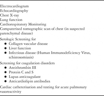
Table 1.

Post-mortem angiography of an infant aged 3 months without cardiopulmonary disease (A), and an infant with bronchopulmonary dysplasia (B). Note the marked reduction of peripheral vascularisation (“diminished background haze”) in the infant with bronchopulmonary dysplasia.
 You have
Access
- 15
- Cited by