Background
Dysfunction of the pulmonary valve causing pulmonary stenosis and/or regurgitation represents a frequent and challenging condition in patients who have undergone surgical correction of congenital cardiac malformations during childhood.Reference Gatzoulis, Balaji and Webber1, Reference Chan, Fyfe, McKay, Sade and Crawford2 With improvement in prenatal diagnosis, perinatal management, cardiac surgery, non-invasive imaging and intensive care, the number of patients surviving to adulthood has increased significantly over the last decades.Reference Marelli, Mackie, Ionescu-Ittu, Rahme and Pilote3 Surgical revision of the dysfunctional right ventricular outflow tract, therefore, has become one of the most common operations performed in adults with congenitally malformed hearts. Such repair often involves placement of a valved conduit from the right ventricle to the pulmonary arteries. Commonly used conduits are pulmonary or aortic homografts, porcine aortic valved conduits, and bovine jugular venous veins. Although surgical placement can be performed with a very low mortality, the lifespan of conduits is limited, being reported to be less than 10 years.Reference Brown, Ruzmetov, Rodefeld, Vijay and Turrentine4–Reference Tweddell, Pelech and Frommelt8 Degeneration usually leads to obstruction of the right ventricular outflow tract, with or without pulmonary regurgitation. Thus, the majority of patients with conduits from the right ventricle to the pulmonary arteries undergo multiple operations during their lifetime. To postpone surgical intervention, stenting of obstructed conduits has been performed using bare metal devices. This leads to successful relief of obstruction, with improvement in haemodynamics, but causes free pulmonary regurgitation.Reference Powell, Lock, Keane and Perry5, Reference Fogelman, Nykanen, Smallhorn, McCrindle, Freedom and Benson9, Reference Peng, McElhinney and Nugent10 Although originally considered to be a rather benign condition,Reference Murphy, Freed, Keane, Norwood, Castaneda and Nadas11 chronic volume overload of the right ventricle has now been shown to cause significant right ventricular dysfunction, exercise intolerance, and arrhythmia, with the risk of sudden death.Reference Gatzoulis, Balaji and Webber1, Reference Frigiola, Redington, Cullen and Vogel12, Reference Carvalho, Shinebourne, Busst, Rigby and Redington13 In 2000, the first pulmonary valve was implanted percutaneously, with the aim of avoiding surgery in a 12-year-old boy who developed dysfunction of his conduit, which would conventionally have necessitated open heart surgery.Reference Bonhoeffer, Boudjemline and Saliba14 In this article, we review the indications, technical aspects, and early and late results of such percutaneous implantation in the setting of the dysfunctional right ventricular outflow tract.
Historical review
Percutaneous implantation of valves represents a fast-growing, albeit very young, field in cardiology. The valve implanted without cardiac arrest, however, was reported more than 50 years ago, when Hufnagel et al.Reference Hufnagel, Harvey, Rabil and McDermott15 inserted a valve for treatment of aortic regurgitation into the descending aorta after clamping the aorta. This pioneering work was performed before the era of cardiopulmonary bypass. Although not quantifiable at that time, the authors reported a reduction of aortic regurgitation by 2/3, assuming that 2/3 of blood flow is distributed to the descending aorta.
In 1992, with recent advances in catheter technology, two separate investigators readdressed this concept, and made attempts to design a non-surgical technique for mechanical and biological replacement of the aortic valve. Andersen and colleaguesReference Andersen, Knudsen and Hasenkam16 mounted porcine aortic valves onto an balloon-expandable stent, and positioned them into the aortic root of other pigs without thoracotomy.Reference Andersen, Knudsen and Hasenkam16 Pavcnik et al.Reference Pavcnik, Wright and Wallace17 approached the concept with a crimpable mechanical valve.Reference Pavcnik, Wright and Wallace17 Their prosthesis was based on the concept of the ball-in-cage Starr-Edwards valve. The device consisted of a ring made of stainless steel wire, and a cage made of a self-expanding stent. The ball was a detachable latex balloon filled with a radiopaque silicone prepolymer system. The self-expanding portion was placed in the ascending aorta through the carotid artery. This exciting approach to percutaneous insertion remains the only description of use of a mechanical valve, undoubtedly due to the limited lifespan in the early experience when valvar competence was maintained for no longer than 1 to 3 hours. The experience of Andersen and his colleaguesReference Andersen, Knudsen and Hasenkam16 was largely overlooked at the time, and the concept came to fruition only 8 years later, concomitant with development of approaches to the pulmonary valve.Reference Bonhoeffer, Boudjemline and Saliba18 The first such procedure in humans was carried out by us in the year 2000. The initial design of a biological tissue valve mounted on an expandable stent intended for transcatheter insertion, as proposed by Andersen and colleagues,Reference Andersen, Knudsen and Hasenkam16 outlined the principle of current devices. It is very similar to the first aortic valve described by Cribier et al.Reference Cribier, Eltchaninoff and Bash19 in 2002. Over the last six years, devices have been implanted percutaneously and experimentally in all four cardiac valves.Reference Boudjemline, Agnoletti and Bonnet20, Reference Boudjemline and Bonhoeffer21 Applications in humans started with the pulmonary valve because of its lower complexity. The bigger challenge of aortic implantations was limited to patients with surgical contraindications. The technical difficulty, and the fragile nature of the population of patients, initially led to relatively high rates of morbidity and mortality,Reference Webb, Chandavimol and Thompson22–Reference Khambadkone, Coats and Taylor24 which have been in rapid decline over the last few years due to evolving technology and better selection of patients. With further progression, and alterations of the devices for replacement of aortic valves, rates of implantation and survival have increased significantly.Reference Webb, Pasupati and Humphries25, Reference Grube, Schuler and Buellesfeld26 The percutaneous implantation of the pulmonary valve has also shown a marked learning curve over the last six years, with proven improvement in safety and efficacy. This led to the first approval of a valve for transcatheter insertion by regulatory bodies in Europe for clinical use at the end of 2006, followed in 2007 by approval of two devices for implantation in aortic position.
Present state and indications
The surgical indications for treatment of the dysfunctional right ventricular outflow tract are probably the most controversial current topic in congenital cardiac disease. Whereas most institutions treat obstruction of the outflow tract according to symptoms, no unified criterions exist for the timing of valvar replacement in the context of pulmonary regurgitation. Recently, finding the right parameter to detect the point of no return in right ventricular volume overload has been described as the Holy Grail of paediatric cardiology, in particular for adults with congenitally malformed hearts,Reference Mertens27 emphasising the complexity of this issue. The detrimental effects of longstanding pulmonary regurgitation have been shown beyond doubt. Due to the limited longevity of surgical interventions, the risk of multiple open-heart operations has also to be taken into account, and influences the clinical management of such patients. Especially when pulmonary regurgitation is asymptomatic, indications for surgery vary in different institutions. With advanced cardiac magnetic resonance imaging, efforts were made to establish volume thresholds as predictors for outcome after placement of conduits. Cut-off points for end-systolic and end-diastolic right ventricular volumes were reported.Reference Oosterhof, van Straten and Vliegen28–Reference Therrien, Provost, Merchant, Williams, Colman and Webb30 The endpoint in these studies was normalisation of right ventricular volumes after correction of pulmonary regurgitation, with or without surgical remodelling of the right ventricular outflow tract. In none of these studies, however, was it possible to show any improvement in right ventricular ejection fraction.Reference Oosterhof, van Straten and Vliegen28, Reference Buechel, Dave and Kellenberger29, Reference Therrien, Siu, McLaughlin, Liu, Williams and Webb31, Reference Vliegen, van Straten and de Roos32 The impact of the timing of replacement of the valve on exercise performance and risk of arrhythmia is also still unknown.
Clinical criterions
The indications for percutaneous implantation in our series were based on the surgical criterions extant at our institution. These are based partially on guidelines,Reference Therrien, Warnes and Daliento33 combined with clinical experience, published studies, and on arbitrarily chosen cut-off points:
• Patients underwent percutaneous implantation for relief of obstruction within the outflow tract if right ventricular pressures were 2/3 of systemic pressures in presence of symptoms. In absence of symptoms, right ventricular pressures had to be 3/4 of systemic pressures. Right ventricular pressures were assessed by echocardiography, systemic pressures by non-invasive blood pressure measurements.
• Indications for treatment of pulmonary regurgitation were moderate-to-severe regurgitation in addition to one of the following: symptoms; moderate-to-severe right ventricular dysfunction; moderate-to-severe right ventricular dilation, with the cut-off point in these patients who were suitable for magnetic resonance imaging being an end-diastolic ratio of left to right ventricular volumes of 1 to 1.5, or impaired exercise capacity, with a peak uptake of oxygen of less than 65% of the predicted values.
Morphological criterions
Apart from clinical indications, patients had to fulfil morphological criterions to be suitable for implantation. Valvar competence of the current device is maintained only up to a diameter of 22 millimetres. Thus, the dimensions of the right ventricular outflow tract at its narrowest point must not exceed this diameter. Importantly, assessment of the maximal diameter throughout the cardiac cycle at the narrowest point is crucial, considering the potential distensibility of the native or patch-extended right ventricular outflow tract. Dimensions should be obtained by cine images taken in 2 planes on magnetic resonance. Three-dimensional reconstructions represent a sum of volumes during the cardiac cycle, and therefore tend to underestimate the dimensions. Balloon sizing is used in case of ambiguous measurements on magnetic resonance, or when this modality is contraindicated.
Because most native or patch-extended outflow tracts exceed this upper threshold, most devices were implanted in conduits. In addition, native outflow tracts are often distensible, and are therefore a less predictable site for implantation. Although not formally representing a criterion for exclusion, deployment of a device should be considered only in rare occasions in the absence of circumferential conduits.
Beside the upper limits of the dimensions of the site for implantation, lower limits also influence outcomes. Preferably, conduits should not be smaller than 14 millimetres. In our experience, devices implanted into small conduits significantly increase the presence of haemodynamically relevant residual gradients, and lead to early re-operation. Nevertheless, percutaneous implantation might successfully postpone surgery for those with small regurgitant conduits in the absence of obstruction. Ultimately, individual decisions have to be made, and should be according to the expectations of the patients and their parents, taking note of surgical therapeutic options in specific patients.
Issues of technique and equipment
The device and delivery system
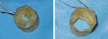
The Medtronic trans-catheter pulmonary valve (Melody™, Medtronic, MN) consists of a bovine jugular venous valve mounted on a platinum iridium stent. Figure 1 shows the device with the leaflets closed and open. The jugular vein carries a central valve within a thinned-down venous wall. This vein is sutured inside an expandable platinum-iridium stent with a length of 34 millimetres. The stent is reinforced at each intersection with gold weld. The venous segment is attached to the stent by continuous polypropylene sutures around the entire circumference and length of the stent. To insure the correct position of the stent onto the delivery system, the outflow end sutures of the device are blue, corresponding to the blue tip of the delivery system. The device is sterilised in glutaraldehyde and isopropyl alcohol, in which it is then packaged.
The percutaneous pulmonary valve.

The valved stent is crimped on a “balloon-in-balloon” front-loading delivery system (Ensemble™, Medtronic, MN). The sheath is made of Teflon, and the lumen of the guidewire is braided. This combination has the advantage of flexibility while maintaining controllability. A retractable sheath avoids displacement of the device during delivery. Prior to implantation, the stent has to be uncovered by pulling back the sheath. Similar to other “balloon-in-balloon” catheters, inflation is performed in series, the inner balloon first, followed by the outer balloon for full deployment. The delivery system is available with an outer balloon of 18, 20 or 22 millimetres.
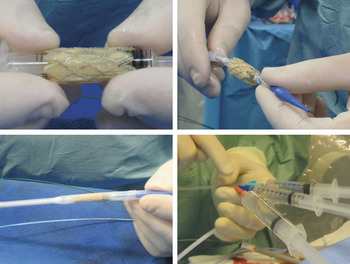
Contrast can be delivered via the retracted sheath from a side port to confirm positioning of the device prior to deployment. Proximally, there are three ports, one for the guidewire (green), one to deploy the inner balloon (indigo) and one to deploy the outer balloon (orange), as shown in Figure 2 (right lower panel).
The valve has been crimped down on a 2 millilitre syringe, mounted on the balloon-in-balloon delivery system, and covered with the outer sheath. The right lower panel shows the 3 ports of the delivery system, one for the guidewire (green), one to deploy the inner balloon (indigo) and one to deploy the outer balloon (orange).

The procedure
All procedures are performed under general anaesthesia. A full aseptic technique to surgical standards is used, and a single dose of broad-spectrum intravenous antibiotics is given for endocarditis prophylaxis. Heparin at 50 international units per kilogram is administered routinely at the beginning of the procedure, and repeated hourly thereafter as required.
Venous access is gained via a right femoral approach. This access provides better comfort for the operator compared to a jugular access. Devices were inserted successfully via jugular access in patients with femoral venous occlusion or azygos venous continuity. A trans-hepatic access, however, should not be recommended in view of the size of the delivery system. Arterial access is obtained for invasive monitoring of blood pressure.
Prior to the procedure, standard right heart catheterisation is performed to assess pressures and saturations. Pressure measurements are made in the right ventricle, pulmonary arteries and aorta with additional measurements, for example in the right and left pulmonary arteries, made as appropriate. A stiff guidewire (0.035 Amplatz ultrastiff, Cook Inc., Bloomington, IN or 0.035 Back-up Meier wire, Boston Scientific, Boston, MA) is positioned into a distal pulmonary artery for subsequent biplane angiography and percutaneous implantation. Subsequently, a Multi-Track™ catheter (NuMed Inc., Hopkinton, NY) is positioned just beyond the expected position of the pulmonary valve. This allows for assessment of the appropriate site for implantation, and quantification of pulmonary regurgitation. While these pre-assessments are carried out, the valved stent is prepared in three sequential saline baths, with 5 minutes in each, to wash off the glutaraldehyde, which is used for sterilization and storing of the valved stent. The device is then loaded onto the delivery system. To ease the down-crimping manoeuvre of the stent, the barrel of a 2 millilitre syringe is used to enable a symmetrical and controlled crimping (Fig. 2; left upper panel), followed by further hand-crimping onto the delivery system. The correct loading should be confirmed by an independent observer to guarantee the correct orientation of the valve. This can be controlled by assuring that the blue suture-line on the device matches the blue tip of the delivery system (Fig. 2; right upper panel). The stent is then covered with the outer sheath during saline flushing. The femoral vein is dilated to 22 French, and the front-loaded delivery system is advanced into the right ventricular outflow tract under fluoroscopic guidance. The proposed position of the stent can be confirmed by injection of contrast. Following uncovering by retracting of the sheath, the device is implanted by successive hand-inflation of the inner and outer balloon. The balloons are deflated and the delivery system withdrawn. Repeat angiography and pressure measurements are made to confirm a positive outcome. In presence of a residual gradient across the right ventricular outflow tract, post-dilation is performed with a high-pressure Mullins balloon (NuMed Inc, Hopkinton, NY) by using an indeflator. Repeat inflation up to 11 atmospheres leads to good relief of obstruction in the majority of cases.
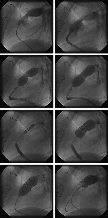
Tortuous, heavily calcified, or steeply originating conduits can complicate negotiation of the delivery system. Looping of the system in the right atrium, and partial uncovering of the device, often allows the delivery system to advance through difficult stenotic lesions. In addition, in presence of complex anatomy, we pre-dilate and/or pre-stent stenotic conduits in order to enable advancement and optimise the result. Furthermore, pre-stenting can be performed within distensible outflow tracts, thereby potentially reducing the risk of embolisation of the device and the occurrence of stent fracture. Clinical importance, risk factors, and management of the latter will be elaborated on in the next section. In Figure 3, we show pre-stenting, implantation, and postdilation of the device in a patient with stenosis of a homograft valve.
Percutaneous implantation of a pulmonary valve in a patient with predominant stenosis of the conduit. The cine angiograms are shown in lateral projection. Top left: the angiogram demonstrates obstruction of the right ventricular outflow tract; Top right: placement of a bare metal stent within the conduit, followed by balloon inflation and complete deployment resulting in relief of obstruction. Third row: positioning and deployment of the Melody device; Bottom left: postdilation of the device with a Mullins high pressure balloon; Bottom right: the final angiogram demonstrates good valvar competence in absence of a residual obstruction.

Acute and long-term outcomes
Immediate procedural outcome
More than 300 implants have been performed to date worldwide. Since the first implant in 2000, the technique has been shown to be feasible and very safe. So far, no procedurally related death has been reported. Successful deployment was achieved in the majority of cases. Procedural complications have occurred, particularly in the early experience.
In our experience of 180 patients, major peri-procedural complications were instability of the device in 2 patients; rupture of a homograft in 3 patients; compression of the main stem of the left coronary artery in one patient, who had previously undergone bare metal stenting; and obstruction of the origin of the right pulmonary artery. Other complications included limited perforation of a distal pulmonary artery resulting in minor bronchial bleeding in 2 patients; partial homograft rupture in 1 patient; and entrapment of the delivery system in the tendinous cords of the tricuspid valve in 2 patients. In 6 patients, major complications required surgical intervention, but none of them led to mortality. Whereas coronary arterial obstruction, and dislodgement of the device in distensible and dilated right ventricular outflow tracts, occurred rather early in our experience, they have since been avoided by three-dimensional imaging prior to the procedure.Reference Khambadkone, Coats and Taylor24, Reference Sridharan, Coats, Khambadkone, Taylor and Bonhoeffer34 Risks for homograft ruptures, however, remain unclear. Although not fatal, resuscitation, auto-transfusion and subsequent open-heart surgery were necessary in one patient. Fortunately, early neurological sequels showed good recovery six months after the procedure. Importantly, in our opinion, opening the chest should not be attempted before termination of bleeding.
Acutely, percutaneous implantation has been proven to reduce significantly right ventricular pressures and the gradient across the outflow tract.Reference Bonhoeffer, Boudjemline and Saliba14, Reference Coats, Khambadkone and Derrick35–Reference Bonhoeffer, Boudjemline and Qureshi37 In addition, diastolic pulmonary arterial pressures rise after deployment, indicating restoration of valvar competence. Angiography prior to and after insertion showed a significant reduction in pulmonary regurgitation.Reference Bonhoeffer, Boudjemline and Saliba14, Reference Coats, Khambadkone and Derrick35–Reference Bonhoeffer, Boudjemline and Qureshi37 There were no patients with more than mild pulmonary regurgitation after the procedure. A symptomatic improvement has been observed after the procedure in the greater majority of our patients. Magnetic resonance imaging performed after the procedure showed a reduction in right ventricular volumes, and an increase in effective right and left ventricular stroke volume.Reference Coats, Khambadkone and Derrick35, Reference Coats, Khambadkone and Derrick36
Follow up
The freedom from moderate or severe pulmonary regurgitation as measured by echocardiography at one year was more than 97%. Pulmonary regurgitation over the follow-up occurred only in those patients who had a recognised endocarditic process. The more common problem was an increase in the pressure gradient across the valved stent. This was predominantly related to early failure of the device. We encountered 2 important complications directly related to the device, namely the “hammock” effect and the appearance of fractures in the stent.
The “hammock” effectReference Khambadkone, Coats and Taylor24 is the incomplete attachment of the venous wall to the stent causing obstruction. In the initial design, the venous wall was sutured only at its extremities to the stent, enabling blood to enter between the venous wall and the inner surface of the conduit. When we recognised this complication, we altered the design of the device so that the venous wall was sutured through the entire length of the stent. Subsequent to this modification, we have not encountered further instances of the hammock effect. Initially, however, we needed to re-intervene in 7 of 21 implantation of the early device. In 3, we inserted a device of the new design, while in 4 we explanted the stent.
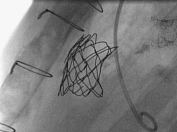
Fracture of stents (Fig. 4) is a well-recognised complication in devices placed in peripheral and central vessels. Stents placed within the right ventricular outflow tract have a high risk of fracturing, presumably due to significant radial and longitudinal deformations. Recently, a prevalence of 40% during a mean follow-up of four years was reported in the context of traditional bare metal stenting, mainly involving Palmaz Schatz laser-cut stainless steel stents.Reference Peng, McElhinney and Nugent10
Incidental fractures of the stent detected on fluoroscopy after percutaneous implantation of the pulmonary valve.

Following percutaneous implantation of the pulmonary valve, 20% of our patients have fractured their stents.Reference Nordmeyer, Khambadkone and Coats38 In the majority of cases, the fractures are clinically silent, with no increase in gradient across the valved stent. In a small proportion of patients, however, we implanted a second valve, with good relief of obstruction. This cohort of patients (5.2%) presented with significant obstruction following the fractures. In one case, fracture led to embolisation of the stent into the right pulmonary artery, the device disintegrating 9 months after implantation. Since this event, we have used careful X-ray monitoring to prevent this complication. Implantation of a second stent, however, is advisable in all cases with loss of integrity, even in the absence of stenosis. We now perform repeated chest radiography and echocardiography during follow-up. Fluoroscopy may provide added benefit by permitting grading of the fractures, and by providing information about the stability of the stent.Reference Nordmeyer, Khambadkone and Coats38 The lack of circumferential calcification increases the risk of fractures, emphasising the impact of distensibility at the site of implantation throughout the cardiac cycle.Reference Nordmeyer, Khambadkone and Coats38
Whereas early failure was treated mainly by implantation of a second device, residual stenosis represents the most common indication for re-operation in our series. This occurred particularly in the early experience, before the use of high-pressure balloons for post-dilation of the deployed stent. Other reasons for re-operation were endocarditis and outgrown conduits, problems seen also with surgically placed conduits.
Of 180 patients in whom we have implanted pulmonary valves, 4 have died. In 2 of these patients, both of whom presented in cardiogenic shock and multi-organ failure, percutaneous implantation was performed as a palliative strategy. The first had a history of 5 previous operations, deteriorated 6 weeks after a technically successful procedure, and died from a chest infection. The second presented in multi-organ failure and severe fluid overload, with critical supravalvar aortic stenosis and obstruction of the right ventricular outflow tract. Despite successful deployment of the valve, and dilation of severe re-aortic stenosis, the patient died of pulmonary oedema 24 hours after the procedure.
Fatal arrhythmia was the cause for the other 2 deaths late after implantation. The first patient, who had repaired pulmonary atresia, died suddenly 35 months after implantation. At autopsy, the valved stent was appropriately seated in the conduit, and had shown good function throughout the follow-up. The second patient, with double outlet right ventricle, had a long history of right ventricular pressure overload. After several previous unsuccessful balloon dilations of the obstructed conduit, he underwent percutaneous insertion of a valve. Despite a good haemodynamic result, with a low velocity on Doppler across the stent at latest follow-up, he died 8 months after insertion of the valve. No reason other than arrhythmia could be found.
Place in clinical management
Percutaneous implantation of a pulmonary valve was granted the Conformité Européene marking by regulatory bodies within Europe at the end of 2006. This new percutaneous strategy, therefore, is now available to a broader population, and will be used more commonly. In our opinion, percutaneous insertion of a pulmonary valve represents the non-invasive treatment of choice in patients with dysfunction of their right ventricular outflow tracts, and should be considered superior to insertion of bare metal stents. This procedure is not considered to be a surrogate for surgical revision of the right ventricular outflow tract, but a therapeutic option for prolonging function of the conduit. This should then result in a reduction of open-heart surgery over the lifespan of patients with congenitally malformed hearts.
Future outlook
Although significant improvement has been achieved in early and late outcomes after percutaneous implantation of pulmonary valves, the risk of stent fractures and homograft rupture are not yet sufficiently understood. Further investigations are necessary to avoid these complications. At present, the device can be implanted safely only in patients with circumferential conduits that do not exceed 22 millimetres in diameter. In particular, the large population of patients who underwent repair by means of a transjunctional patch, and have severe right ventricular dysfunction due to pulmonary regurgitation, cannot currently benefit from this technique. Development of new devices that can down-size the site of implantation, or hybrid approaches incorporating minimally invasive surgery without cardio-pulmonary bypass, may provide potential alternative options.Reference Boudjemline, Agnoletti, Bonnet, Sidi and Bonhoeffer39–Reference Boudjemline, Schievano and Bonnet41 Ultimately, evolution of the treatment of the dysfunctional right ventricular outflow tract can only be achieved by close co-operation between cardiologists, surgeons, specialists in imaging, and bio-medical engineers.