Flecainide is a class IC antiarrhythmic drug the overdose of which is associated with an approximately 10% mortality rate. Reference Cheung and Man1 Flecainide overdose is difficult to treat because of the drug’s high oral bioavailability (90%), high volume of distribution (8–9 L/kg), long half- life (up to 23 hours), poor ability to be dialysed, lack of an antidote, and rapid onset of shock and arrhythmias (as early as 30 minutes after ingestion). Reference Cheung and Man1–Reference Gotz, Pohle and Barckow3 I present the management of a life-threatening flecainide overdose of an infant that had a protracted course due to the exposure to the drug in both the fetal and neonatal periods.
Case presentation
As a fetus, the patient had supraventricular tachycardia and hydrops fetalis. During pregnancy, the infant’s mother had been given flecainide. He was born at 31 weeks gestation weighing 1490 g. He was having atrioventricular reentry tachycardia right after birth.
The patient had been discharged at 3 months of age on oral propranolol (4 mg/kg/day) and flecainide (3 mg/kg/day). Unfortunately, the parents gave flecainide 20 mg/kg/day for a week after discharge.
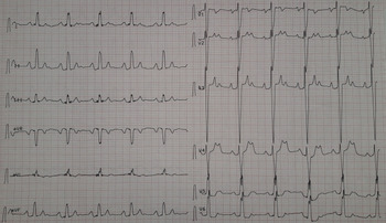
The infant presented to the emergency department with tachypnoea. His heart rate was 60 beats per minute, and blood pressure was 77/50 mmHg. He was admitted to the ICU. His electrocardiogram revealed second-degree (2:1) atrioventricular block (Fig 1). His laboratory values were as follows: pH: 7.46 pCO2: 39 mmHg, lactate: 1.5 mmol/L, Na: 137 mmol/L, AST: 70 U/L, ALT: 77 U/L. An echocardiogram revealed normal left ventricular systolic function.
Second degree (2:1) atrioventricular block.

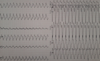
He was initially treated with a NaHCO3 infusion of 1 meq/kg for 6 hours and with an intravenous lipid emulsion of 3 g/kg/day. He developed progressive QRS widening and first-degree atrioventricular block after 12 hours. From then on, for 7 hours his heart rhythm showed variability with ventricular bigeminy, second-degree atrioventricular block, and ventricular tachycardia (Fig 2).
Ventricular tachycardia.

On the second day of his admission, the electrocardiogram mostly revealed wide QRS interval changing to second-degree atrioventricular block for short periods of time. The NaHCO3 infusion was continued as well as intermittent NaHCO3 boluses as needed to combat ventricular tachycardia.
On the third day, QRS interval widening reached 160 ms with a heart rate of 146 beats per minute. His echocardiography showed asynchronous contraction between the ventricles. A lidocaine infusion was started at a rate of 50 μg/minute which succeeded in converting the rhythm to sinus after 1 hour. The lipid infusion was stopped.
On the fourth day, the lidocaine infusion rate was decreased to 30 μg/minute and for 20 hours his electrocardiogram showed sinus rhythm. However, ventricular tachycardia recurred. The lidocaine infusion rate was increased to 50 μg/minute, restoring sinus rhythm.
On the fifth day, ventricular tachycardia recurred while lidocaine infusion was continuing. The serum lactate increased to 4.3 mmol/L. Although his rhythm showed variations from sinus rhythm to ventricular tachycardia at certain intervals for different periods of times, wide QRS tachycardia was the predominant rhythm so esmolol was loaded and infused at a rate of 200 μg/kg/minute. This therapy restored sinus rhythm. The infusion rate was decreased to 100 μg/kg/minute as sinus rhythm was restored for 12 hours and a Wolf-Parkinson–White pattern was observed.
On the sixth day, lidocaine and esmolol infusions were stopped 4 hours after oral propranolol (4 mg/kg/day) was given. The patient was discharged after 2 days. The patient remained conscious throughout the ordeal. His haemodynamic status remained stable as well as without any hypotensive events. He was fed by his mother regularly.
Discussion
Flecainide delays phase 0 depolarisation through its high-affinity binding for open-state sodium channels, which results in the slowing of conduction in the atria, ventricles, and His-Purkinje system along with an increased refractory period in the ventricular tissue. Reference Roden and Woosley4 Symptoms of toxicity include noncardiac manifestations, such as nausea, vomiting, and seizures, as well as cardiac manifestations, such as bradycardia, widening of the QRS complex, and ventricular tachycardia. Reference Koppel, Oberdisse and Heinemeyer5
My case had a protracted course of rhythm instability when compared to other case reports. The youngest report of flecainide intoxication in literature is a 3-day-old premature neonate with permanent junctional reciprocating tachycardia which was managed successfully by the reversal of the sodium blockade with intravenous sodium bicarbonate and supportive care. Reference Karmegaraj, Menon, Prabhu and Vaidyanathan6 A 4-month-old infant with renal tubular acidosis type IV was treated with extracorporeal membrane oxygenation due to flecainide toxicity. Reference Bhimani, Rao, Nadorlik, Saarel and Aziz7 The patient acutely decompensated with hypotension as the rhythm degenerated into sustained wide complex tachycardia at rates of 225–240 beats/min. Sinus rhythm was restored after 36 hours.
Another 18-day-old, 2-week premature patient had a wide-complex tachycardia with left bundle branch morphology with associated pallor and poor capillary refill due to twofold dosing of flecainide. Reference Jang, Hoffman and Nelson8 The patient had received 8 mg every 8 hours instead of the prescribed dose of 4 mg at the same interval.
I did not perform gastric lavage as he was receiving an excessive dose of flecainide for 7 days. It is also notable that the patient was being exposed to the drug in the fetal period as well as receiving high doses (8 mg/kg/day) in the neonatal period. This long exposure may explain why he had a protracted course due to the effects of flecainide overdose. Detecting serum flecainide levels would have been helpful to show the response to the attempts of clearance and the various electro-mechanical manifestations. However, our laboratory did not have the ability to detect serum flecainide levels.
Salerno et al used a canine model of ventricular dysrhythmia induced by flecainide and treated with sodium bicarbonate. Reference Salerno, Murakami and Johnston9 I used intermittent infusions as well as intravenous pushes to combat wide QRS interval leading to ventricular tachycardia. Alkalisation with sodium bicarbonate to achieve a pH goal of 7.5–7.55 is thought to decrease the ionised form of flecainide that usually binds to sodium channels. Reference Bhimani, Rao, Nadorlik, Saarel and Aziz7 Lipid infusion removes the lipid-soluble drugs from serum. Reference Karmegaraj, Menon, Prabhu and Vaidyanathan6 Because there is no established treatment protocol on how to infuse lipids in these cases, I chose to give the treatment for 3 days.
Conclusion
Based on limited case reports as well as experimental evidence, sodium bicarbonate is the mainstay of medical therapy. Antiarrhythmic and intravenous fat emulsions are also other treatment options. When all these options fail, the early use of extracorporeal membrane oxygenation may be considered.
Acknowledgements
None.
Financial support
This research received no specific grant from any funding agency, commercial, or not-for-profit sectors.
Conflict of interest
None.
Ethical standards
As this work is a case presentation and does not involve human and/or animal experimentation, ethical standards statement was omitted.