Idiopathic ventricular tachycardia is a clinical entity without any structural heart disease detected by conventional diagnostic evaluations. The right ventricular outflow tract is the predominant site of origin for idiopathic right ventricular tachycardia in children.Reference Fukuhara, Sumitomo and Nakamura 1 We present a case series of idiopathic ventricular tachycardia arising from unusual sites of the right ventricle below the level of the right ventricular outflow tract and causing tachycardia-induced cardiomyopathy in children, which were eventually cured by radiofrequency catheter ablation.
Materials and methods
A total of three children were identified, who presented with ventricular tachycardia originating from unusual sites of the right ventricle. The clinical data were retrospectively collected and reviewed.
All three patients were excluded from having arrhythmogenic right ventricular cardiomyopathy by careful evaluation of their echocardiograms and MRI results. Patients were brought to the electrophysiology laboratory after written informed consent had been obtained. With the patient under general anaesthesia, an electroanatomic map of the right ventricle was made using the CARTO system (Carto 3 EP Navigation system; Biosense Webster, New Brunswick, State of New Jersey, United States of America). A 4-mm-tip mapping/ablation catheter (THERMOCool®/NaviStar®; Biosense-Cordis, New Brunswick, State of New Jersey, United States of America) with or without an irrigated tip was used. Spontaneous ventricular tachycardia was identified in all three patients. Radiofrequency ablation was applied to the site with earliest activity during ventricular tachycardia. Successful catheter ablation was defined as the absence of spontaneous or inducible ventricular tachycardia with isoproterenol infusion for 30–60 minutes after the final radiofrequency ablation.
Anti-arrhythmic agents were discontinued following the successful ablation procedure, and all the patients were closely followed-up for procedure-related complications and recurrence of ventricular tachycardia with 12-lead surface electrocardiography and 24-hour Holter recording. Left ventricular diameter and function were evaluated by echocardiography.
Results
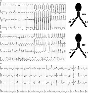
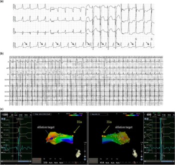
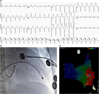
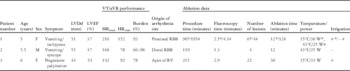
Demographic data are listed in Table 1. All three patients developed symptoms including palpitation, vomiting, tachypnea, or syncope, meanwhile they had decreased left ventricular ejection fraction and enlarged left ventricular end-diastolic diameter by echocardiography evaluation. Tracings of electrocardiography demonstrated analogous left bundle branch block QRS morphology in each patient, with varied axis in the inferior leads and QRS duration from patient to patient (Figs 1a, 2a and 3a). Based on Holter recordings, the incessant natureReference Moore, Patel and Shannon 2 (⩾75% ventricular arrhythmia burden) of ventricular tachycardia or accelerated ventricular rhythm (⩽120 beats/minute) was confirmed in all three patients. Before electrophysiology study, patients 1 and 2 were both treated by intravenous amiodarone, followed by a combination of oral amiodarone and metoprolol for 12 and 18 months, respectively. The follow-up Holter and echocardiography showed significantly decreased ventricular rhythm burden and cardiac chamber with improved left ventricular systolic function; however, 1 month after discontinuation of amiodarone, the incessant ventricular tachycardia recurred with symptoms of palpitation or associated with vomiting. Patient 3 was resistant to propafenone and verapamil.
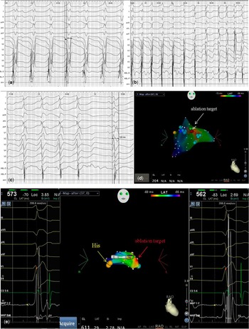
In patient 1, 12-lead ECG demonstrated a fascicular tachycardia with QRS duration of 81 ms, incomplete LBBB morphology, axis left deviation, AV dissociation (sinus P is labelled by S), and retrograde V-A conduction (labelled by P’) before the first radiofrequency ablation ( a ). VT recurred after the first ablation with the QRS morphology changing to complete RBBB pattern, with duration of 118 ms and AV dissociation (sinus P is labelled by arrow), suggesting that the VT origin was slightly proximal to the ablation site ( b ). The normal sinus rhythm has complete RBBB, with QRS duration of 123 ms after the second ablation ( c ). AV=atrioventricular; ECG=electrocardiography; LB=left bundle; LBBB=left bundle branch block; RB=right bundle; RBBB=right bundle branch block; VT=ventricular tachycardia.

In patient 2, 12-lead ECG before the ablation demonstrated an accelerated ventricular rhythm with QRS duration of 123 ms, complete LBBB, and normal axis. There was AV dissociation (sinus P is labelled by arrow) with two conducted beats (labelled S). Note that the conducted beat had a shorter QRS duration of 100 ms ( a ). The intra-cardiac recording showed that the ablation terminated VT gradually, accompanied by narrow sinus beats changing to complete RBBB morphology ( b ). The 3D electrical anatomical map shows the earliest activation of VT with RBBP in the distal region, with reversed His-RBBP interval of −22 ms ( c ). AV=atrioventricular; ECG=electrocardiography; LBBB=left bundle branch block; RBBB=right bundle branch block; RBBP=right bundle branch potential; VT=ventricular tachycardia; 3D=three dimensional.

In patient 3, 12-lead ECG revealed persistent VT with a heart rate of 122 beats/minute. The QRS complexes had complete LBBB morphology, with the duration of 113 ms. The axis of VT was directed superiorly and leftwards, and late R-wave transition at V5 was present. The 1:1 V-A retrograde conduction was identifiable (arrow) ( a ). The ablation target of VT was at the apex of the right ventricle revealed by the fluoroscopy ( b ) and 3D electrical anatomic map ( c ), respectively. ECG=electrocardiography; LBBB=left bundle branch block; VT=ventricular tachycardia; 3D=three dimensional.

Patient demographics and electrophysiology data.

LVDd=left ventricular end-diastolic diameter; LVEF=left ventricular ejection fraction; RBB=right bundle branch; RV=right ventricle; VT=ventricular tachycardia; aVR=accelerated ventricular arrhythmia; * and #=first and second RF procedures, respectively; +=with irrigation (saline flow rate 17 ml/minute); −=without irrigation
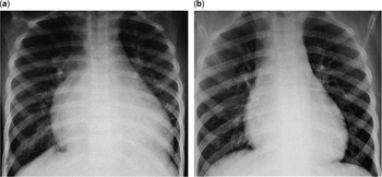
The sites of successful termination of ventricular tachycardia were the proximal right bundle branch, distal right bundle branch, and apex of the right ventricle in patients 1, 2, and 3, respectively (Figs 4a–e, 2b and c, and 3b and c). In patient 1, ventricular tachycardia recurred 4 hours after the first ablation, but with right bundle branch block morphology of the QRS complex (Fig 1b). A repeat procedure was performed 1 month later, and final successful ablation was achieved at a more proximal site within the right bundle branch region. Patients 1 and 2 developed complete right bundle branch block after the successful ablation (Fig 1c). No complications occurred, and there has been no recurrence of ventricular tachycardia after the final ablation during follow-up at 6, 3, and 18 months in patients 1, 2, and 3, respectively. All three patients have remained free of symptoms and have achieved normalisation of left ventricular size and systolic function (Fig 5).
In patient 1, the intra-cardiac recording showed that the earliest activation with RBBP was 32 ms earlier than the onset of surface QRS ( a ). The first ablation in the vicinity of the proximal RBB initially changed the QRS morphology to the RBBB pattern, and thereafter slowed down and terminated VT. The sinus rhythm had a complete RBBB with QRS duration of 120 ms ( b – d ). During the second procedure, the 3D mapping of VT was resumed, and RBBP was found again at the site of ablation target, preceding His potential of 13 ms ( e ). RBB=right bundle branch; RBBB=right bundle branch block; RBBP=right bundle branch potential; VT=ventricular tachycardia; 3D=three dimensional.

In patient 1, chest X-ray on first admission showed significantly increased cardiothoracic ratio ( a ), and repeat chest X-ray 3 months after the second ablation showed normalisation of cardiac size ( b ).

Discussion
The majority of idiopathic right ventricular tachycardia arises from the right ventricular outflow tract. In one adult study, of the 278 patients who underwent ablation for idiopathic right ventricular tachycardia or ventricular premature depolarisations, 90% of ventricular arrhythmias arose from the right ventricular outflow tract, followed by 5% from the tricuspid valve annulus region, and 2.9 and 2.5% from the basal and apical right ventricle segments, respectively.Reference Van Herendael, Garcia and Lin 3 Very rare cases of ventricular tachycardia not originating from the right ventricular outflow tract have been described in children.Reference Moniotte, Triedman and Cecchin 4 , Reference Sosa and Scanavacca 5 We describe a series of children with left bundle branch block morphology ventricular tachycardia originating from unusual sites in the right ventricle, resulting in tachycardia-induced cardiomyopathy. We demonstrate that radiofrequency catheter ablation can be performed safely with successful resolution of arrhythmia and impaired ventricular function.
Classically, fascicular tachycardia originates from a left ventricular fascicle. In contrast, tachycardia directly arising from the right bundle branch is very rare.Reference Moniotte, Triedman and Cecchin 4 – Reference Seethala, Mezu and Němec 7 The origins of ventricular tachycardia at the proximal and distal right bundle branch in patients 1 and 2, respectively, were confirmed by the careful mapping of the distribution of entire right bundle branch potential along the septum during an electrophysiology study. In patient 1, the narrow QRS of left bundle branch block morphology, atrioventricular dissociation, and a pre-systolic right bundle branch potential preceding the QRS complex were very suggestive of ventricular tachycardia from the right His-Purkinje system. The retrograde His-right bundle branch potential conduction distinguished it from junctional rhythm with left bundle branch block and bundle branch re-entry tachycardia. Incremental atrial pacing can overdrive the ventricle with narrow QRS complex until atrioventricular block occurs, which excludes the existence of an atriofascicular accessory pathway of the Mahaim fibre.Reference Chen, Gu and Yang 6 – Reference Sternick, Sosa and Timmermans 8 Moreover, the evidence for this diagnosis was the transition of a narrow QRS ventricular tachycardia with incomplete left bundle branch block to a wider QRS tachycardia with a right bundle branch block pattern after our first ablation. Complete right bundle branch block was also present in sinus rhythm, which indicates that the ablation site was the right bundle branch distal to the site of ventricular tachycardia. The second ablation successfully terminated the ventricular tachycardia and resulted in a normal sinus rhythm with right bundle branch block. In patient 2, the QRS complex of ventricular tachycardia was relatively wider, which may be due to the distal right bundle branch origin ventricular tachycardia propagating its activation more quickly over the myocardium to the left side than capturing the left fascicle by retrograde conduction.Reference Chen, Gu and Yang 6 The idiopathic ventricular tachycardia arising from the apex was first reported in 2008,Reference Navarrete 9 and, to our knowledge, there has been no paediatric case published in the literature thus far. The electrocardiography characteristics of apical ventricular tachycardia had late pre-cordial R-wave transition at V5-V6, negative QRS complexes in all inferior leads, and positive QRS in aVL lead in contrast with ventricular tachycardia originating from the right ventricular outflow tract.Reference Van Herendael, Garcia and Lin 3 , Reference Navarrete 9
Tachycardia-induced cardiomyopathy has been defined as a myocardial dysfunction that is wholly or partially reversible after control of the responsible tachyarrhythmia.Reference Moore, Patel and Shannon 2 , Reference Arya, Haghjoo, Davari and Sadr-Ameli 10 Few reports have described tachycardia-induced cardiomyopathy secondary to ventricular tachycardia in children. The study representing the largest known series of children with tachycardia-induced cardiomyopathy showed that among 81 patients the tachycardia subtype of ventricular tachycardia only accounts for 7.0%.Reference Moore, Patel and Shannon 2 Among the three children we described, the impaired left ventricular function was induced by the incessant ventricular tachycardia, the likely mechanism of which appeared to be increased automatically because of its focal origin and chronotropic response. Amiodarone and β-blocker therapy were used to slow the ventricular rate, reduce the ventricular tachycardia burden, and partially improve the cardiac function in two of them, and eventually catheter ablation was a good option for arrhythmia and tachycardia-induced cardiomyopathy treatment.
In conclusion, clinicians should be aware that idiopathic right ventricular tachycardia can arise from unusual sites in children besides the right ventricular outflow tract and induce cardiomyopathy. Catheter ablation is a safe and effective option for such arrhythmias.
Acknowledgement
None.
Financial Support
This work was supported by the general programme of Shanghai municipal commission of health and family planning (grant number 201440631). This study was approved by the Ethics Committee of Children's Hospital of Fudan University.
Conflicts of Interest
None.