Introduction
The pinewood nematode (PWN), Bursaphelenchus xylophilus (Steiner & Buhrer, Reference Steiner and Buhrer1934; Fuchs, Reference Fuchs1937), is the causative agent of pine wilt disease (PWD). Indigenous in North America, this nematode is widespread in natural coniferous forests in Canada and the USA (Sutherland & Peterson, Reference Sutherland, Peterson, Futai, Togashi and Ikeda1999) without representing an economic problem to native pine forests. In non-native forest ecosystems, namely in Asian countries such as Japan, China, South Korea and Taiwan (for a review see Vicente et al., Reference Vicente, Espada, Vieira and Mota2012), the PWN is one of the most feared pests and pathogens due to its impact on forest health, natural ecosystem stability and international trade (Vicente et al., Reference Vicente, Espada, Vieira and Mota2012). The effects of the PWN on the global ecosystem and natural biodiversity are irreversible (Kiyohara & Bolla, Reference Kiyohara and Bolla1990; Suzuki, Reference Suzuki2002). The PWN and several other Bursaphelenchus species have been intercepted from packaging and wood products in several countries, e.g. Austria (Tomiczek et al., Reference Tomiczek, Braasch, Burgermeister, Metge, Hoyer and Brandstetter2003), China (Gu et al., Reference Gu, Braasch, Burgermeister, Brandstetter and Zhang2006), Finland (Tomminen et al., Reference Tomminen, Halik and Berglahl1991) and Germany (Braasch et al., Reference Braasch, Tomiczek, Metge, Hoyer, Burgermeister, Wulfert and Schonfeld2001), and, more recently, in packaging wood imported from countries considered free of this pest, such as Brazil, Belgium, Italy and Spain (Gu et al., Reference Gu, Braasch, Burgermeister, Brandstetter and Zhang2006). This has increased the need to avoid the spread of this pathogen outside North America.
Bursaphelenchus xylophilus was reported for the first time in Europe in Portugal in 1999, from dead maritime pine (Pinus pinaster) trees located in the Setúbal Peninsula, 30 km SE of Lisbon (Mota et al., Reference Mota, Braasch, Bravo, Penas, Burgermeister, Metge and Sousa1999). Until 2008, the PWN was confined to the Setúbal Peninsula, but despite all efforts made by the Portuguese authorities, the disease spread to new areas in 2008 (Rodrigues, Reference Rodrigues, Mota and Vieira2008), and was recently detected in Madeira Island, 1000 km SW of continental Portugal (Fonseca et al., Reference Fonseca, Lopes, Cardoso, Pestana, Abreu, Nunes, Mota and Abrantes2010; Vicente et al., Reference Vicente, Espada, Vieira and Mota2012), as well as in Spain (Abelleira et al., Reference Abelleira, Picoaga, Mansilla and Aguin2011; Robertson et al., Reference Robertson, Arcos, Escuer, Merino, Esparrago, Abelleira and Navas2011). Thus, it is of vital importance to determine the origin of the new isolates and understand their routes of spread, in order to prevent further infections in Europe. Molecular markers displaying enough genetic polymorphism could be useful for understanding the epidemiology of this disease and its dissemination pathways. DNA-based techniques developed during the past decades provide an attractive solution to nematode identification. In spite of that, determination of intraspecific variability is difficult. Due to the high copy number in individual cells, lack of recombination and strict maternal inheritance, mitochondrial genes (mtDNA) are known to be an excellent marker for the study of phylogenetic relationships.
In recent years, mitochondrial genes have been used as markers to study intraspecific variation (Madani et al., Reference Madani, Subbotin, Ward, Li and De Boer2010) and the genetic relationships among Peruvian and Canadian populations of Globodera pallida in order to identify the origin of new populations (Picard et al., Reference Picard, Sempere and Plantard2007; Plantard et al., Reference Plantard, Picard, Valette, Scurrah, Grenier and Mugniéry2008; Madani et al., Reference Madani, Subbotin, Ward, Li and De Boer2010). For example, mtDNA cytochrome b (cytb) gene sequences allowed the determination of the origin of new G. pallida populations (Plantard et al., Reference Plantard, Picard, Valette, Scurrah, Grenier and Mugniéry2008), which makes this a possible marker to be tested in the study of B. xylophilus intraspecific variability.
PWN is an unusual plant parasite, as it is a parasite of the aboveground parts of trees, migrating through plant tissues and never entering the soil. It is a fungal feeder that uses an insect vector, a cerambycid beetle, particularly Monochamus spp. (Akbult & Stamps, Reference Akbult and Stamps2011), for transmission from one tree to another, implying that it should contain a set of parasitic genes distinct from those of the major plant parasitic nematodes (Kikuchi, Reference Kikuchi, Zhao, Futai, Sutherland and Takeuchi2008). The only other example of a similar plant parasite within this genus (containing nearly 100 species) is B. cocophilus, which parasitizes coconut and palm trees in Central and South America, and is the causal agent of ‘red ring disease’.
Plant cell walls are mainly made up of cellulose and, consequently, cellulases (endo-β-1,4-glucanases) are cell wall degrading enzymes, produced by many plant pathogenic nematodes, such as B. xylophilus (Smant et al., Reference Smant, Stokkermans, Yan, De Boer, Baum, Wang, Hussey, Gommers, Henrissat, Davis, Helder, Schots and Bakker1998; Jones et al., Reference Jones, Furlanetto and Kikuchi2005; Zhao &Lin, Reference Zhao and Lin2005; Kikuchi et al., Reference Kikuchi, Aikawa, Kosaka, Pritchard, Ogura and Jones2007). Previous studies of Ma et al. (Reference Ma, Lu, Liang and Zhang2011), using RNA interference to analyse the function of the endo-β-1,4-glucanase gene of B. xylophilus, show that the propagation and dispersal ability of this nematode decreased when the endo-β-1,4-glucanase gene was silenced. Bursaphelenchus xylophilus uses a stylet to penetrate the plant cell wall. This stylet is associated with oesophageal glands, which produce secretions containing cellulases. Endogenous cellulase genes have been identified in obligatory plant-parasitic nematodes, such as Heterodera, Globodera and Meloidogyne species (Smant et al., Reference Smant, Stokkermans, Yan, De Boer, Baum, Wang, Hussey, Gommers, Henrissat, Davis, Helder, Schots and Bakker1998; Rosso et al., Reference Rosso, Favery, Piotte, Arthaud, De Boer, Hussey, Bakker, Baum and Abad1999; Goellner et al., Reference Goellner, Smant, De Boer, Baum and Davis2000). The proteins encoded by these genes belong to the glycosyl hydrolase family (GHF), which in B. xylophilus are more similar to fungal GHF cellulases, suggesting acquisition by horizontal gene transfer from fungi (Jones et al., Reference Jones, Furlanetto and Kikuchi2005; Kikuchi et al., Reference Kikuchi, Aikawa, Kosaka, Pritchard, Ogura and Jones2007; Xie et al., Reference Xie, Cheng, Shi, Zhang, Dai, Cheng and Luo2009). Sequence analysis of the cellulase genes allowed species identification as well as intraspecific variability evaluation within the genus Bursaphelenchus (Cheng et al., Reference Cheng, Cheng, Xu and Xie2008, Reference Cheng, Dai, Xiao and Xie2010).
The aim of the present study was to characterize Portuguese populations of the PWN, B. xylophilus, using cytb and cellulase gene sequences. These sequences were used for comparative phylogenetic analyses of PWN populations collected from different geographic locations from continental Portugal and Madeira Island. Cytb and cellulase sequences were compared with sequences from B. xylophilus from other countries, such as USA, Japan, China and South Korea, in order to understand the possible origin of the new outbreaks of the disease in continental Portugal and Madeira Island.
Materials and methods
Nematode sampling and cultures
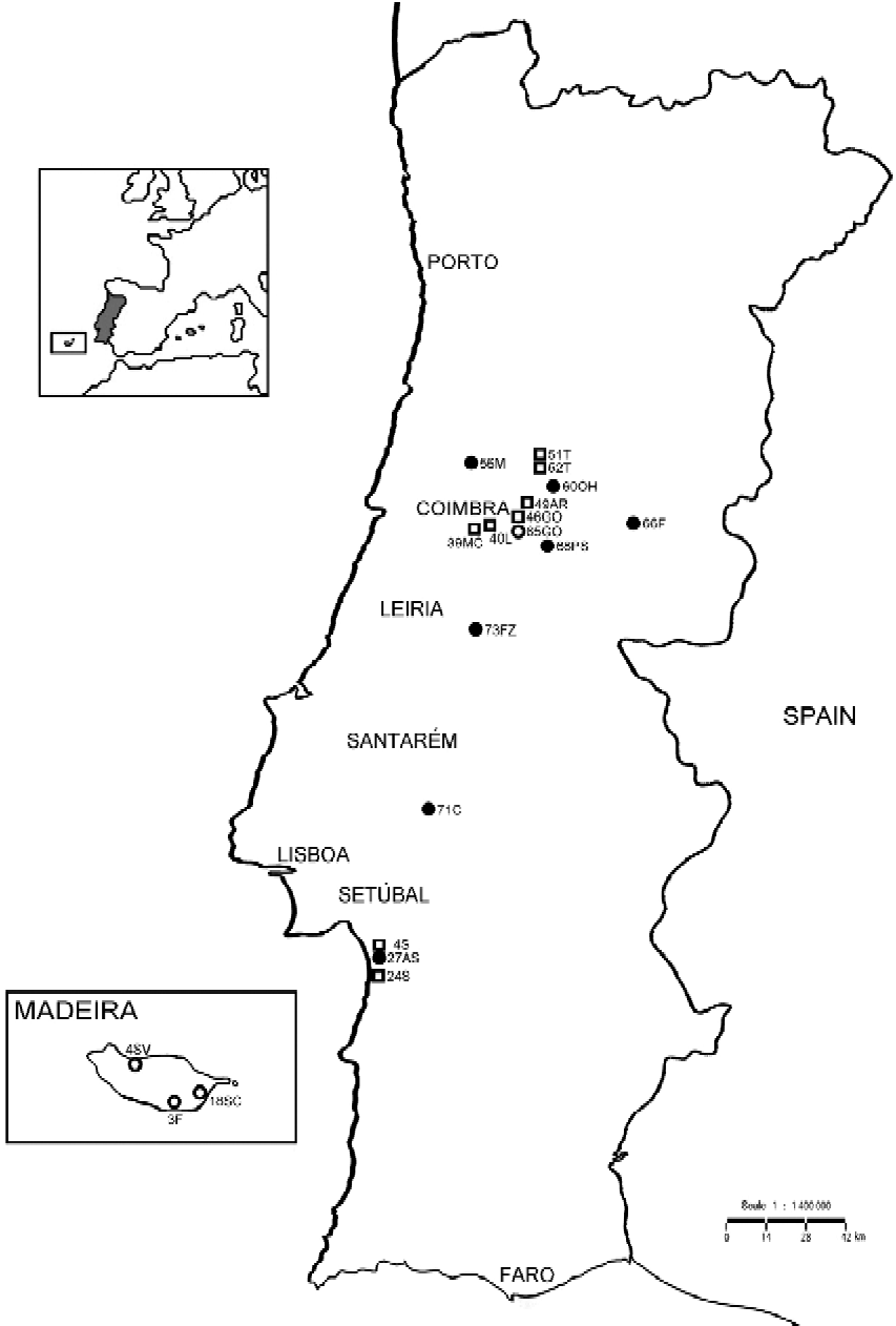
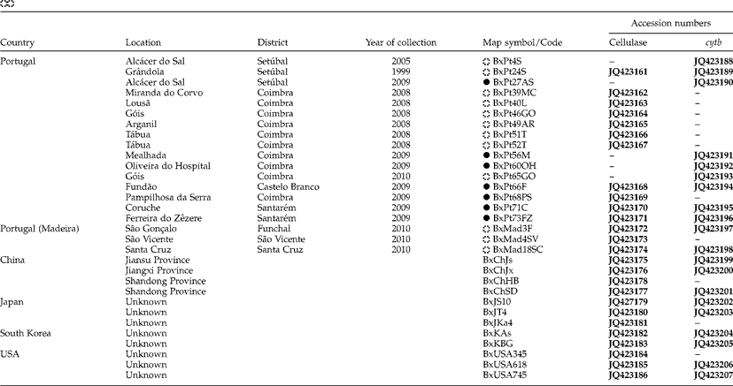
The present study used 19 B. xylophilus isolates from Portugal (continental and Madeira Island) (fig. 1) which were compared with several foreign isolates: three US isolates, two South Korean, three Japanese and four Chinese (table 1). Madeira Island isolates were provided by the Nematology Lab, IMAR-CMA (Department of Life Sciences, University of Coimbra), and continental Portugal isolates were provided by AFN (National Forest Authority) from the national surveys conducted every year. One B. mucronatus (BmPt0) Portuguese isolate was also used. Each isolate was obtained from a wood sample collected from one tree. Nematode cultures were maintained in the laboratory in barley grown with Botrytis cinerea (Y. Mamiya, pers. comm.). Nematodes were extracted using a modified Baermann funnel technique (Southey, Reference Southey1986). Prior to DNA extraction, nematodes were collected and concentrated in a 1.5-ml microtube (Eppendorf) in a minimal amount of water.
Map of the geographical distribution of B. xylophilus in continental Portugal and Madeira Island, showing the sampling sites of isolates collected until 2008 and in 2009–2010. Isolate codes and symbols are shown in table 1 (until 2008 □, 2009 ●, 2010 ○).

List of B. xylophilus isolates, location, year of collection, map symbol/code and accession numbers of cellulase and cytb sequences (until 2008 □, 2009 ●, 2010 ○). Accession numbers for sequences determined in the present study are shown in bold.
–, No sequence available.
DNA extraction
Genomic DNA was extracted from a suspension of 50 μl with more than 10,000 nematodes from each isolate. DNA was extracted with the JETQUICK Tissue DNA Spin Kit extraction kit (Genomed, Löhne, Germany), according to the manufacturer's protocol. DNA was used for sequence analysis of cytb and cellulase genes. Nematode DNA was kept at − 20°C for further use.
Sequencing of cytochrome b and cellulase genes
For cytb amplification, 50 μl polymerase chain reactions (PCR) contained 1 × PCR buffer (Fermentas, Vilnius, Lithuania), 1.5 mm MgCl2 (Fermentas), 0.2 mm each deoxyribonucleotide (dNTP) (Fermentas), 0.4 μm of each primer (STABvida, Lisboa, Portugal), 0.05 units of recombinant Taq DNA polymerase (Fermentas) and 5 μl template DNA (10–20 ng). Primers were designed based on the full mitochondrial sequence of B. xylophilus (isolate BxPt15SC, accession number JQ429761; A. Amorim, pers. comm.): BxCYB F (5′-AAA TAG AAT TTT TGT TTT TGG TAT-3′) and BxCYB R (5′-TGT GAA AAA TCT ATA AGA CCT AAA CTT-3′). Primers were designed to amplify the entire cytb gene sequence. The PCR program consisted of an initial denaturation step of 3 min at 95°C, followed by 30 cycles of denaturation at 95°C for 30 s, annealing at 44°C for 30 s and extension at 72°C for 1 min 30 s. The final extension step was performed at 72°C for 5 min. PCR products were electrophoresed in 1% agarose gels, stained with ethidium bromide and sequenced by STABvida, Portugal.
For amplification of the cellulase gene, primers ENG00s (5′-TCT AAA ATG AAG TCT CTT GTG-3′) and ENG00a (5′-AGT CCT CTA AGC ATC GTC-3′) (Kikuchi et al., Reference Kikuchi, Jones, Aikawa, Kosaka and Ogura2004) were used. PCR amplifications were conducted in 50 μl PCR reactions containing 1 × PCR buffer (Fermentas), 2 mm MgCl2 (Fermentas), 0.2 mm each dNTP (Fermentas), 10 pmol of each primer (STABvida), 1.25 units of recombinant Taq DNA polymerase (Fermentas) and 5 μl template DNA (10–20 ng). The PCR program consisted of an initial 3 min denaturation at 95°C, followed by 30 cycles of denaturation at 94°C for 1 min, annealing at 50°C for 1 min and extension at 72°C for 1 min. The last step was performed at 72°C for 5 min. PCR products were visualized in 1% agarose gels stained with ethidium bromide, purified with MinElute Gel Extraction Kit (Qiagen, Hilden, Germany) and quantified using Quant-iT dsDNA High-Sensitivity Assay Kit (Invitrogen, Paisley, UK). Gel-purified PCR products were ligated into pGEM-T Easy Vector (Promega, Madison, Winconsin, USA) and transformed into Escherichia coli strain JM109 competent cells (Promega) according to the manufacturer's instructions. Plasmids were purified from two or three positive clones using the GeneJET Plasmid Miniprep Kit (Fermentas) and the cellulase gene was obtained by restriction with the endonuclease EcoRI (Fermentas). The gel band corresponding to the cellulase gene was purified using MinElute Gel Extraction Kit (Qiagen) and sequenced by STABvida, Portugal.
Phylogenetic analysis of cytochrome b and cellulase sequences
Multiple sequence alignments of cytb and cellulase nucleotide sequences were assembled using the ClustalW algorithm as implemented in BioEdit version 7.1.3.0 (Hall, Reference Hall1999), under default alignment parameters. Using MEGA5 version 5.05 (Tamura et al., Reference Tamura, Peterson, Peterson, Stecher, Nei and Kumar2011) phylogenetic relationships between isolates were reconstructed by neighbour-joining (Saitou & Nei, Reference Saitou and Nei1987) and maximum likelihood (ML) methods, using nucleotide sequences. The best-fitting evolutionary model of nucleotide substitutions was determined. Gaps or indels were treated using the complete deletion option in MEGA5. Clades from the ML tree were supported by bootstrap analysis (Felsenstein, Reference Felsenstein1985) with 100 replicates.
Results
Phylogenetic analysis of the cytochrome b gene
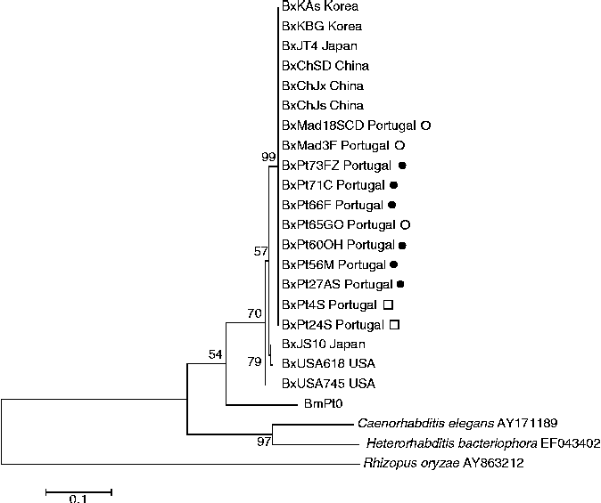
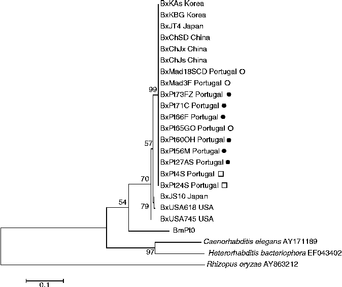
A PCR product of approximately 1200 bp, containing the partial sequence of cytb was obtained and sequenced (accession numbers in table 1) using 11 Portuguese isolates of B. xylophilus, nine foreign isolates from China, Japan, South Korea and USA, and one B. mucronatus (BmPt0 accession number JQ655730) Portuguese isolate. Other nematodes, such as Caenorhabditis elegans and Heterorhabditis bacteriophora were also included in the analysis and the fungus Rhizopus oryzae (accession numbers in table 2) was used as outgroup. Only a few polymorphisms were found among B. xylophilus isolates. One major cluster was obtained that includes all Portuguese and foreign isolates (bootstrap value of 70%), with the exception of US isolates (BxUSA618 and BxUSA745) and a Japanese isolate (BxJS10) (fig. 2) (bootstrap support of 99%). However, the similarities of these three isolates with the Portuguese and foreign isolates from the major cluster, still varies between 98 and 99%. No differentiation was obtained between geographical isolates from Portugal. All Portuguese isolates group with Asian isolates from China, South Korea and Japan (with the exception of BxJS10).
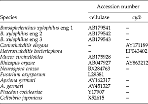
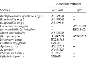
Cellulase and cytb accession numbers of reference organisms/strains used for phylogenetic analysis.
Maximum likelihood cytb phylogenetic tree based on nucleotide sequences of 20 isolates of B. xylophilus and reference strains (alignment length 387 bp). Percentage bootstrap is indicated on internal branches (100 replicates); scale bar indicates 0.1 substitutions per site. The Tamura & Nei model (Tamura & Nei, Reference Tamura and Nei1993) was used. Symbols represent isolates collected until □, 2008; ●, 2009 and ○, 2010.
Phylogenetic analysis of the cellulase gene
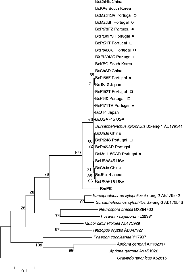
Fourteen Portuguese and 12 foreign isolates from China, Japan, South Korea and USA of B. xylophilus, as well as one B. mucronatus (BmPt0) Portuguese isolate, were used to generate a PCR product of approximately 950 bp, containing the partial sequence of the cellulase gene that was sequenced. Accession numbers are listed in table 1. Phylogenetic analyses were performed by comparing our sequences with others available in public databases, such as those from B. xylophilus eng 1, eng 2 and eng 3, from fungi (R. oryzae, Mucor circinelloides, Neurospora crassa, Fusarium oxysporum) and from insects (Phaedon cochleariae, Apriona germari); the bacterial sequence from the bacterium Cellvibrio japonicus was used as an outgroup (table 2). One major cluster was obtained which contains all B. xylophilus isolates (fig. 3) (bootstrap support of 96%), sharing similarities between 98 and 99%. Inside this major cluster, there is another one, which includes all Portuguese isolates collected since the first detection in 1999 until the recent (2010) detection in Madeira Island (bootstrap 65%) and also isolates from China (BxChJs, BxChSD, BxChHB), South Korea (BxKAs, BxKBG), Japan (BxJT4, BxJS10) and USA (BxUSA745). There is no clear geographic differentiation between Portuguese isolates collected until 2008 (during the time that the disease was restricted to Setúbal Peninsula) and after this date (new outbreaks in the centre and north of Portugal and in Madeira Island). Isolates BxPt66F and BxJS10 group together inside the previous cluster as well as BxPt24S, BxPt49AR and BxChJs. Grouping with the major cluster, are isolates BxUSA345, BxUSA618, BxJKa4 and BxChJx, the most distant related isolates being BxJKa4 and BxUSA618. The presence of isolates from China, Japan and USA in different branches of the phylogenetic tree points to the existence of intraspecific variability. Bx-eng-1 sequence groups with Portuguese and foreign isolates (sequence identity of 99–100%). Grouping apart from this cluster there are two sequences, Bx-eng-2 and Bx-eng-3 that probably represent different cellulase genes, as already proposed by Kikuchi and co-authors (2004). Bursaphelenchus xylophilus cellulase sequences share similarities of between 49 and 62% with the fungi R. oryzae and M. circinelloides, and of 49–66% with N. crassa and F. oxysporum. The similarity between B. xylophilus and fungal cellulases (49–66%) is higher than that between B. xylophilus and the bacterium C. japonicus sequences (36–41%).
Maximum likelihood cellulase phylogenetic tree based on nucleotide sequences of 26 isolates of B. xylophilus and reference strains (alignment length 787 bp). Percentage bootstrap is indicated on internal branches (100 replicates); scale bar indicates 0.1 substitutions per site. The Kimura two-parameter model (Kimura, Reference Kimura1980) was used. Symbols represent isolates collected until □, 2008; ●, 2009 and ○, 2010.
Discussion
Following the first report (Mota et al., Reference Mota, Braasch, Bravo, Penas, Burgermeister, Metge and Sousa1999), B. xylophilus was successfully restricted to the Setúbal Peninsula until 2008. In 2008, the nematode was detected in new outbreak spots in central and northern regions of the country (Rodrigues, Reference Rodrigues, Mota and Vieira2008) and, more recently, in one of the islands of the Madeira archipelago (Fonseca et al., Reference Fonseca, Lopes, Cardoso, Pestana, Abreu, Nunes, Mota and Abrantes2010).
The first study conducted to understand the genetic diversity of Portuguese populations was performed by Vieira et al. (Reference Vieira, Burgermeister, Mota, Metge and Silva2007) using 24 isolates from the initial affected area. In this study, using random amplification of polymorphic DNA (RAPD) analysis, the authors showed that there was almost no genetic diversity among Portuguese isolates and that these grouped with a Chinese isolate instead of an isolate from the USA.
In a recent study using the inter-simple sequence repeats (ISSR) technique and intergenic spacer (IGS) sequences, 34 isolates representing the different geographic locations of B. xylophilus in Portugal, from 1999 to 2010, were analysed, to understand the pathways of spread and the introduction point of the disease (Valadas et al., Reference Valadas, Laranjo, Barbosa, Espada, Mota and Oliveira2012). This study was the first to use a significant number of B. xylophilus isolates from continental Portugal and Madeira Island as well as foreign isolates. The ISSR technique proved to be useful in determining genetic diversity of Portuguese B. xylophilus isolates.
ISSR fingerprints showed high genetic variability among Portuguese isolates, except for those obtained prior to 2008. Until 2008, B. xylophilus populations found in continental Portugal showed low genetic diversity, pointing to a single introduction, probably from Asia, in agreement with the results of Vieira et al. (Reference Vieira, Burgermeister, Mota, Metge and Silva2007). In contrast, recent populations from continental Portugal (2009–2010) and Madeira show high genetic diversity, suggesting multiple introductions from different origins.
In the present study, cytb and cellulase genes were used, with the aim of understanding the genetic diversity of Portuguese populations, comparing them with foreign isolates from Japan, China, South Korea and USA. Our results agree with previous studies (Vieira et al., Reference Vieira, Burgermeister, Mota, Metge and Silva2007; Valadas et al., Reference Valadas, Laranjo, Barbosa, Espada, Mota and Oliveira2012) in suggesting a possible Asian origin for Portuguese isolates.
Due to the high copy number in individual cells, the lack of recombination and the strict maternal inheritance, mitochondrial DNA (mtDNA) seems to be a potential marker to study genetic diversity and characterize populations from the same organism (Madani et al., Reference Madani, Subbotin, Ward, Li and De Boer2010). In order to characterize populations and infer the origin of Portuguese isolates, the cytb gene was used in the present study. Bursaphelenchus xylophilus populations were characterized and, although the intraspecific diversity was very low, it was possible to confirm the previously suggested similarity of Portuguese and Asian isolates (fig. 2) (Vieira et al., Reference Vieira, Burgermeister, Mota, Metge and Silva2007; Valadas et al., Reference Valadas, Laranjo, Barbosa, Espada, Mota and Oliveira2012). However, the low resolution obtained with cytb prompted us to use the cellulase gene, in an attempt to find a better genetic marker.
In the cellulase phylogenetic tree (fig. 3), Portuguese and foreign isolates group together in the same large cluster, which also includes the Bx-eng-1 sequence, previously obtained by Kikuchi and collaborators (2004). Inside this major clade, it is possible to observe some genetic diversity among isolates, showing that cellulase sequences are more discriminative to study intraspecific variations than cytb sequences. However, no clear grouping of Portuguese isolates according to their geographic location or year of collection is observed. The clustering of Portuguese isolates with different Asian isolates may suggest more than one introduction in Portugal of the PWN of Asian origin.
Figure 3 shows that B. xylophilus cellulases are more similar to the fungal cellulases from R. oryzae and M. circinelloides than to cellulases from insects or bacteria. This large group clusters first with B. mucronatus and then with Bx-eng-2 and Bx-eng-3, which seem to code for different cellulase genes (Kikuchi et al., Reference Kikuchi, Jones, Aikawa, Kosaka and Ogura2004). Previous phylogenetic studies had already suggested that cellulase genes from nematodes may have been acquired from different microbial donors (Dieterich & Sommer, Reference Dieterich and Sommer2009). Cellulases from plant parasitic Tylenchida, such as Globodera, Heterodera and Meloidogyne (cyst and root-knob nematodes) are from the GHF5 family of cellulases and seem to have been acquired by lateral gene transfer (LGT) from bacteria (Kikuchi, Reference Kikuchi, Zhao, Futai, Sutherland and Takeuchi2008). On the other hand, our results agree with other studies, which have suggested that B. xylophilus might have acquired a different family of cellulases, GHF45, from fungi by LGT (Kikuchi et al., Reference Kikuchi, Jones, Aikawa, Kosaka and Ogura2004). This may be related to the fact that this species is solely a fungal feeder, transmitted by insect vectors to dead or dying trees during oviposition. Although B. xylophilus, as well as cyst and root-knob nematodes, belongs to the Tylenchida, their cellulases are not directly related. GHF45 cellulases are exclusively produced by Bursaphelenchus species.
This study was the first to use cellulase and cytb genes to characterize PWN populations. However, contrary to other studies, our results suggest that cellulase is a better marker to study genetic diversity among populations of B. xylophilus.
The recent release of the full B. xylophilus genome sequence (Kikuchi et al., Reference Kikuchi, Cotton, Dalzell, Hasegawa, Kanzaki, McVeigh, Takanashi, Tsai, Assefa, Cock, Otto, Hunt, Reid, Sanchez-Flores, Tsuchihara, Yokoi, Larsson, Miwa, Maule, Sahashi, Jones and Berriman2011) may unravel new molecular markers to study intraspecific variability to determine pathways of disease spread and follow the evolution of introduced isolates. Furthermore, recent studies on mitochondrial DNA sequencing (A. Amorim, pers. comm.) should be considered to evaluate the potential of mtDNA and its different genes for phylogenetic and biodiversity studies.
Acknowledgements
This research was supported by the EC 7th 324 Framework project REPHRAME (KBBE.2010.1.4-09, ‘Analysis of the potential of the pine wood nematode (Bursaphelenchus xylophilus) to spread, survive and cause pine wilt in European coniferous forests in support of EU plant health policy’) and by a national project from the Portuguese National Forestry Authority (AFN, Autoridade Florestal Nacional), ‘O nemátode da madeira do pinheiro (NMP), Bursaphelenchus xylophilus’. This work was also funded by FEDER (Funds through the Operational Programme for Competitiveness Factors) – COMPETE and National Funds through FCT (Fundação para a Ciência e a Tecnologia) under the Strategic Project PEst-C/AGR/UI0115/2011. We would like to thank Dr Bouang Zhao, Nanjing Forestry University, China; Dr Hyerim Han, Korea Forest Research Institute, South Korea; Dr K. Futai, Kyoto University, Japan; Dr Robin Giblin-Davis, University of Florida, USA and Isabel Abrantes, Coimbra University, Portugal, for providing nematode cultures of B. xylophilus. We thank Filipe Pereira, Cláudia Moreira, Barbara van Asch and António Amorim for providing sequence data of B. xylophilus cytochrome b. We would like to thank Pedro Barbosa, for providing the map used in fig. 1. This paper is a portion of the PhD dissertation of the first author, who is supported by a doctoral scholarship from FCT (SFRH/BD/22086/2005).