Introduction
An early and reliable way to detect dementia with Lewy bodies (DLB) is important to ensure safe management and aid in planning for future care. Extrapyramidal motor symptoms in DLB patients make them more functionally disabled than patients with Alzheimer's disease (AD) with similar cognitive scores (McKeith et al., Reference McKeith2006). The core features of DLB, such as visual hallucination and fluctuating cognitive dysfunction, have been highly correlated with the psychological and behavioral problems (Ricci et al., Reference Ricci2009). DLB patients also display sensitivity to typical and atypical neuroleptics, which can lead to a potentially fatal complication (McKeith et al., Reference McKeith, Fairbairn, Perry, Thompson and Perry1992). Patients with DLB have a more impaired quality of life than do patients with AD who have a similar level of cognitive impairment, and nearly double the cost of care (Boström et al., Reference Boström, Jönsson, Minthon and Londos2007). At present, the diagnostic criteria for DLB display high specificity, but low sensitivity (McKeith et al., Reference McKeith2006).
The Montreal Cognitive Assessment (MoCA) is a brief global cognitive screening tool designed to address some of the limitations of the Mini-mental State Examination (MMSE) and to detect mild cognitive impairment (MCI) or mild dementia (Nasreddine et al., Reference Nasreddine2005). This test assesses a broader range of cognitive domains than the MMSE, and displays greater sensitivity than the MMSE (Julayanont et al., Reference Julayanont, Phillips, Chertkow, Nasreddine and Larner2013). The MoCA examines multiple domains of cognitive function, such as short-term memory recall, visuospatial ability, executive function, attention–concentration–working memory, language, and orientation to time and place. The MMSE remains the most commonly used cognitive screening instrument and has been translated into multiple languages. Yet, compared with the MMSE, the MoCA has higher sensitivity and specificity to differentiate participants with mild AD from individuals with normal cognition (Nasreddine et al., Reference Nasreddine2005). In addition, the MoCA is also a suitably accurate brief test than the MMSE to screen all levels of cognition in Parkinson's disease (PD), PD-MCI, and idiopathic rapid eye movement sleep behavior disorder-MCI (Dalrymple-Alford et al., Reference Dalrymple-Alford2010; Gagnon et al., Reference Gagnon, Postuma, Joncas, Desjardins and Latreille2010). The mixed performance of the MMSE and its subscores relative to AD and DLB has been reported in the literature; one such example is that DLB has worse attention and construction subscores but a better memory subscore than AD (Ala et al., Reference Ala, Hughes, Kyrouac, Ghobrial and Elble2002; Hanyu et al., Reference Hanyu2006). The MMSE score loss in pure DLB is less severe than in cases with a significant AD pathology (Nelson et al., Reference Nelson2009). The practical suggestions on differentiating DLB from AD by using subscores derived from the MMSE, such as Ala scores, modified Ala scores, the qualitative scoring pentagons copy test, or triplet orientation score, have not yet been generally acknowledged (Ala et al., Reference Ala, Hughes, Kyrouac, Ghobrial and Elble2002; Palmqvist et al., Reference Palmqvist, Hansson, Minthon and Londos2009; Caffarra et al., Reference Caffarra2013). No published report has described the performance characteristics of DLB as reflected in this increasingly used screening instrument, the MoCA.
This study was designed to analyze and examine the sensitivity, specificity, and threshold scores of the MoCA and MMSE that best correspond to a clinical diagnosis of DLB. The performance of the MoCA in normal participants and mild-to-moderate AD or DLB patients is also discussed.
Methods and participants
Sixty-seven AD, 36 DLB, and 62 healthy participants without dementia (NC), aged 60 to 90, were invited to participate in this study. Participants with AD and DLB were recruited from neurologic or geropsychiatric clinics. Detailed history taking, physical, neurological and mental status examinations, laboratory examinations, and brain imaging were performed for all participants. All the AD participants were diagnosed with probable AD according to the Diagnostic and Statistical Manual of Mental Disorders, Fourth Edition, and the National Institute of Neurological and Communicative Disorders and Stroke/Alzheimer's Disease and Related Disorders Association criteria (McKhann et al., Reference McKhann, Drachman, Folstein, Katzman, Price and Stadlan1984). The diagnosis of DLB for the participants was made based on the criteria of the consensus consortium on DLB (McKeith et al., Reference McKeith2005). The NC participants were recruited from the community. They all underwent a series of screening questions, physical, neurological and mental status examinations, and the Activity of Daily Living Inventory test. None showed evidence of neurological or psychiatric diseases.
All NC, AD, and DLB participants took the MoCA and MMSE tests at the same time, and the latter two groups were also evaluated with the Clinical Dementia Rating (CDR) scale. The CDR ratings of the AD and DLB participants were 0.5, 1, or 2. For those with 12 years or fewer of formal education, following the standard procedure of the MoCA rating scale, one point was added to the MoCA score (Nasreddine et al., Reference Nasreddine2005). The institutional review board of Tainan Hospital approved the study protocol before it was initiated.
We used generalized Cochran–Mantel–Haenszel tests to assess the differences in participant demographic characteristics among the study groups. We used receiver operating characteristics (ROC) curve analysis with area under the curve (AUC), sensitivity, specificity, positive predictive value (PPV), and negative predictive value (NPV) to evaluate the performance of the MoCA and MMSE in the clinical diagnosis of AD and DLB. The Mann–Whitney U test and multiple regression analysis were used to compare and analyze the different neuropsychological test results among the study groups with/without adjusting pre-specified confounding factors, such as age, gender, education, and CDR. All statistical analyses were conducted using SAS (v9.2, SAS Inc., Cary, NC) and SPSS (V17.0, SPSS Inc., Chicago, IL).
Results
Participant characteristics and MoCA and MMSE performance in each group
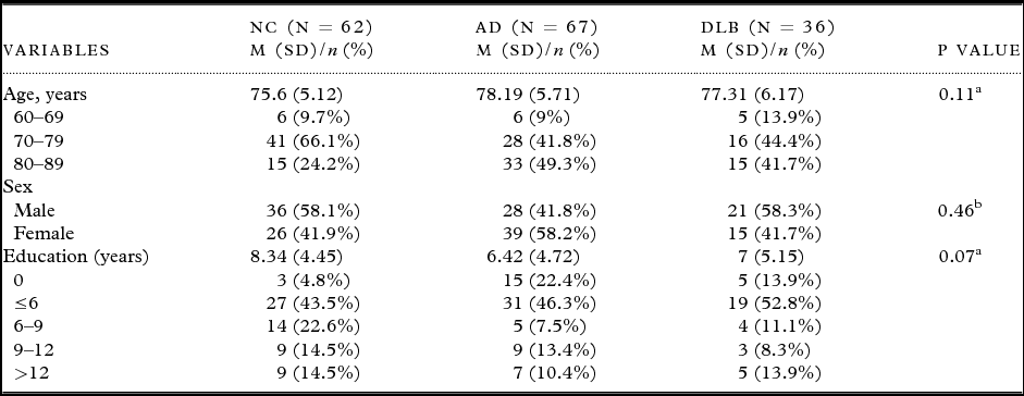
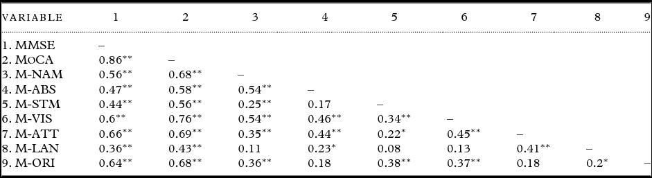
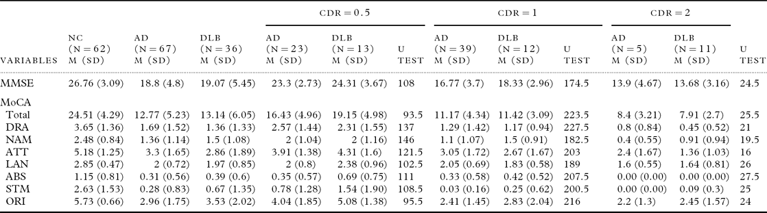
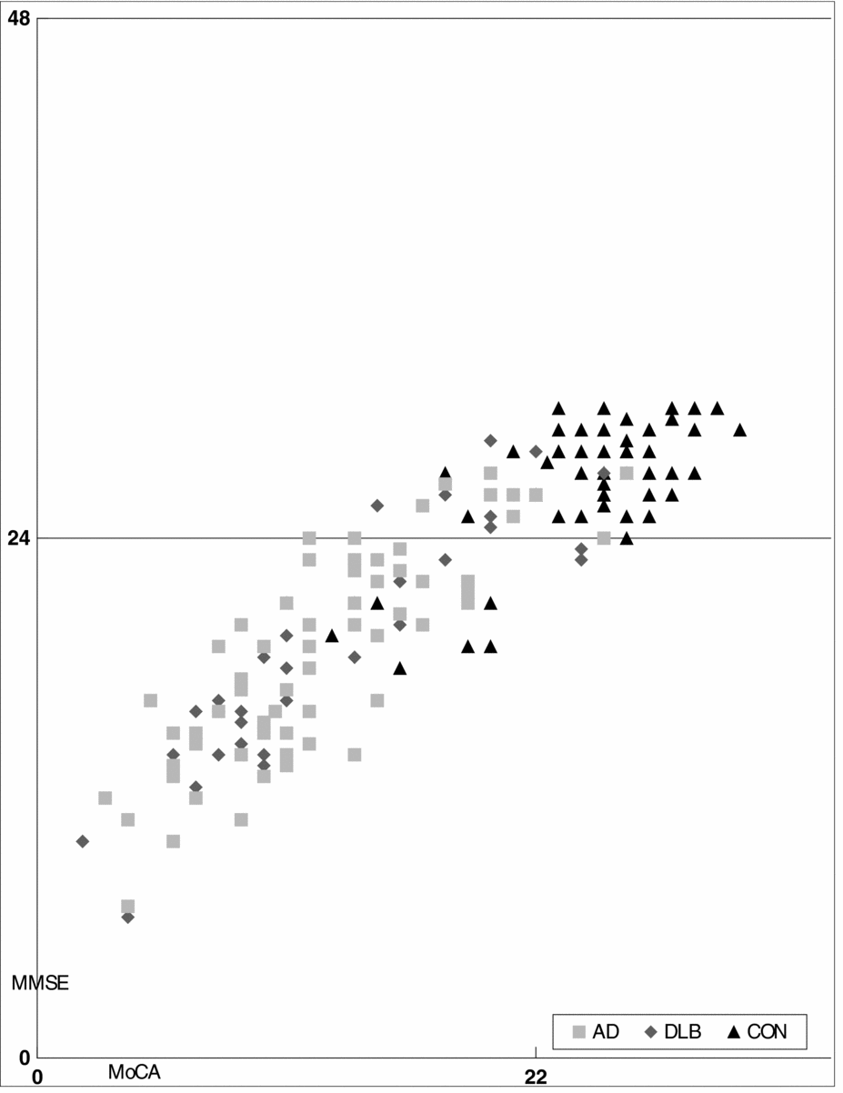
The diagnosis of AD or DLB remained the same after a two-year follow-up at the clinic. No significant difference was noted in mean age, sex ratio, and years of education among the NC, AD, and DLB groups (Table 1). The MoCA and the MMSE were found to be good neuropsychological instruments for detecting dementia. The scatter plot of the MoCA and MMSE scores of the three groups showed they were consistent with each other (Figure 1). The Pearson correlation coefficients also indicated that the total scales of the MoCA and MMSE and most subscales of the MoCA were highly correlated (p < 0.01; Table 2). The majority of the NC group scored in the normal range, and the majority of the AD and DLB participants scored in the abnormal range of both tests. In all, 9% (6/67) of the AD and 19.4% (7/36) of the DLB participants scored in the normal range of the MoCA and in the abnormal range of the MMSE, and only 5.6% (2/36) of the DLB participants scored in the normal range of the MMSE and in the abnormal range of the MoCA. The mean of the total MoCA score in the NC, AD, and DLB groups, respectively, was 24.51, 12.77, and 13.14 (Table 3). The MoCA and MMSE total scores and all subscales of the MoCA of the AD and DLB patients showed no difference (p = 0.016) at different stages of severity.
The demographic data of NC, AD, and DLB

NC = participants without dementia; AD = Alzheimer's disease; DLB = dementia with Lewy bodies.
aUsing one-way ANOVA;bUsing Cochran–Mantel–Haenszel tests.
Pearson correlation coefficients of the MMSE and MoCA in total and subscale scores
M- = item of MoCA; NAM = naming; ABS = abstraction; STM = short-term memory; VIS = visuospatial and executive function; ATT = attention; LAN = language; ORI = orientation; MoCA = Montreal Cognitive Assessment; MMSE = Mini-Mental State Examination.
*p < 0.05; **p < 0.01.
MMSE, MoCA scores in NC, AD, and DLB three groups (with the Mann–Whitney U test)
DRA=drawing; NAM=naming; ATT=attention; LAN=language; ABS=abstraction; STM=short-term memory; ORI=orientation; MoCA=Montreal Cognitive Assessment; MMSE=Mini-Mental State Examination; CDR=Clinical Dementia Rating.
Note that none of the Mann--Whitney U test is significant.
Scatter plot of the MoCA and MMSE scores for AD, DLB, and NC groups.
Sensitivity, specificity, and cut-off points of the MoCA and MMSE in DLB and AD participants
When calculating an ROC curve and AUC to compare the MoCA in distinguishing the AD, DLB, and NC participants, the cut-off point of AD dementia was 21/22 with sensitivity of 95.5% and specificity of 82.3% (PPV: 85.3%; NPV: 94.4%) (Figure 2). Likewise, the cut-off point for DLB was 22/23 with sensitivity of 91.7% and specificity of 80.6% (PPV: 73.3%; NPV: 94.3%) (Figure 3). The AUC of the MoCA for predicting AD was 0.945 (95% CI: 0.908–0.983), and that for predicting DLB was 0.932 (95% CI: 0.885–0.978). For the MMSE, the cut-off points of AD and DLB from NC were both 24/25, with sensitivity of 88.1% and specificity of 85.5% (PPV: 86.8%; NPV: 86.9%) for AD, and sensitivity of 77.8% and specificity of 85.5% (PPV: 75.7%; NPV: 86.9%) for DLB. The AUC of the MMSE for predicting AD was 0.920 (95% CI: 0.875–0.965), and for predicting DLB was 0.895 (95% CI: 0.831–0.959).
ROC curve analysis of the MMSE and MoCA to detect AD.
ROC curve analysis of the MMSE and MoCA to detect DLB.
Comparison of the performance of the MoCA and MMSE in each group
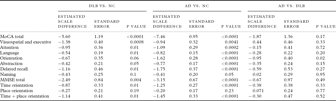
After controlling for sex, age, and education in AD CDR 0.5, DLB CDR 0.5, and the NC, comparisons of the MoCA and MMSE showed that the scores of the NC group were significantly higher than those of the DLB or AD group (Table 4). In all subscales of the MoCA, the NC group was significantly different from the AD or DLB group, except the orientation and naming items showed no significant difference between the NC and DLB groups. In the time orientation item of the MMSE, the DLB group had lower scores than the NC group, but in the place orientation of the MMSE, there was no difference between these two groups. In the moderate stage, CDR = 2, the naming item scores of the MoCA differed between the AD and DLB groups, with the DLB group having higher scores than the AD group (p = 0.0421).
Under controlling age, sex, and education, the comparison of the MMSE, MoCA in NC (62 cases), AD CDR 0.5 (23 cases), and DLB CDR 0.5 (13 cases) (three groups comparison)
NC = participants without dementia; AD = Alzheimer's disease; DLB = dementia with Lewy bodies; MoCA = Montreal Cognitive Assessment; MMSE = Mini-Mental State Examination.
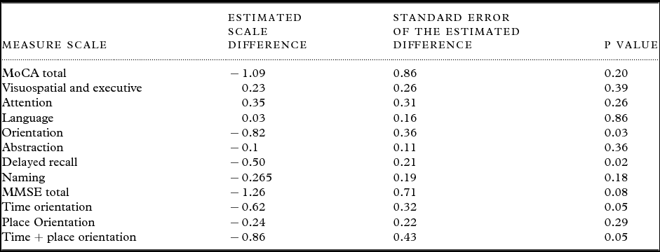
In pair comparisons of the AD and DLB groups, after adjusting for CDR, age, gender, and education, the total scores of the MoCA and MMSE showed no significant difference (Table 5). Nonetheless, in the orientation and short-term memory subscales of the MoCA, the AD group had lower scores than the DLB group, revealing a significant difference between these two groups.
Under controlling age, sex, education, and the CDR stage, the comparison of MMSE, MoCA in AD and DLB all cases
MoCA = Montreal Cognitive Assessment; MMSE = Mini-Mental State Examination.
Discussion
Cut-off score of the MoCA in the DLB group
Similar to previous studies, this study provides evidence that both the MoCA and the MMSE produce good discrimination between both forms of dementia and the healthy elderly group. The results of this study show that the cut-off point of DLB is slightly higher than that of AD, at 22/23 and 21/22, respectively. The previously suggested cut-off point of AD on the MoCA was 26, with sensitivity of 100% and specificity of 87% (Nasreddine et al., Reference Nasreddine2005). The data provided by Luis et al. suggested that a score of 23 might be superior (Luis et al., Reference Luis, Keegan and Mullan2009). In addition, a recent study with 150 hospital-based patients, with ages ranging from 20 to 87 years, shows that downward adjustment of the MoCA cut-off point to 20/30 maximized test accuracy and improved specificity (95%) for some loss of sensitivity (63%) (Larner, Reference Larner2011). Considering the cultural differences in Asia, a Korean study suggested a cut-off value of 22/23 for the MoCA-K in screening AD, with sensitivity of 98% and specificity of 84% (Lee et al., Reference Lee2008). A recent study of the MoCA in northern Taiwan showed 21/22 to be the best score to discriminate AD from NCs (Tsai et al., Reference Tsai, Lee, Wang, Shia, Nasreddine and Fuh2011). A study of a large body of population-based normative data for the MoCA, including 2,653 ethnically diverse participants, showed the MoCA total scores were lower than previously published normative data, with a mean of 23.4 at all ages (Rossetti et al., Reference Rossetti, Lacritz, Cullum and Weiner2011). The mean score on the MoCA for those aged between 70 and 80 was 21.2, though there were only 79 samples (Rossetti et al., Reference Rossetti, Lacritz, Cullum and Weiner2011). Age should be considered, since their results showed the MoCA scores were weakly but significantly inversely correlated with age, and also, their results showed those with more years of education obtained higher MoCA scores (Rossetti et al., Reference Rossetti, Lacritz, Cullum and Weiner2011). Hence, the possible causes of the different AD cut-off scores of the MoCA between different countries were probably the different number of years of education and the average age. In the samples from Korea and Taiwan, the years of education were less than those in the Canadian data, with averages of 7–10 and 10–13 years, respectively (Ferman et al., Reference Ferman2006; Lee et al., Reference Lee2008; Tsai et al., Reference Tsai, Lee, Wang, Shia, Nasreddine and Fuh2011). The cultural factor should also be considered; for example, some of our NC participants were unable to recognize the rhino on the picture.
There is as yet no suggested cut-off score for the MoCA for screening DLB. A cut-off score of 22/23 was suggested by this study, with sensitivity of 92%, which is higher than the MMSE with sensitivity of 78% in detecting DLB. The cut-off score of the MoCA is slightly higher for DLB than for AD. People with early-stage DLB usually present with attention, motor, or psychiatric changes rather than impaired memory function. DLB has more impairment in function and in quality of life than AD, even when the level of dementia is similar (McKeith et al., Reference McKeith2006; Boström et al., Reference Boström, Jönsson, Minthon and Londos2007). The more functional impairment of DLB makes for higher CDR scores than for AD, though the cognitive function of DLB is more preserved. Meanwhile, as at the very mild stage, the NC group had higher scores than the AD group, and compared to DLB, there were no differences in orientation and naming on the MoCA in pair comparisons controlling for age, gender, and educational factors. Therefore, one could imply that, with an identical demographic basis, the cut-off score of the MoCA in DLB should not be less than that for AD.
Performance on the MoCA items in the DLB, AD, and NC groups
Based on the AUCs of DLB and AD, the MoCA seems to be a more sensitive instrument than the MMSE in screening out AD and DLB from the NC group (Figures 2 and 3). In pair comparisons of AD and DLB in these two tests, and controlling for age, gender, years of education, and CDR, the orientation and short-term memory scores of the two groups on the MoCA items showed a difference. Consistent with past studies, a relative preservation of short-term memory and orientation were noted in early DLB, compared to AD (Shimomura et al., Reference Shimomura1998; Kraybill et al., Reference Kraybill2005; Crowell et al., Reference Crowell, Luis, Cox and Mullan2007; Palmqvist et al., Reference Palmqvist, Hansson, Minthon and Londos2009). The relative preservation of verbal memory and the naming task results revealed that verbal skills in DLB are also better than in AD; this is supported by preliminary studies and the naming item results of the MoCA at CDR 0.5 and 2 in this study (Johnson et al., Reference Johnson, Morris and Galvin2005; Kraybill et al., Reference Kraybill2005).
Previous findings have shown that DLB patients have more severe impairment in visual perception and attention than AD patients (Kraybill et al., Reference Kraybill2005; Crowell et al., Reference Crowell, Luis, Cox and Mullan2007). The attention item of the MoCA did not show a difference between these two groups. The calculation constituted half scores on the attention item in the MoCA, and this might be the reason why this item was not able to discriminate between DLB and AD.
Similar to previous MMSE studies, the orientation item of the MoCA showed a difference between these two forms of dementia (Palmqvist et al., Reference Palmqvist, Hansson, Minthon and Londos2009; Andersson et al., Reference Andersson, Zetterberg, Minthon, Blennow and Londos2011). As in the very mild stage, the orientation item of the MoCA in DLB, especially temporal orientation, was not significantly different from that of non-dementia cases, while in AD, this item was significantly lower than that of non-dementia cases. Unlike the MMSE, the MoCA orientation item emphasizes the time domain, which gives rise to different presentations between DLB and AD. In another study, DLB had better locational orientation scores, but the samples included more severe CDR and fewer very mild CDR cases than in our study (Shimomura et al., Reference Shimomura1998). More severe CDR means more impairment of locational orientation.
DLB patients have been reported to have more visuospatial/constructional deficits than AD patients (Johnson et al., Reference Johnson, Morris and Galvin2005; Crowell et al., Reference Crowell, Luis, Cox and Mullan2007). However, another study comparing community-based dementia patients with autopsy-verified AD patients, those with a DLB pathology or both, showed no differences in visual constructional ability between groups (Kraybill et al., Reference Kraybill2005). Using visuospatial ability as a way to discriminate DLB from AD also has been criticized by Ballard et al. (Reference Ballard, O’Brien and Tovee2002), since the fluctuating cognition might influence this presentation (Ballard et al., Reference Ballard, O’Brien and Tovee2002). Past one pentagon drawing study, the scoring system with a six-point hierarchical scale developed by Bourke et al. (Reference Bourke, Castleden, Stephen and Dennis1995) supports the significantly worse pentagon coping of DLB than AD (Cormack et al., Reference Cormack, Aarsland, Ballard and Tovée2004). A new method with qualitative scoring of the MMSE pentagon test developed recently also showed DLB had a significantly lower pentagon score than AD (Caffarra et al., Reference Caffarra2013). The pentagon drawing only scored 1 among the 30 MMSE scores, so it is hard to discriminate the difference between DLB and AD (Cormack et al., Reference Cormack, Aarsland, Ballard and Tovée2004). The visuospatial and executive item of the MoCA mixes clock drawing, cubic construction, and alternative trail-making, which might obscure the focus of the visuospatial/construction ability.
Limitations
Because of the low sensitivity of the clinical diagnosis and the lower prevalence of DLB than AD, we had a smaller sample size of DLB than AD in this study. Most autopsy-proven DLB displays a concomitant AD pathology. For example, around 25% of autopsied AD cases had cortical limbic Lewy bodies in sufficient quantity to be classified as mixed DLB/AD, and most of them did not clinically manifest the distinctive symptoms of DLB (McKeith et al., Reference McKeith1996). The co-occurrence of an AD and DLB pathology has been confirmed (Dickson, Reference Dickson2001), and this makes the clinical diagnosis more difficult. Though the revised clinical consensus criteria of DLB gave greater diagnostic weight to clinical features suggestive of DLB, the sensitivity and specificity have not yet been explored (McKeith et al., Reference McKeith2005). Hence, the clinically diagnosed probable AD or DLB cases in our study were possibly misclassified, rendering the cognitive test results somewhat different from those of studies with autopsy-verified cases.
Conclusions
The diagnosis of DLB is still primarily based upon clinical history, neuroimaging study, and laboratory evaluation. The neuropsychological profile might play a role in the differential diagnosis of DLB and other dementias. The results of this study showed that the MoCA is a more sensitive instrument than the MMSE to screen AD or DLB patients from non-dementia cases. Modifying the short-term memory, visuospatial/executive and attention items in the MoCA might afford an increased probability of quickly screening AD from DLB.
Conflict of interest
None.
Description of authors’ roles
C. S.-M. Wang was responsible for the design of the study, interviewing the patients, and drafting the paper. M.-C. Pai supervised the project and paper writing. P.-L. Chen was responsible for the statistical design and analysis, and assisted in revising the paper. P.-F. Chien performed the neuropsychological tests and collected the data. N.-T. Hou and Y.-C. Huang interviewed the patients and assisted in collecting the data. All authors read and approved the final version of this paper.
Acknowledgments
This study was supported by grants from the Department of Health, Executive Yuan, Taiwan. The authors gratefully acknowledge the technical assistance of Wan-Jung Tsai and Pei-Chia Hung as well as secretarial assistance from Li-Hui Wang.