Introduction
Retinoic acid (RA) plays a vital role in embryonic development. Both excess and insufficient RA levels lead to severe congenital defects in the eyes, as well as the skull, limbs, brain, heart, and other systems and structures (Ross et al., Reference Ross, McCaffery, Drager and De Luca2000). Studies in zebrafish (Danio rerio) have demonstrated that excess RA during optic primordia formation leads to duplication of the ventral retina (Hyatt et al., Reference Hyatt, Schmitt, Marsh-Armstrong and Dowling1992). Conversely, using citral, a retinaldehyde dehydrogenase (RALDH) inhibitor, to reduce endogenous RA results in eyes lacking a ventral retina (Marsh-Armstrong et al., Reference Marsh-Armstrong, McCaffery, Gilbert, Dowling and Drager1994). Studies examining RA effects in mice have had to employ double and triple gene knockouts since mice have three RALDH enzymes, whereas the RALDH enzymes in zebrafish are readily inhibited by water-soluble biochemical agents that block RA synthesis (Molotkov et al., Reference Molotkov, Molotkova and Duester2006; Kumar et al., Reference Kumar, Sandell, Trainor, Koentgen and Duester2012). The relatively large size of its eyes and well-documented embryogenesis make zebrafish an ideal candidate for studying the role of RA during eye development.
Microphthalmia is a congenital ocular deformation characterized by abnormally small eyes, with or without structural abnormalities. It is clinically juxtaposed with anophthalmia, a more severe condition in which ocular components are absent. Studies in both human and animal models have associated microphthalmia with genes that are involved in vitamin A metabolism. For example, STRA6(STimulated by Retinoic Acid gene 6) is proposed to play a role in the pathogenesis of anophthalmia/microphthalmia (A/M).
STRA6 encodes a transmembrane receptor for retinol-binding protein, which mediates cellular uptake of retinol. Once channeled into the cell, retinol is oxidized to retinal, which is in turn oxidized to RA. A mutation in STRA6 will disrupt vitamin A metabolism and ultimately reduce levels of RA. Clinical research has identified variants of STRA6 in patients with A/M (White et al., Reference White, Lu, Metlapally, Katowitz, Kherani, Wang, Tran-Viet and Young2008). In particular, truncating mutations in STRA6 link to Matthew–Wood syndrome, a rare congenital disorder characterized by microphthalmia as well as heart and lung defects (Golzio et al., Reference Golzio, Martinovic-Bouriel, Thomas, Mougou-Zrelli, Grattagliano-Bessieres, Bonniere, Delahaye, Munnich, Encha-Razavi, Lyonnet, Vekemans, Attie-Bitach and Etchevers2007).
In animal models, microphthalmia has been experimentally induced by reducing levels of RA. Isken et al. (Reference Isken, Golczak, Oberhauser, Hunzelmann, Driever, Imanishi, Palczewski and von Lintig2008) used an antisense morpholino oligonucleotide to knock down STRA6 in developing zebrafish. Injected embryos developed a microphthalmia, curved body axis, cardiac edema, and craniofacial defects (Isken et al., Reference Isken, Golczak, Oberhauser, Hunzelmann, Driever, Imanishi, Palczewski and von Lintig2008). These multisystem malformations are similar to those observed in children suffering from Matthew–Wood Syndrome. Chicken embryos also develop microphthalmia at specific developmental stages when RA signaling is inhibited (Sen et al., Reference Sen, Harpavat, Peters and Cepko2005). Additionally, morpholino knockdown of bcox, a key enzyme in vitamin A biogenesis in zebrafish, resulted in a small eye and photoreceptor defects (Biehlmaier et al., Reference Biehlmaier, Lampert, von Lintig and Kohler2005). We have found that zebrafish embryos treated with RA inhibitors [citral or diethylaminobenzaldehyde (DEAB)] just before bud formation [i.e., ∼9 h postfertilization (hpf)] develop microphthalmia. Inhibiting RA synthesis slightly later, on the other hand, results in a different phenotype, namely an absence of the ventral retina (Marsh-Armstrong et al., Reference Marsh-Armstrong, McCaffery, Gilbert, Dowling and Drager1994). Our results provide further evidence that RA deficiency plays a role in the pathogenesis of microphthalmia, and also that small alterations in the timing of the RA deficiency result in quite different phenotypes.
Materials and methods
Zebrafish care
AB, α-actin-GFP, and RGYn fish strains were maintained under standard conditions at 28.5°C on a 10-h dark and 14-h light cycle (Higashijima et al., Reference Higashijima, Okamoto, Ueno, Hotta and Eguchi1997; Westerfield, Reference Westerfield2000). The RGYn fish utilizes retinoic acid response element (RARE)-driven promoters to express enhanced yellow fluorescence protein (EYFP) (Perz-Edwards et al., Reference Perz-Edwards, Hardison and Linney2001). To work with embryos at the desired developmental stage, zebrafish tanks were kept in a modular lighting system that alters the fish’s day/night cycles. The light box was set to automatically turn on and off at 12:15 am and 2:15 pm, respectively. Embryos were staged in hpf and days postfertilization (dpf) based on morphological features. Embryos were raised in E3 buffer (5 mM NaCl, 0.17 mM KCl, 0.33 mM CaCl2, 0.33 mM MgSO4) at 28.5°C. All animal husbandry and experiments were approved and conducted in accordance with guidelines set forth by the Institutional Animal Care and Use Committee of Harvard University and Western University of Health Sciences and adhered to the ARVO Statement for the Use of Animals in Ophthalmic and Vision Research.
DEAB treatment
A stock solution of 40 mM DEAB (VWR, Radnor, PA) was made by dissolving 0.071 g of DEAB in 10 ml of dimethyl sulfoxide (DMSO) and kept at room temperature. DEAB is a potent partially competitive inhibitor of aldehyde dehydrogenases, the enzymes that convert retinal to RA in vivo as well as in vitro (Russo et al., Reference Russo, Hauguitz and Hilton1988; Mahmoud et al., Reference Mahmoud, Potter, Colvin, Hilton and Mezey1993). The optimal concentration of DEAB and the appropriate embryonic stage for biochemical perturbation were determined through a series of experimental trials. 8, 9, and 10 hpf embryos were treated for 2 h with different DEAB concentrations in E3 buffer: 10, 50, 100, 200, and 400 μM. Control embryos were treated with equivalent DMSO-treated E3 buffer. After treatment, embryos were washed three times with E3 buffer and allowed to grow for the duration of the study. Demonstration of DEAB’s effectiveness at inhibiting RA synthesis was accomplished by incubating 100 μM DEAB with 60 hpf RGYn embryos for 2 h. The treated embryos were then embedded in methylcellulose and oriented with the lateral side up for examination using a fluorescence microscope with an fluorescein isothiocyanate (FITC) filter. RGYn embryos express an RARE-driven EYFP construct. In the presence of RA, a fluorescence signal is detected. In the absence of RA, the signal is eliminated. Treating RGYn embryos at 8–10 hpf was not beneficial since the RARE EYFP construct is not expressed at that time (Perz-Edwards et al., Reference Perz-Edwards, Hardison and Linney2001).
Eye measurement
Lateral images of zebrafish eyes were taken using a Motic 1000 camera (Motic North America, Richmond, Canada) attached to a stereomicroscope and analyzed using ImageJ. Each eye was measured in the dorsal to ventral (vertical) and anterior to posterior (horizontal) plane. To more accurately demonstrate changes to the eye size, the surface area (SA) of each eye was calculated using a modified SA formula for an ellipse: 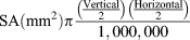 ${\rm{SA }}({\rm{mm}}^2 ){\rm{\pi }}{{\left( {{{{\rm{Vertical}}} \over 2}} \right)\left( {{{{\rm{Horizontal}}} \over 2}} \right)} \over {1,000,000}}$. Horizontal body length measurements were similarly measured on the lateral axis.
Histology
Embryos were fixed in 4% paraformaldehyde in phosphate buffered saline (PBS) at 4°C overnight. Embryos were rinsed and stored in 100% methanol at −20°C. Embryos were dehydrated in ethanol and embedded in Epon resin. Embryos were bathed in propylene oxide twice, 15 min each time. Samples were infused with 1:1 propylene oxide and Epon and mixed for 2 h at room temperature. Embryos were then placed in 1:3 propylene oxide and Epon and mixed overnight at room temperature. The next day, embryos were transferred into appropriately labeled wells in an embedding mold. The embedding mold containing embryos was placed in an oven at 65°C for approximately 1 day, to polymerize the resin.
Embedded embryos were coronally sectioned using a Sorvall JB-4 microtome. One-micrometer sections of the eye were transferred to a 25 × 75 × 1 mm glass slide and left to dry on a hot plate. Sections were stained according to a modified Lee’s methylene blue basic fuchsin dye procedure and then examined using a Zeiss light microscope (Carl Zeiss, Thornwood, NY) to locate the position of the desired section. Slides were covered with a coverslip using DPX (Distrene, Plasticiser, Xylene) mounting solution. Sections were imaged on a Leica light microscope with a RetigaExi camera (QImaging, Surrey, Canada). Presence of an optic nerve (ON) was used as the criterion to select appropriate central sections for analysis.
Visual background adaptation
Petri dishes containing 5.5 dpf larvae were placed in a small box to condition them to 20 min of darkness, and dorsal view images of the larvae taken immediately. The larvae were then conditioned to 15 min of bright light, and images captured in the same manner (Neuhauss et al., Reference Neuhauss, Biehlmaier, Seeliger, Das, Kohler, Harris and Baier1999; Muto et al., Reference Muto, Orger, Wehman, Smear, Kay, Page-McCaw, Gahtan, Xiao, Nevin, Gosse, Staub, Finger-Baier and Baier2005).
Optokinetic reflex
Experiments were done using a modified optokinetic reflex (OKR) stimulator system, consisting of computer, binocular microscope, video camera, projector, screen, and computer-generated stimulation pattern (Stujenske et al., Reference Stujenske, Dowling and Emran2011). The computer was equipped with LabView (National Instruments, Austin, TX), custom-built software that provided both the stimulus and the image and video capture. Computer-generated stimuli, which allowed for continuous variation of velocity, were composed of alternating vertical black and white stripes.
Under dim light, 5.5 dpf larvae were placed in a small Petri dish containing methylcellulose. Using the image and video capture in LabView, a box was made around the head of each larva to capture responses of the eyes for the duration of the stimulus. The stimulus was programmed to run for 2 min, during which the width of the stripes as well as the speed and the direction of the stimulation pattern were manipulated to elicit responses from the larvae. Captured videos of the eyes were analyzed using Tracker 4.0 (Brown & Cox, Reference Brown and Cox2009).
Electroretinogram
Experiments were performed on 5.5 dpf larvae at room temperature in the mid afternoon. Anesthetized larvae were placed laterally on a filter paper covered with 2% agarose and perfused with Ringer’s solution. Ringer’s solution was maintained at pH 7.8 by continuously gassing it with 97% O2 and 3% CO2.The eye was positioned, with the cornea facing upward, at the center of the stimulus light spot (diameter = 5 mm) generated by a halogen light source. To record electroretinogram (ERG) responses, a glass electrode with a tip diameter of 2–6 μm containing a chloride-coated silver wire and filled with Ringer’s solution was gently inserted under the surface of the eye near the cornea. ERGs were amplified at 1,000 total gain and low-pass filtered at 300 Hz. Stimulus flash duration was controlled by an electromechanical shutter and a customized LabView macro. Software-gathered data generated the ERG waveforms for each individually tested eye.
Statistics
Microsoft Excel 2010 and SPSS v13 were used for statistical analysis. t-tests and analysis of variance (ANOVA) were used for treatment comparisons. Regression analysis and the Fisher’s Exact test were used for survival analysis. Significance was set at 0.05.
Results
DEAB treatment at 9 hpf increases mortality and induces microphthalmia
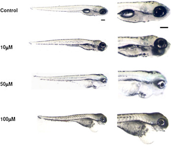
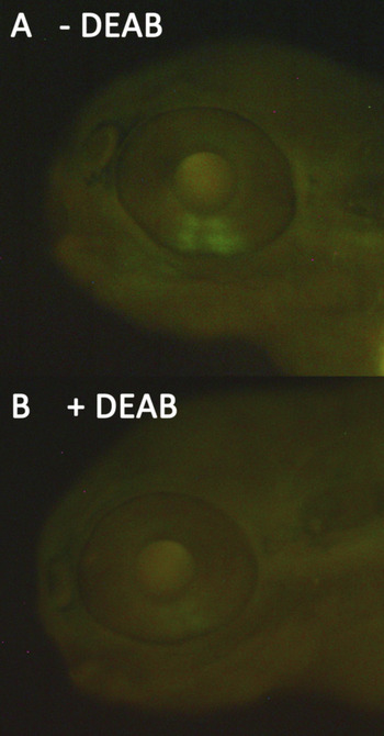
Zebrafish embryos treated with 10, 50, 100, 200, or 400 μM DEAB at 9 hpf for 2 h showed variable survivability. Embryos treated with 400 μM DEAB failed to develop and died by 24 hpf. Treatment with 200 μM resulted in severe cardiac edema and significant mortality within the first few days after treatment. DEAB treatments between 10 and 100 μM resulted in relatively high survival rates, and eye size was almost always reduced as compared to controls (Fig. 1). The 100 μM DEAB treatments resulted in the most consistent phenotype and were used for subsequent experiments. Treatment of RGYn embryos with 100 μM DEAB dramatically reduced RA activity as evidenced by the decreased EYFP signal (Fig. 2).
(Color online) Effect of DEAB at different concentrations. 9 hpf embryos were treated with different concentrations of DEAB: 10, 50, and 100 μM. At 5.5 dpf, morphological changes induced by RA deficiency appear to be increasingly severe as DEAB concentration increases. At all concentrations, DEAB-treated larvae show an abnormal swim bladder and a shorter body axis. Larvae treated with 10 μM appear relatively normal. Larvae treated with 50 μM showed cardiac edema and smaller eyes. Larvae treated with 100 μM showed prominent cardiac edema, curved body axis, and microphthalmia. Lateral view. Scale bar =200 μm.

(Color online) DEAB treatment inhibits RA signaling. 60 hpf RGYn embryos were treated with 100 μM DEAB for 2 h. (A) Embryo at 60 hpf before DEAB treatment. The green fluorescence is indicative of RA activity in the ventral retina activating the RARE EYFP construct in the RGYn fish and viewed through an FITC filter. (B) The same embryo after a 2-h incubation with DEAB beginning at 60 hpf. Expression of the RARE-driven EYFP construct is greatly reduced.

Treating 9 hpf embryos with 100 μM DEAB for 2 h did reduce their survivability somewhat. At 36 hpf, 62.8% of DEAB-treated embryos (n = 972) had survived compared to 85.2% of controls (n = 399). That 15% of the controls did not survive could be accounted for by natural death and/or unintended impact from experimental procedures such as injury from pipette suction during washes. When the survival rate of control larvae is normalized to 100%, the survival rate of DEAB-treated larvae normalizes to 73.6%. Embryos surviving past 36 hpf usually remained alive up to 6 dpf, at which point most of them died due to rupture of an edematous heart. Less than 3% of DEAB-treated larvae survived to adulthood, and none of these long-term survivors showed noticeably smaller eyes or cardiac edema.
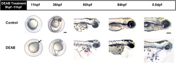
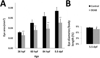
A small percentage (<25%) of DEAB-treated embryos developed a transient microphthalmia with little or no cardiac edema. The vast majority of the 100 μM DEAB-treated embryos (>75%), on the other hand, developed a progressive and persistent microphthalmia along with cardiac edema (Figs. 3 and 4). Starting at 84 hpf, the small eye phenotype became quite pronounced. By this stage, other typical characteristics of RA deficiency were observed in DEAB-treated larvae: lack of a touch response, shortened body axis, curly tail, reduced forebrains, enlarged yolk sac, and motor problems such as the inability to swim. By 5.5 dpf, the microphthalmia is quite dramatic in DEAB larvae (Fig. 4). The eye size of DEAB-treated larvae is just 77% of that of controls. Although the body length of DEAB larvae is also reduced at 5.5 dpf, eye diameter relative to body length is nevertheless significantly smaller in DEAB-treated animals as compared to control larvae (Fig. 4B).
(Color online) RA deficiency perturbs eye development. At 11 hpf, 2 h after treatment, DEAB-treated embryos show signs of abnormal development (black arrow). At 36 hpf, they appear developmentally delayed, showing less pigmentation, and their eyes are not as well defined as the controls. By 60 hpf, microphthalmia is quite noticeable in DEAB-treated embryos. Cardiac edema is also quite evident. At 84 hpf, DEAB-treated embryos continue to have smaller eyes, cardiac edema, and have an overall smaller body size. These differences become quite pronounced by 5.5 dpf, at which stage DEAB-treated embryos’ eyes are smaller and rounder than control eyes, even when adjusting for the difference in body size. Lateral view. Scale bar = 200 μm.

RA deficiency significantly reduces eye size. (A) Control and DEAB eyes continue to grow throughout larval development. DEAB eyes have a significantly smaller SA as compared to controls beginning at 36 hpf and continuing through 132 hpf (5.5 dpf). 36 hpf (Control, n = 14; DEAB, n = 48); 60 hpf (Control, n = 14; DEAB, n = 22); 84 hpf (Control, n = 14; DEAB, n = 35); and 5.5 dpf (Control, n = 21; DEAB, n = 57). (B) The eye diameter, relative to body length, in DEAB-treated larvae is smaller compared to controls at 5.5 dpf (Control, n = 6; DEAB, n = 15). For all data measures, error bars represent the standard deviation of the mean. *P = 0.003; **P << 0.001, t-test.

Retinal development is perturbed in DEAB-treated larvae
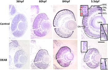
Histological analysis confirmed microphthalmia but also revealed abnormal retinal development in DEAB-treated larvae at 36, 60, and 84 hpf (Fig. 5). Transverse sections at 36 hpf showed that retinas from DEAB-treated embryos were discernibly smaller and the retinal cell population was reduced.
(Color online) Retinal histology of controls versus DEAB eyes. At 36 hpf, DEAB sections show an ON but cell density is reduced. By 60 hpf, controls have proper retinal lamination with five layers: ganglion cell layer (GCL), inner plexiform layer, inner nuclear layer (INL), outer plexiform layer, and outer nuclear layer (ONL). 60 hpf DEAB retinas lack these well-defined retinal layers. By 84 hpf, DEAB retinas appear significantly smaller than age-matched controls and retinal cell layers are not well organized. At 5.5 dpf, DEAB retinas show distinct retinal cell layers with normal stratification. Top left inset: Red arrows identify the pink-stained outer segments extending from purple-stained photoreceptor inner segments to the retinal pigment epithelium (RPE) in control 5.5 dpf retinas. Similar stained structures are not identified in DEAB-treated retinas. Bottom right inset: The elongated cells of the ciliary marginal zone found in controls and DEAB-treated fish. Black arrows point to the elongated proliferative cells. Transverse sections selected represent the typical retina seen within each age group: 36 hpf (Control, n = 4; DEAB, n = 6); 60 hpf (Control, n = 5; DEAB, n = 4); 84 hpf (Control, n = 7; DEAB, n = 9); and 5.5 dpf (Control, n = 8; DEAB, n = 11). Dorsal is up. Scale bar = 100 μm.

By 60 hpf, the retinal cells in controls had begun to differentiate and to migrate creating a laminated structure. However, at this stage, retinas from the DEAB treatment group were developmentally delayed; both the inner and outer plexiform layers were minimally developed, although a small ON was apparent, indicating some ganglion cell differentiation. By 84 hpf, control retinas had developed further, whereas retinas from DEAB-treated fish remained smaller and showed variation in cell layer lamination. Whereas most retinas from DEAB-treated fish at 84 hpf showed distinctive layers of cells, there were some that still had negligible lamination. Although not as well defined, the ganglion cell layer (GCL), inner nuclear layer (INL), and outer nuclear layer (ONL) are present in this retina. Further, photoreceptors and bipolar cells could be distinguished by their elongated oval shape and circular shape, respectively. Finally, by 5.5 dpf, retinas from DEAB-treated fish remained significantly smaller in size but had distinctive cell layers with appropriate layer stratification. The outer plexiform layer (OPL) and inner plexiform layer (IPL) as well as ganglion cells, bipolar cells, and photoreceptors cells were clearly visible.
DEAB-treated larvae have no behavioral or physiological visual response
To test the responsivity of DEAB-treated larvae to light, we first examined their visual background adaptation (VBA) response. VBA is a neuroendocrine camouflage response that matches pigment distribution in the body to the environment’s light levels (von Frisch, Reference von Frisch1911; Hogben & Slome, Reference Hogben and Slome1931). Dark-adapted fish disperse their melanin pigment and thus appear dark. Light-adapted fish aggregate melanin granules and thus appear pale. Blind fish, unable to detect light, appear dark under any condition, whereas fish with impaired vision have been shown to have gradations of melanin dispersal that corresponds to the degree of visual impairment (Neuhauss et al., Reference Neuhauss, Biehlmaier, Seeliger, Das, Kohler, Harris and Baier1999).
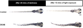
After 20 min of darkness, 5.5 dpf control larvae appeared dark. After 15 min of light exposure, they appeared transparent. 5.5 dpf DEAB-treated larvae on the other hand did not exhibit a VBA response, appearing dark under both dark and light conditions (Fig. 6). VBA depends on the retina and pineal pathways and does not appear to require the optic tectum based on evidence in zebrafish showing that neither unilateral nor bilateral ablations of the tectal neuropil permanently eliminated VBA responses (Roeser & Baier, Reference Roeser and Baier2003; Zhang et al., Reference Zhang, Song, Thompson, Madonna, Millhauser, Toro, Varga, Westerfield, Gamse, Chen and Cone2010). Thus, abolishment of the VBA suggests that DEAB larvae may be visually impaired and possibly have impaired pineal functionality as well—though this latter point was never specifically examined.
(Color online) DEAB larvae do not show a VBA response at 5.5 dpf. After 20 min in the dark, control larvae disperse their melanin granules, appearing dark. After 15 min of light exposure, they adapt by aggregating melanin granules, becoming translucent. DEAB-treated larvae failed to adjust their melanin dispersal in response to changes in ambient light level, appearing dark under both conditions. Dorsal view. Scale bar = 200 μm.

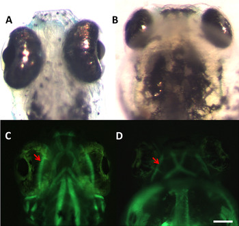
The OKR is a reflexive eye movement in response to gradient motion. All vertebrates display this visually guided behavior. The purpose of this reflex was to compensate for body and head motion and stabilize visual images on the retina. It also allows animal’s eyes to follow moving objects, while its head remains stationary. This reflexive behavior develops rapidly in zebrafish, making it possible to test larvae as young as 5 dpf, at which time the OKR is fully developed (Brockerhoff et al., Reference Brockerhoff, Hurley, Janssen-Bienhold, Neuhauss, Driever and Dowling1995; Neuhauss et al., Reference Neuhauss, Biehlmaier, Seeliger, Das, Kohler, Harris and Baier1999; Huang & Neuhauss, Reference Huang and Neuhauss2008). The OKR serves as an advantageous behavioral paradigm to study vision because it is robust and does not require training. A response is easily elicited by moving vertical stripes through the animal’s visual field. The eyes follow the stripes with a smooth pursuit movement, followed by a rapid eye movement (saccade) in the opposite direction (Emran et al., Reference Emran, Rihel, Adolph, Wong, Kraves and Dowling2007). 5.5 dpf DEAB-treated larvae failed to show any OKR responses. (n = 11). However, we also observed that whereas the extracellular muscles developed in these fish, they did not appear to attach correctly to the eyes in DEAB-treated fish (Fig. 7), perhaps explaining the lack of OKR responses in these fish.
(Color online) Extraocular muscles are present but not in the correct location relative to the eyes after RA deficiency. (A and B) Control and DEAB-treated embryos at 5.5 dpf viewed from the ventral side. (C and D) Fluorescence images showing the muscles tagged with an α-actin-GFP construct in the same Control and DEAB-treated embryos. The oblique muscles (red arrows) of DEAB embryos develop but are behind the eye compared to Control oblique muscles, which are correctly attached to the eye. Scale bar = 100 μm.

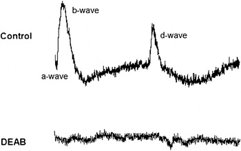
To determine if the absence of both OKR and VBA response was a direct consequence of retinal impairment, ERG recordings were performed with larvae at 5.5 dpf, at which age ERG responses are readily elicited from zebrafish retinas (Brockerhoff et al., Reference Brockerhoff, Hurley, Janssen-Bienhold, Neuhauss, Driever and Dowling1995). The ERG, commonly used in diagnosis of retinal diseases, is a mass electrical response of the outer retina to brief flashes of light. A typical zebrafish ERG recording consists of three main components: a-, b-, and d-waves. The a-wave originates from the photoreceptors, the b-wave from ON bipolar cells, and the d-wave mainly from OFF bipolar cells (Sieving et al., Reference Sieving, Murayama and Naarendorp1994).
Intact eye recordings indicated that retinas from DEAB-treated larvae did not generate any ERG components (Fig. 8). Control recordings showed typical zebrafish ERGs, in which the b-wave, significantly larger than the d-wave, is preceded by a small but distinguishable a-wave (Dowling, Reference Dowling2012). The absence of an a-wave indicates that photoreceptors from DEAB-treated larvae do not respond to light.
DEAB larvae do not have an ERG response at 5.5 dpf. In zebrafish, a normal ERG shows three waves: the a-wave originates from the photoreceptor cells, b-wave from the ON bipolar cells, and d-wave from the OFF bipolar cells. In DEAB eyes, the ERG lacks all three components, as shown by a relatively flat line (n = 9).

Discussion
Earlier work demonstrated that exogenous RA given between 10 and 11 hpf induces excess ventral retina formation, whereas inhibition of RA synthesis by citral, another RA synthesis inhibitor, at the 9–12 somite stage of zebrafish development prevents ventral retina formation. Extending this work, we found that inhibiting RA at a slightly earlier time point, by just 1 h, leads to the development of microphthalmia, a delay in retinal development. Our initial findings were presented using citral as in earlier studies (Cameron & Dowling, Reference Cameron and Dowling2009). However, due to DEAB’s selectivity for the retinal dehydrogenase pathway and greater embryo survival, we elected to use it for this study (Russo et al., Reference Russo, Hauguitz and Hilton1988; Mahmoud et al., Reference Mahmoud, Potter, Colvin, Hilton and Mezey1993; Perz-Edwards et al., Reference Perz-Edwards, Hardison and Linney2001). We have independently demonstrated that DEAB effectively eliminated RA synthesis, albeit in slightly older embryos (Fig. 2). It should be noted that RA signaling, as measured by the fluorescence in the RGYn embryos, was not completely eliminated due to existing RA; DEAB only inhibits its synthesis and does not degrade RA already present.
We inhibited RA synthesis using DEAB for 2 h, just as the optic primordium is forming (9 hpf), and examined the developing eye for several days after. DEAB-treated larvae had statistically smaller eyes compared to controls beginning at 36 hpf and continuing to at least 5.5 dpf (Figs. 3 and 4). This observation supports the well-established link between vitamin A deficiency (VAD) and congenital ocular malformations (Mark et al., Reference Mark, Ghyselinck and Chambon2006). The multisystem abnormalities observed in DEAB-treated fish also substantiate the finding of Wilson et al. (Reference Wilson, Roth and Warkany1953) that vitamin A-deficient rat fetuses present a large array of congenital malformations affecting the ocular, cardiac, respiratory, and urogenital systems, collectively referred to as fetal VAD syndrome.
The DEAB phenotype resembles human syndromic microphthalmia, which is typically associated with craniofacial dysmorphic features, heart and vascular malformations, skeletal and limb anomalies, and gut and brain defects (Golzio et al., Reference Golzio, Martinovic-Bouriel, Thomas, Mougou-Zrelli, Grattagliano-Bessieres, Bonniere, Delahaye, Munnich, Encha-Razavi, Lyonnet, Vekemans, Attie-Bitach and Etchevers2007). In particular, severe microphthalmia and cardiac edema observed in DEAB-treated larvae are similar to the symptoms observed in patients suffering from Matthew–Wood syndrome (Golzio et al., Reference Golzio, Martinovic-Bouriel, Thomas, Mougou-Zrelli, Grattagliano-Bessieres, Bonniere, Delahaye, Munnich, Encha-Razavi, Lyonnet, Vekemans, Attie-Bitach and Etchevers2007; Pasutto et al., Reference Pasutto, Sticht, Hammersen, Gillessen-Kaesbach, Fitzpatrick, Nurnberg, Brasch, Schirmer-Zimmermann, Tolmie, Chitayat, Houge, Fernandez-Martinez, Keating, Mortier, Hennekam, von der Wense, Slavotinek, Meinecke, Bitoun, Becker, Nurnberg, Reis and Rauch2007). This correlation has also been demonstrated by the study of Isken et al. (Reference Isken, Golczak, Oberhauser, Hunzelmann, Driever, Imanishi, Palczewski and von Lintig2008), which showed that STRA6-deficient zebrafish embryos developed microphthalmia and heart edema. Similar to DEAB-treated fish, STRA6 knockdown fish had normal retinal lamination at 4 dpf (Isken et al., Reference Isken, Golczak, Oberhauser, Hunzelmann, Driever, Imanishi, Palczewski and von Lintig2008).
Since the expression of STRA6 is directly regulated by RA signaling and is required for cellular uptake of retinol to synthesize RA, it is not surprising that STRA6 knockdown embryos develop the same symptoms as the RA-deficient larvae in our study. Ironically, Isken et al. (Reference Isken, Golczak, Oberhauser, Hunzelmann, Driever, Imanishi, Palczewski and von Lintig2008) suggested that microphthalmia as well as cardiac and craniofacial abnormalities are caused by RA excess in surrounding tissues rather than RA deficiency. Our data disagree with this view.
DEAB effectively down regulates the synthesis of RA, thereby challenging the assertion of Isken et al. (Reference Isken, Golczak, Oberhauser, Hunzelmann, Driever, Imanishi, Palczewski and von Lintig2008) on the role of extracellular excess RA (Russo et al., Reference Russo, Hauguitz and Hilton1988). The ocular, cardiac, and other development malformations in DEAB-treated fish were undoubtedly induced by RA deprivation not RA excess. Given the well-established association between VAD and ocular defects, the DEAB phenotype presents a more appropriate model for clinical microphthalmia.
While we have not presented evidence examining the mechanism behind the observed microphthalmia, the proliferating ciliary marginal zone is present in DEAB-treated zebrafish larvae (Fig 5, lower inset) (Wehman et al., Reference Wehman, Staub, Meyers, Raymond and Baier2005). Nevertheless, the rate of eye growth was statistically slower for DEAB-treated fish (ANOVA P << 0.001) suggesting that proliferation may be impaired. It is also possible that changes in apoptosis similar to those seen in the chick could account for the reduced eye size (Sen et al., Reference Sen, Harpavat, Peters and Cepko2005).
Several published studies have attested to the involvement of RA regulation in photoreceptor differentiation and maturation (Hyatt et al., Reference Hyatt, Schmitt, Fadool and Dowling1996; Biehlmaier et al., Reference Biehlmaier, Lampert, von Lintig and Kohler2005; Prabhudesai et al., Reference Prabhudesai, Cameron and Stenkamp2005; Khanna et al., Reference Khanna, Akimoto, Siffroi-Fernandez, Friedman, Hicks and Swaroop2006). The top inset images shown in Fig. 5 suggest that outer segment development in the DEAB larvae may be impaired and could explain the complete lack of an ERG response. This argument fits well with the historical evidence linking RA signaling and photoreceptor development. Even the small group of DEAB-treated larvae that did not develop microphthalmia had reduced visual function as measured by the OKR and ERG. The OKR saccades per minute were reduced 25% compared to controls (P = 0.01; n = 6 for each group), and the ERG amplitudes (both a- and b-wave) from these larvae were also reduced compared to controls, though the sample size was insufficient for statistical significance (n = 3). Taken together with the complete lack of visual response in their microphthalmia counterparts, RA inhibition is likely affecting the retina’s function beginning at the photoreceptor level. However, the exact effect on the photoreceptors needs elucidation using molecular techniques and electron microscopy to identify the cellular changes.
Retinal cell fate specification follows a specific timeline (Cepko et al., Reference Cepko, Austin, Yang, Alexiades and Ezzeddine1996). Although we treated our embryos before the eye anlage forms, our results suggest that RA plays a vital regulatory role to a host of genes controlling eye development. Once this regulatory process is hampered—in our case by inhibiting RA synthesis—the downstream long-term consequences can be quite severe. In this case microphthalmia, a dysfunctional vision system and, ultimately, death are the results.
The mechanism of the persistent microphthalmia in DEAB-treated fish is not clear. First, RA synthesis is presumed to only be inhibited for a brief time using our protocol. DEAB is a competitive inhibitor, and once DEAB is oxidized to 4-diethylaminobenzoic acid (which happens quickly), it loses its ability to bind to the active site on RALDH (Russo et al., Reference Russo, Hauguitz and Hilton1988). When the DEAB solution is removed and the embryos washed in fresh E3, recovery of RA synthesis should quickly ensue—though we never specifically tested this. Second, this period of inhibition overlaps with the time when RA inhibition was shown to cause the loss of ventral retina tissue (Marsh-Armstrong et al., Reference Marsh-Armstrong, McCaffery, Gilbert, Dowling and Drager1994). Marsh-Armstrong et al. (Reference Marsh-Armstrong, McCaffery, Gilbert, Dowling and Drager1994) found that citral treatment beginning at 10 hpf was sufficient to prevent ventral retina formation, and yet, we never saw this phenotype treating at 9 hpf. We did however reproduce the ventral retina absence phenotype by treating our fish with DEAB an hour later (10 hpf). We found similar results using citral but chose to use DEAB for this study because of its specific interaction with the RALDH enzymes (Mahmoud et al., Reference Mahmoud, Potter, Colvin, Hilton and Mezey1993).
Deficiency of RA at slightly different times in zebrafish can cause quite different phenotypes. We conclude, therefore, that the regulation of RA synthesis and signaling is tightly controlled during embryonic development. Despite many published studies exploring and clarifying the role that RA plays in early eye development, the gene targets for RA are still largely unknown (Adler & Canto-Soler, Reference Adler and Canto-Soler2007; Duester, Reference Duester2008a,Reference Duesterb). The use of comprehensive gene expression analysis tools, such as microarrays, has been useful in understanding the genes involved in eye development (Cameron et al., Reference Cameron, Gentile, Middleton and Yurco2005; Leung & Dowling, Reference Leung and Dowling2005; Leung et al., Reference Leung, Ma and Dowling2007). Similar tools may prove beneficial toward our understanding of RAs gene regulatory role during eye development and explaining the causes of microphthalmia, alterations in dorsal/ventral patterning, and other eye and visual defects.
Acknowledgments
The authors thank Drs. Yong Li, Farida Emran, Jonathan Matsui, and Jian Liu for constructive discussions; Dr. Elwood Linney for the RGYn transgenics, and Joseph Stujenske for assistance with the electrophysiology. This work was supported by The Harvard College Research Program (HGL) and the National Institutes of Health EY000811 (JED) and T32-EY07145(DJC).