Wiskott-Aldrich syndrome is a rare X-linked recessive inherited disease that is characterized by immune deficiency, eczema, and thorombocytopaenia.Reference Perry, Spector and Schumann1 An association with vasculitis, or aneurysmal formation, has been reported in occasional cases.Reference McCluggage, Armstrong, Maxwell, Ellis and McCluskey2 As far as we are aware, only a few adult patients have been surgically treated thus far for aortic aneurysms.Reference Johnston, Unsworth, Dwight and Kennedy3–Reference Bernabeu, Josa and Nomdedeu6 We describe here our own experience with a 7-year old boy.
Case report
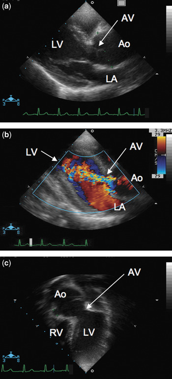
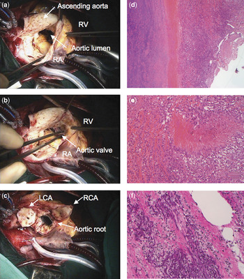
Our patient, a 7-year-old boy, was diagnosed with Wiskott-Aldrich syndrome at the age of 5 years. Since then, he has been followed at our institute. His medical history was typical for the syndrome, including recurrent infections of the respiratory tract, pneumonias, and a pulmonary abscess due to infection with Haemophilus influenzae, eczema, and a cerebral haemorrhage owing to thorombocytopaenia. Splenectomy was performed at the age of 5 years, after which his platelet count returned to normal. In addition to the phenotype of Wiskott-Aldrich, he was also found to have large lymphocytic leukaemia, presumably following prolonged infection with cytomegalovirus in infancy. His immunological phenotype has been described elsewhere.Reference Boztug, Baumann and Ballmaier7 At that time, a routine chest X-ray suggested presence of an ascending aortic aneurysm. His pulse pressure was 107/45 mmHg in the right arm, and 101/36 mmHg in the right leg. Computed tomography was performed, which confirmed the diagnosis. The ascending aorta was dilated to 40 mm, representing 241% of the normal value for his age, which is 16.6 plus or minus 1.8 mm.Reference Rose, Castillo and Wessel8 Echocardiography demonstrated dilation of the aortic root (Fig. 1a, c), and moderate aortic regurgitation (Fig. 1b). Left ventricular volume was slightly increased, but ventricular function was preserved, with a fractional shortening of 42%. An elective operation was performed using cardiopulmonary bypass under moderate hypothermia at 27 degrees Celsius. After the ascending aorta was cross-clamped and incised vertically, inspection from inside and outside revealed a diffusely infiltrated, thickened, and dilated aortic wall (Fig. 2a). Myocardial protection was achieved by selective administration of cold crystalloid cardioplegia. The aortic valve was trifoliate, and showed no inflammatory reaction, but inflammation of the aortic wall had spread to the aortic root (Fig. 2b). Because of this, an aortic valve-sparing operation was not considered to be an option. The aortic leaflets were excised, and the proximal portion of the ascending aorta was transected at the sinutubular junction. After mobilization of the aortic root, coronary arterial buttons were excised (Fig. 2c), and the root was replaced with a 21 mm Biovalsalva™ prosthesis. The period of aortic cross-clamping was 66 minutes, and cardiopulmonary bypass lasted for 105 minutes. After cardiopulmonary bypass, human leukocyte antigen compatible pooled platelets were transfused to maintain his platelet count over 150,000/l. The postoperative course was uneventful. He was extubated on the same day, and returned to the haemato-oncologic ward on the 9th postoperative day. Histological examination of the aorta revealed a markedly thickened wall, showing aortitis with inflammatory infiltration (Fig. 2d). There were several necrotic areas in the medial layer, which were infiltrated by macrophages (Fig. 2e). Use of an elastic stain revealed marked destruction of the elastic fibres of the aortic wall (Fig. 2f). After a period of 3 months, he received bone marrow transplantation from a matched related donor, and was discharged 5 months after the operation. The echocardiogram at discharge showed a competent prosthetic valve, a left ventricle of normal size, and fractional shortening of 37%.
Preoperative transthoracal echocardiograpy and colour Doppler imaging in our patient with Wiskott-Aldrich syndrome. Panel a shows a parasternal long-axis view demonstrating the presence of aortic root dilatation. In Panel b, Colour Doppler in the parasternal short-axis view shows the moderate aortic regurgitation. Panel c is a four chamber view showing the aortic root dilation. Abbreviations: LA, left atrium; LV, left ventricle; RV, right ventricle; Ao, ascending aorta; AV, aortic valve.

Intra-operative and histologic photographs of the aortic wall. Panel a shows that the ascending aortic wall was diffusely infiltrated, thickened, and dilated. In Panel b, it can be seen that the leaflets of the aortic valve showed no inflammatory reaction, but the wall of the aortic root was diffusely infiltrated and thickened. Panel c shows the resected aortic valve after excision of the coronary arterial buttons. The wall of the ascending aorta was transected at the sinutubular junction. Panel d is a low power micrograph of a section stained with haemotoxylin and eosin, showing the extensive lymphocytic and histiocytic infiltrate and the destruction of elastic fibres of the aortic wall. The high power micrograph, shown in Panel e, demonstrates the focal necrotizing vasculitis, while Panel f, again at high power, has been stained to show marked destruction of elastic fibres within the aortic wall. Abbreviations: RA, right atrium; RV, right ventricle; RCA, right coronary artery; LCA, left coronary artery.

Discussion
Life expectancy of patients with Wiskott-Aldrich syndrome has been improved by splenectomy and/or bone marrow transplantation.Reference Mullen, Anderson and Blaese9 Such patients, who are surviving longer now, appear to be at a higher risk of lympho-proliferative diseases and vasculitis. Vasculitis and aneurysmal formation are rare but life-threatening complications. The risk of death from aneurysmal rupture is increased due to thrombocytopaenia and impaired platelet function. Surgical management of these patients is considered more complex than the general population because of their higher probability of infectious and bleeding complications. Involvement of the aorta is even more rarely reported. To the best of our knowledge, surgical management thus far has been reported only in 4 young adults.Reference Johnston, Unsworth, Dwight and Kennedy3–Reference Bernabeu, Josa and Nomdedeu6 Our patient, aged 7 years, presented with an ascending aortic aneurysm and moderate aortic regurgitation, and required replacement of his aortic root. Despite the risks described above, he recovered without any complications. Our experience shows, therefore, that patients with aortic aneurysms in the setting of Wiskott-Aldrich syndrome can be managed surgically even at a young age. Careful subsequent observation is mandatory because of the risk of further dilation of the native aorta. As for the surgical procedure, Vricella and colleaguesReference Vricella, Williams and Revekes10 recommended valve-sparing operations for children, but we could not use this option because of the extreme inflammation of the aortic wall. To the best of our knowledge, ours is the first report on a child with Wiskott-Aldrich syndrome surviving replacement of the aortic root and subsequent bone marrow transplantation.
Acknowledgements
We thank Dr Bredt Martin from institute for Pathology, Hannover Medical School, Hannover, Germany, directed by Prof. Hans-Heinlich Kreipe, for providing histologic findings and photos. We also thank Dr Harald Bertram and Dr Dietmar Boethig from Division of Pediatric Cardiology and Intensive Care Medicine, and Dr Ulrich Baumann from Division of Pediatric Pulmonology and Neonatology, for their help in preparation of the manuscript.