LMNA mutations could induce a variety of genetic disorders referred to as laminopathies which can manifest clinical features affecting many organs, including the skeletal and cardiac muscle, adipose tissue, nervous system, cutaneous tissue, and bone.Reference Jacob and Garg1 And the manifestations of patients with LMNA cardiomyopathy are usually various and complex with a high incidence of atrial fibrillation, atrioventricular block, stroke, heart failure, and even sudden cardiac arrest.Reference Worman and Bonne2 Here, we present a Chinese family with dilated cardiomyopathy caused by the LMNA mutation (p. R429C) and described its electrocardiogram and imaging features.
Case report
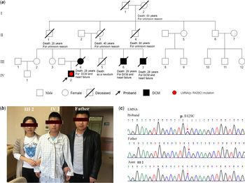
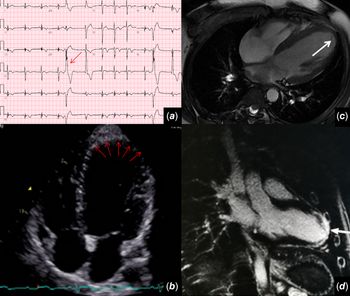
A 14-year-old boy was admitted for intermittent chest tightness upon physical or emotional stress for 3 years. The symptoms exacerbated for 2 weeks accompanied with palpitation and dyspnoea. His past medical history was unremarkable, without skeletal muscle disease, pre-mature ageing, or metabolic disorders (Fig 1A and B). His mother and nearly half of his maternal relatives died at an early age (Fig 1A). On admission, his vital signs were stable and physical examination was positive for an enlarged left cardiac dullness border. Laboratory tests were otherwise normal except a slightly elevated hypersensitive troponin T (0.047ng/mL). The electrocardiogram was striking with T wave change and pre-mature ventricular beats whose origin was considered as left ventricular apex (Fig 2A). Twenty-four-hour Holter electrocardiogram indicated frequent pre-mature ventricular beats (3% of total heartbeats). Echocardiography 1 year ago reported a left ventricular end-diastolic diameter of 33 mm, and ejection fraction was estimated at 79%. Repeat echocardiography after admission showed a left ventricular end-diastolic diameter of 48 mm and an ejection fraction of 52%, with a rounded and thinning left ventricular apex (Fig 2B). Coronary artery CT angiography showed no abnormality in origin, course, or atherosclerotic change. Cardiac magnetic resonance showed a slightly enlarged left ventricular chamber with a significantly thinning, delayed enhanced apical cap, and adjacent wall, suggesting fibrosis at these areas (Fig 2C and D). Whole-exome sequencing and subsequent Sanger sequencing revealed a novel mutation in LMNA (p. R429C) (Fig 1C). The diagnosis of dilated cardiomyopathy was made by combining the characteristic of electrocardiogram and cardiac magnetic resonance and confirmed by genetic testing. The patient was treated with metoprolol and perindopril. He reported alleviation of symptoms at 1-year follow-up and is still on our follow-up list.
Pedigree of the described Chinese family of dilated cardiomyopathy. A, Family tree. DCM = dilated cardiomyopathy; B, Proband and family members; C, Sanger sequencing verified the LMNA p. R429C mutation in the proband.

Examinations of the proband. A, ECG shows diffuse T wave change and premature ventricular beats (red arrow); B, Echocardiography shows a rounded, thinning and akinetic left ventricular apex (red arrows); C, Cardiac MRI confirms a rounded and thinning LV apex (white arrow) and D, delayed enhancement at the apical cap and adjacent wall.

Discussion
Dilated cardiomyopathy is one of the most common causes of heart failure and sudden cardiac death in adolescents.Reference Cowan, Kinnamon, Morales, Salyer, Nickerson and Hershberger3 Different genetic mutations may result in different onset ages and severity of dilated cardiomyopathy. Lamins A and C, encoded by LMNA gene, are nuclear intermediate filament proteins that form one of the major structural components of the lamina network, which underlies and mechanically supports the nuclear envelope.Reference Kawakami, Ogimoto and Tokunaga4 LMNA mutations cause a variety of inherited diseases referred to as laminopathies which are associated with a wide spectrum of disease phenotypes, ranging from skeletal muscle disease, pre-mature ageing, metabolic disorders, and cardiac abnormalities.Reference D’Ambrosio, Petillo and Torella5 Herein, we reported a novel LMNA mutation-induced familial dilated cardiomyopathy and described its imaging features. A better understanding of the molecular bases and imaging characteristics of laminopathies is of vital importance to make more accurate definitions and timely diagnosis for this kind of patients. The current mutation site, LMNA c.C1285T (p. R429C), in this patient suggested that carrying the mutant gene may lead to early onset of dilated cardiomyopathy, and early involvement of left ventricle may start from the apex, in contrast with the diffuse hypokinetic and enlarged left ventricle. Besides, the disease progress is a gradual and continuous process, and symptom onset and electrocardiogram alterations may occur prior to structural and functional change. This may explain why the initial echocardiogram of this patient was largely normal and non-diagnostic 1 year ago but revealed marked motion and contour abnormality on repeat examination and confirmed by MRI. Therefore, serial echocardiography is necessary in suspected patients, especially for those with a positive family history. Moreover, it implies the need for a multidisciplinary approach in the early recognition of these malignant disorders and how the figure of the cardio-myo-geneticist could play a vital role in facilitating the diagnostic process and addressing the adoption of appropriate prevention measures.
Acknowledgements
We thank all subjects for supporting this scientific research.
Financial Support
This work was funded by the Capital Health Research and Development of Special [NO. Z191100006619007], Beijing Municipal Administration of Hospitals Clinical Medicine Development of Special Funding Support [NO. ZYLX201831], and Beijing Municipal Administration of Hospitals’ Ascent Plan [NO. DFL20190902].
Conflict of Interest
None.
Ethical Standards
The authors assert that this report complies with the ethical standards of the Helsinki convention, and consent for this publication has been granted by the patient’s family.