Introduction
In domestic animals, in vitro embryo production (IVP) has improved significantly over the years. Despite the encouraging results, blastocyst rates are still low ranging from 25 to 40% (Rizos et al., Reference Rizos, Ward, Duffy, Boland and Lonergan2002). Studies have demonstrated that in vitro-produced embryos differ from their in vivo counterparts in terms of morphology (Boni et al., Reference Boni, Tosti, Roviello and Dale1999; Driver et al., Reference Driver, Peñagaricano, Huang, Ahmad, Hackbart, Wiltbank and Khatib2012), metabolism (Khurana and Niemann, Reference Khurana and Niemann2000b), developmental competence (Leibfried-Rutledge, Reference Leibfried-Rutledge1999), survival following cryopreservation (Khurana and Niemann, Reference Khurana and Niemann2000a), gene expression (Niemann and Wrenzycki, Reference Niemann and Wrenzycki2000; Pavani et al., Reference Pavani, Baron, Correia, Lourenço, Bettencourt, Sousa and da Silva2016), and pregnancy rates (Hasler, Reference Hasler2000). These differences are partly due to the suboptimal conditions provided by the in vitro maturation (IVM) environment that disrupts the balance between cytoplasmic and nuclear maturation essential for oocyte competence (Ríos et al., Reference Ríos, Buschiazzo, Mucci, Kaiser, Cesari and Alberio2015). Moreover, IVM conditions may not be providing the ideal conditions for proper cumulus cell (CC) signalling with the oocyte (Tesfaye et al., Reference Tesfaye, Ghanem, Carter, Fair, Sirard, Hoelker, Schellander and Lonergan2009). The creation of suitable IVM conditions is, therefore, essential to the improvement of oocyte competence and the resulting blastocyst development (Makarevich and Markkula, Reference Makarevich and Markkula2002; Balasubramanian and Rho, Reference Balasubramanian and Rho2007; Warzych et al., Reference Warzych, Peippo, Szydlowski and Lechniak2007; Korhonen et al., Reference Korhonen, Kananen, Ketoja, Matomäki, Halmekytö and Peippo2010).
IVM media have been previously supplemented with different growth factors (Zhang et al., Reference Zhang, Hansen and Ealy2010; Machado et al., Reference Machado, Caixeta, Sudiman, Gilchrist, Thompson, Lima, Price and Buratini2015), including epidermal growth factor (EGF) (Park and Lin, Reference Park and Lin1993; Park et al., Reference Park, Iga and Niwa1997), as well as known antioxidants (Dalvit et al., Reference Dalvit, Llanes, Descalzo, Insani, Beconi and Cetica2005; Anchordoquy et al., Reference Anchordoquy, Anchordoquy, Testa, Sirini and Furnus2015; Torres et al., Reference Torres, Muñoz, Urrego, Echeverry and Lopez2016) to improve oocyte competence and embryonic development in bovine. EGF in addition to follicle-stimulating hormone (FSH) induce oocyte meiotic resumption, CC expansion, and oocyte competence in a variety of species (Lonergan et al., Reference Lonergan, Carolan, Van Langendonckt, Donnay, Khatir and Mermillod1996; Abeydeera et al., Reference Abeydeera, Wang, Cantley, Rieke, Prather and Day1998; De La Fuente et al., Reference De La Fuente, O’Brien and Eppig1999) in vitro. The mechanism by which FSH and EGF promote oocyte maturation is still unclear (Farin et al., Reference Farin, Rodriguez, Alexander, Hockney, Herrick and Kennedy-Stoskopf2007). The addition of growth factors has been demonstrated to increase granulosa cell (GC) proliferation, as well as decrease apoptosis and enhance antrum formation (Mao et al., Reference Mao, Smith, Rucker, Wu, McCauley, Cantley, Prather, Didion and Day2004; Palmerini et al., Reference Palmerini, Nottola, Tunjung, Kadowaki, Bianchi, Cecconi, Sato and Macchiarelli2016).
The addition of antioxidants has been proven to mitigate cell damage that results from the oxidative stress encountered in in vitro conditions, enhancing oocyte quality and blastocyst development (Dalvit et al., Reference Dalvit, Llanes, Descalzo, Insani, Beconi and Cetica2005; Anchordoquy et al., Reference Anchordoquy, Anchordoquy, Testa, Sirini and Furnus2015; Torres et al., Reference Torres, Muñoz, Urrego, Echeverry and Lopez2016). Different types of antioxidants, such as resveratrol, l-carnitine, quercetin, cysteamine, vitamin C among others, have been tested in gametes and embryos of many species with variable degrees of success (Wang et al., Reference Wang, Tian, Zhang, He, Ji, Li, Tan and Liu2014; Sovernigo et al., Reference Sovernigo, Adona, Monzani, Guemra, Barros, Lopes and Leal2017; Jiang et al., Reference Jiang, Yao, Zhao, Gao, Jin, Li, Yan and Xu2019; Piras et al., Reference Piras, Ariu, Falchi, Zedda, Pau, Schianchi, Paramio and Bogliolo2020; El-Sanea et al., Reference El-Sanea, Abdoon, Kandil, El-Toukhy, El-Maaty and Ahmed2021).
Melatonin (Mel) is well known for regulating circadian rhythm and reproductive seasonality (Reiter et al., Reference Reiter, Tan and Korkmaz2009). Its production has been demonstrated not only by the pineal gland but also in other organs including the ovaries (Itoh et al., Reference Itoh, Ishizuka, Kuribayashi, Amemiya and Sumi1999). High levels of Mel have been found in follicular fluid of different species (Rönnberg et al., Reference Rönnberg, Kauppila, Leppäluoto, Martikainen and Vakkuri1990; Shi et al., Reference Shi, Tian, Zhou, Wang, Gao, Zhu, Zeng, Tian and Liu2009), which suggests its important role in the growth and maturation of the mammalian oocyte, possibly through their receptors found on GCs or on oocytes directly (Tamura et al., Reference Tamura, Nakamura, Korkmaz, Manchester, Tan, Sugino and Reiter2009; Sampaio et al., Reference Sampaio, Conceição, Miranda, Sampaio and Ohashi2012). Mel has also been described as a potential scavenger of reactive oxygen species (ROS) and antioxidant in a variety of species (Manda et al., Reference Manda, Anzai, Kumari and Bhatia2007; Mehrzadi et al., Reference Mehrzadi, Safa, Kamrava, Darabi, Hayat and Motevalian2017). Its addition in IVM, in vitro fertilization (IVF) and in vitro culture (IVC) media have been studied in different species to decrease the amount of ROS produced by the damaging in vitro conditions and attain a better embryo development rate (Ishizuka et al., Reference Ishizuka, Kuribayashi, Murai, Amemiya and Itoh2000; Abecia et al., Reference Abecia, Forcada and Zúñiga2002; Shi et al., Reference Shi, Tian, Zhou, Wang, Gao, Zhu, Zeng, Tian and Liu2009; El-Raey et al., Reference El-Raey, Geshi, Somfai, Kaneda, Hirako, Abdel-Ghaffar, Sosa, El-Roos and Nagai2011; Kim et al., Reference Kim, Park, Kim, Choi, Cho, Lee, Cha, Kim, Lee and Yoon2013).
Studies combining growth factors and oxidative stress modulators have been carried out previously in a variety of species (Singhal et al., Reference Singhal, Prasad, Singh, Prasad and Gupta2009; Córdova et al., Reference Córdova, Morató, Izquierdo, Paramio and Mogas2010; Shabankareh and Zandi, Reference Shabankareh and Zandi2010; Guimarães et al., Reference Guimarães, Pereira, Diógenes and Dode2016) with promising results obtained when a combination of these two classes of supplements was added into the IVM medium (Córdova et al., Reference Córdova, Morató, Izquierdo, Paramio and Mogas2010; Lott et al., Reference Lott, Anchamparuthy, Mcgilliard, Mullarky and Gwazdauskas2011; Anchordoquy et al., Reference Anchordoquy, Anchordoquy, Testa, Sirini and Furnus2015; Ríos et al., Reference Ríos, Buschiazzo, Mucci, Kaiser, Cesari and Alberio2015). The positive effects of EGF (Lonergan et al., Reference Lonergan, Carolan, Van Langendonckt, Donnay, Khatir and Mermillod1996; De La Fuente et al., Reference De La Fuente, O’Brien and Eppig1999) and Mel (Do et al., Reference Do, Shibata, Taniguchi, Nii, Nguyen, Tanihara, Takagi and Otoi2015; Zhao et al., Reference Zhao, Min, Du, Hao, Liu, Qin, Wang and Zhu2015) when supplemented separately in IVM medium have been studied previously, but to our knowledge, their combined effect has not yet been determined in bovine.
The evaluation of oocyte competency has previously been carried out by examining its morphology; however, this parameter has not been a real indicator for its capacity to develop into a blastocyst. More recently, gene expression analysis in CCs has been used as an approach to identify oocyte quality markers and to decipher the crosstalk between the oocyte and its surroundings (Assidi et al., Reference Assidi, Dufort, Ali, Hamel, Algriany, Dielemann and Sirard2008). Although it has been very well demonstrated that a proper oocyte’s in vitro maturation is an unquestionable prerequisite to the formation of good quality embryos, it is known that IVM conditions are still not providing the necessary environment for successful oocyte maturation, which could be related to inadequate signalling from the CCs that is reflected downstream in reduced in vitro developmental rates (Tesfaye et al., Reference Tesfaye, Ghanem, Carter, Fair, Sirard, Hoelker, Schellander and Lonergan2009). Therefore, improvements of IVM protocols have the potential to affect further embryo quality, as demonstrated for changes observed in gene expression at the blastocyst stage after different in vitro maturation and culture protocols (Knijn et al., Reference Knijn, Wrenzycki, Hendriksen, Vos, Herrmann, van der Weijden, Niemann and Dieleman2002).
In this study we evaluated the effects of IVM medium supplementation with two factors (EGF and Mel) that have been shown to influence developmental competence and embryo quality. The main aim of this work was to determine if supplementation with EGF and/or Mel could improve the developmental competence of heifer oocytes (determined by expression of gene candidates related to the acquisition of oocyte competency) and whether there are downstream effects on parthenogenetic blastocyst development in vitro (determined by the expression of candidate genes related to apoptosis, methylation, glucose metabolism and transport, pluripotency, and antioxidant capacity).
Materials and methods
Collection of cumulus–oocyte complexes (COCs)
Prepubertal heifer ovaries [lack of visible corpus luteum (CL) or dominant follicle (DF)] were brought from a local slaughterhouse to the laboratory in saline solution at 34°C. Ovaries were then washed and antral follicles (3 mm to 6 mm diameter) were aspirated into previously warmed Ham’s F-10 solution supplemented with 2% fetal bovine serum (FBS; Gibco Life Technologies, Canada). After sedimentation of the follicular fluid, COCs were recovered and selected using a stereomicroscope.
Groups for study
-
Group Control: IVM base protocol (please refer to section on ‘In vitro maturation’).
-
Group EGF: IVM supplemented with 10 ng/ml of EGF; Sigma-Aldrich, E4127).
-
Group EGF+Mel: IVM supplemented with 10 ng/ml EGF; Sigma-Aldrich, E4127) and 50 ng/ml melatonin (Sigma-Aldrich, M5250).
-
Group Mel: IVM supplemented with 50 ng/ml of melatonin (Sigma-Aldrich, M5250).
The specific concentrations of EGF and Mel were chosen based on previous research by Kang et al., (Reference Kang, Koo, Kwon, Park, Jang, Kang and Lee2009).
The different treatment groups were evaluated as follows: assessment of oocyte developmental competence using candidate genes specific to CCs, determination of development rate of parthenotes, and assessment of blastocyst quality using candidate genes previously described in the literature.
In vitro maturation (IVM)
Oocytes were subjected to IVM as described by Mastromonaco et al. (Reference Mastromonaco, Favetta, Smith, Filion and King2007) with small modifications. Briefly, COCs were washed three times in TCM199 medium + HEPES and then matured in one of the treatment groups (please refer to the preceding section) in 80-μl drops (20–25 COCs/drop) using a base medium of TCM199 + 10% fetal calf serum (FCS) supplemented with 1 μg/ml of estradiol (Sigma-Aldrich, E2758-1G), 25 μg/ml of bovine FSH (bFSH; Folltropin-V, Vetoquinol) and 1 μg/ml of bovine luteinising hormone (bLH; NIH, Washington DC, USA). Only oocytes with three or more layers of CCs and homogenous cytoplasm were randomly assigned to the different experimental groups. All IVM treatments were done under silicone oil (Paisley Products, Scarborough, ON, Canada) at 38.5°C in 5% CO2 for 24 h.
Parthenogenetic activation and in vitro culture
After IVM, CCs were removed from oocytes by pipetting in a 0.3 mg/ml hyaluronidase solution for 4 min. Metaphase II oocytes, with a visible first polar body, homogenous dark cytoplasm, and without signs of degeneration, were selected for parthenogenetic activation as previously described by Miranda et al. (Reference Miranda, Bressan, De Bem, Merighe, Ohashi, King and Meirelles2012). Briefly, matured oocytes were activated with ionomycin solution (5 µM; Sigma, I0634) and incubated with 6-DMAP (2 mM; Sigma-Aldrich, D2629) diluted in supplemented SOF as described by Mastromonaco et al. (Reference Mastromonaco, Favetta, Smith, Filion and King2007) for 3 h in a 5% O2, 5% CO2, and 89.5% N2 humidified gas atmosphere at 38.5°C.
After activation presumptive 15–20 parthenotes were cultured in vitro in 25 μl of SOF medium as described by Mastromonaco et al. (Reference Mastromonaco, Favetta, Smith, Filion and King2007) in a 5% O2, 5% CO2 and 89.5% N2 humidified gas atmosphere at 38.5°C for 7 days. Cleavage rate was assessed 48 h post-activation and blastocysts were visually determined at days 6 and 7 of culture, in all groups. On day 7, only expanded blastocysts were chosen, snap frozen in liquid nitrogen, and stored at −80°C for further mRNA expression analysis.
Gene expression analysis in CCs and blastocysts
After IVM, a pool of 50 COCs (n = 50 per replicate/IVM treatment) was denuded by gentle pipetting and cumulus cells (CCs) were retrieved. CCs were placed in RNase DNase-free Eppendorf tubes, washed in 0.1% phosphate-buffered saline–polyvinyl alcohol (PBS–PVA), snap frozen and stored at −80°C until RNA extraction. RNA was then extracted using the RNeasy Micro Kit (Qiagen, #74004) and DNase treated using an RNase-free DNase set (Qiagen, #79254) as per the manufacturer’s instructions. RNA was then resuspended in 15 µl of RNase DNase-free water and quantified using a NanoDrop 2000c spectrophotometer (Thermo Scientific, Canada). All samples had an acceptable 260/280 ratio of absorbance between 1.8 and 2.2. Then, 500 ng of RNA was reverse transcribed using the one-step protocol with qScript™ cDNA SuperMix (Quanta Biosciences, Canada) following the manufacturer’s instructions. After reverse transcription, each CC sample was diluted to obtain a concentration of 2.5 ng/μl and stored at −20°C until needed.
Day 7 fully expanded parthenotes from four biological replicates were pooled and picked up from the culture medium, washed in 0.1% PBS–PVA, snap frozen in RNase DNase-free Eppendorf tubes and stored at −80°C until RNA extraction. Each replicate contained a pool of 10–12 expanded embryos to minimize individual variations. RNA was extracted using the RNeasy Micro Kit (Qiagen, #74004), and DNase treated with RNase-free DNase set (Qiagen, #79254) as per manufacturer’s instructions. RNA was finally resuspended in 8 µl of RNase DNase water and then quantified using a NanoDrop 2000c spectrophotometer (Thermo Scientific, Canada). All samples had an acceptable 260/280 ratio of absorbance between 1.8 and 2.2. All RNA was then retrotranscribed using the one-step protocol with qScript™ cDNA SuperMix (Quanta Biosciences, Canada) as per the manufacturer’s instructions. Each sample was diluted to 0.125 embryo equivalent/µl and stored at −20°C.
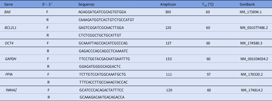
For CCs we analyzed the expression of genes (Table 1) considered predictive of developmental competence (pentraxin-3, PTX3; Gremlin 1, GREM1; tumour necrosis factor-alpha, TNFAIP6; hyaluronan synthase 2, HAS2) and genes related to the antioxidant status (copper zinc superoxide dismutase, CuZnSOD; glutathione peroxidase 1, GPX1). Every gene was evaluated as relative quantification to the two most stable housekeeping genes PPIA and YWHAZ. qPCR experiments were performed with three or four separate replicates from each treatment.
Primer sequences to analyze transcript abundance in cumulus cells

In blastocysts, the expression levels of three genes considered important for blastocyst quality were determined (Table 2). Two genes were related to apoptosis (BCL2 associated X, BAX; B-cell lymphoma 2, BCL-2) and one gene related to pluripotency (octamer-binding transcription factor 4, OCT4). Every gene was evaluated as relative quantification to the three most stable housekeeping genes GAPDH, YWHAZ and PPIA.
Primer sequences to analyze blastocyst quality

A standard curve sample was diluted in a two-fold series to generate a seven-point standard curve. RT-qPCR for the samples of interest (analyzed by triplicates) and standard curves were performed in a 10-µl final reaction volume per well (in a 96 qPCR well plate, Thermo Scientific AB-0900). Final volume per well contained 5 µl of Sso Fast™ EvaGreen® PCR Supermix (Bio-Rad, #1725200), 1 µl of 5 µM forward and reverse primers, 2 µl of RNase DNase-free H2O and 2 µl of cDNA (2.5 ng/μl) substrate. The reaction was performed using real-time PCR on a MyiQCycler apparatus (Bio-Rad Laboratories) according to the manufacturer’s instructions. Conditions of amplifications were an initial step of 95°C (3 min), followed by 40 cycles of 95°C (30 s), 60°C (30 s), and 72°C (20 s), followed by the acquisition of the melting curve (fluorescence acquisition every 0.5°C).
All primers for RT-qPCR were designed using Primer3 software (www.primer3plus.com) and ordered from Sigma-Aldrich (Canada). For each gene, primers were optimized in a pool of CC independent cDNA samples and only those with an efficacy within the range 95–105% and a correlation coefficient of at least 0.98 were used for gene expression. Samples of interest were within the quantification range of the standard curve. Agarose gel electrophoresis (1.5%) was performed using ethidium bromide and visualized by exposure to ultraviolet light to evaluate single PCR product synthesis and size.
Immunofluorescence
Oct-4 protein levels were analyzed in embryos collected on day 7 of IVC by immunofluorescence following the protocol proposed by Dyce et al. (Reference Dyce, Liu, Tayade, Kidder, Betts and Li2011) with some modifications described below. Blastocysts were washed twice with PBS and fixed in 4% paraformaldehyde in PBS for 20 min. Cells were then washed three times in PBS with 0.1% Tween 20 and incubated for 10 min in the same solution followed by 20 min in PBS with 1% Triton X-100. Next, embryos were blocked for 2 h on PBS with 5% bovine serum albumin (BSA) and 0.05% Triton X-100 (blocking solution) followed by incubation with primary antibody, anti-Oct-4 A (sc-9081; 1:200), overnight at 4°C. Cells were then washed in a blocking solution (PBS with 5% BSA and 0.05% Triton X-100) and incubated with 1:400 Alexafluor594-conjugated goat anti-rabbit IgG for 1 h at room temperature. This was followed with a blocking solution wash and incubation with Hoechst 33342 stain for 10 min, followed by washing three times with PBS–PVA.
Some blastocysts were not incubated with primary antibody (negative controls) and all embryos were mounted using anti-fading fluorescent mount medium (Vectashield) and visualized using an Olympus FV1200 confocal microscope with laser wavelengths of 350 nm for Hoechst 33342 and 543 nm for propidium iodide (PI) at ×20 magnification. Z-stacks were taken for each embryo at a depth of 2 μm. In total, 18 embryos were analyzed, nine in each experimental group (Control and EGF+Mel). Images were analyzed using ImageJ software and fluorescence densitometry was calculated, subtracting the background value, and relatively compared throughout the groups of study (Betts et al., Reference Betts, Bain and Madan2014).
Statistical analysis
Activated oocyte, cleavage and blastocyst rates were analyzed using the chi-squared (χ2) test. Analysis of mRNA relative quantification of genes for cumulus cells (CCs) and blastocysts was carried out using the 2−ΔΔCt method (Livak and Schmittgen, Reference Livak and Schmittgen2001). Qbase+ by biogazelle qPCR analysis software was used (www.qbaseplus.com). GeNorm software was used to determine the most stable housekeeping genes. Kruskal–Wallis and Mann–Whitney non-parametric tests were performed to establish differences among conditions. The BAX/BCL2 ratio was calculated by dividing each individual unit of BAX by those of BCL2 in each sample. Fluorescence densitometry was estimated using ImageJ software and relatively compared between the treatments using a t-test. GraphPad Prism 5 software was used for all analyses. In all cases, differences at P < 0.05 were considered statistically significant.
Results
Expression of developmental competence markers in cumulus cells
Using candidate genes suggested by previous microarray and qPCR studies (Assidi et al., Reference Assidi, Dufort, Ali, Hamel, Algriany, Dielemann and Sirard2008; Tesfaye et al., Reference Tesfaye, Ghanem, Carter, Fair, Sirard, Hoelker, Schellander and Lonergan2009; Zhang et al., Reference Zhang, Hansen and Ealy2010), we selected four differentially expressed genes from CCs associated with oocyte competence (PTX3, TNFAIP6, GREM1 and HAS2). We also assessed mRNA expression of enzymes related to antioxidant capacity, CuZnSOD and GPX1, among the different treatments. Gene expression results are shown in Figure 1.
mRNA expression of cumulus cells markers in COCs exposed to different IVM media. (A) TNFAIP6: tumour necrosis factor-alpha; (B) GREM1: Gremlin 1; (C) HAS2: hyaluronan synthase 2; (D) PTX3: pentraxin-3; (E) GPX1: glutathione peroxidase 1; (F) CuZnSOD: Cu-Zn superoxide dismutase. Data were normalized to the most stable housekeeping genes PPIA and YWHAZ. a,b,cDifferent superscripts indicate differences (P < 0.05) between treatments. Non-parametric Mann–Whitney test was used (P < 0.05). EGF, epidermal growth factor; Mel, melatonin.

mRNA expression levels of TNFAIP6, PTX3 and HAS2 were not affected by the IVM treatment (P > 0.05). However, GREM1 mRNA expression was significantly upregulated in the EGF group compared with the Control (P < 0.05). GREM1 mRNA levels in Mel and EGF+Mel groups, despite being elevated, were not different from any other group (P > 0.05). With regards to the genes related to the antioxidant system, no differences were found in GPX1 gene expression among the different IVM treatments (P > 0.05), but gene expression of CuZnSOD was downregulated in CCs matured in medium supplemented with EGF and/or Mel compared with the Control (P < 0.05), but not significantly for EGF alone (P > 0.05).
Parthenogenesis and embryo development
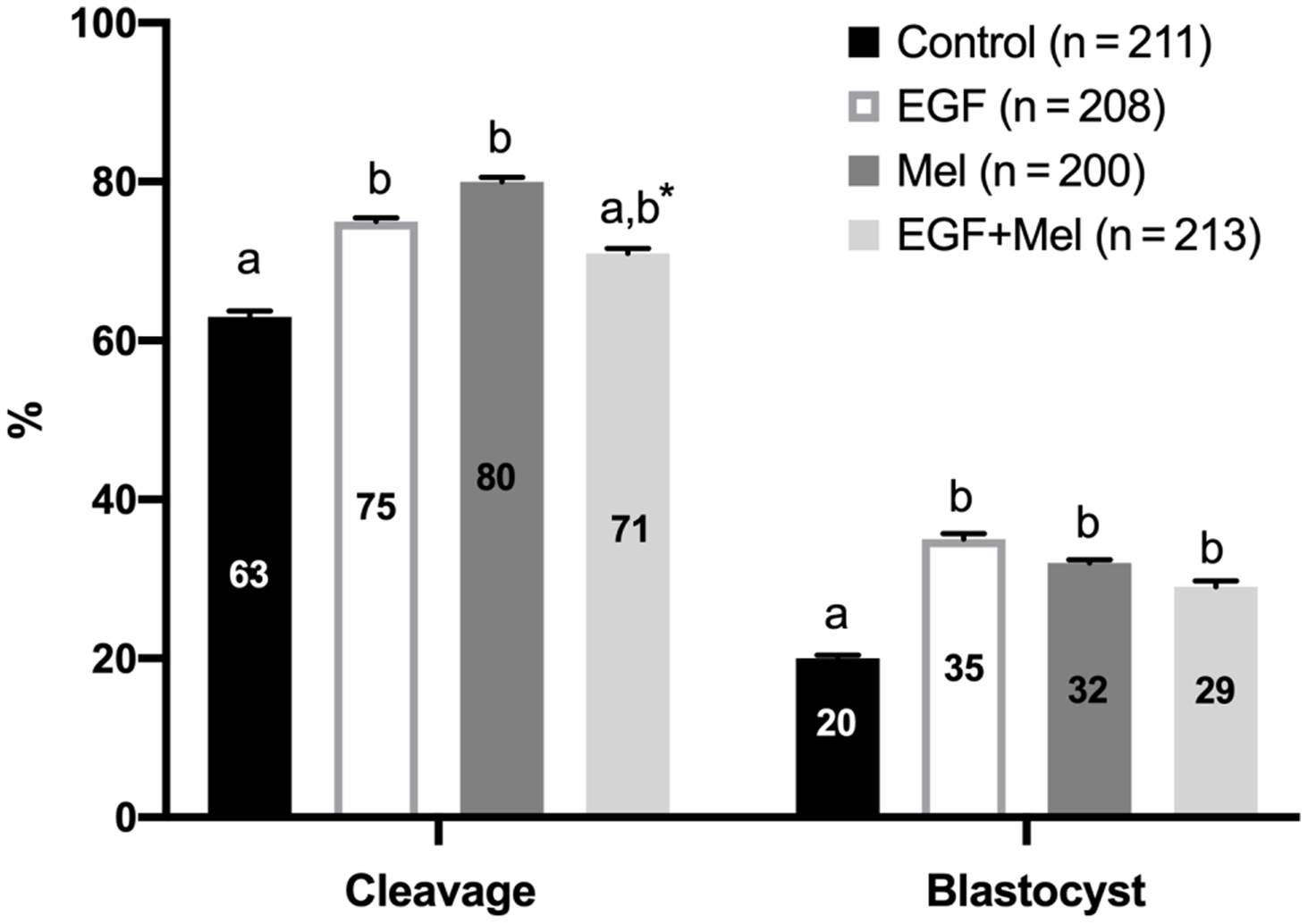
The parthenogenetic embryo development rates, including oocyte activation are shown in Figure 2. With a total number of 1096 oocytes used for IVM, 832 oocytes showed the first polar body after 22 h of IVM (75.9% of nuclear maturation) without any effect on the treatment groups (P > 0.05). After parthenogenesis, cleavage rates were significantly increased in EGF and Mel treatments compared with the Control group (P < 0.05), whereas EGF+Mel showed a tendency (P = 0.07) compared with the Control group to be similar to EGF or Mel alone. Blastocyst rates were elevated for all treatment groups (P < 0.05) compared with the Control group but did not differ among them (P > 0.05).
Parthenogenetic embryo development rates of bovine oocytes in vitro matured with EGF and melatonin. Percentage [error bars; standard error of the mean (SEM)] of cleavage and blastocyst were calculated regarding to the total of activated/mature oocytes per group (n) after seven replicates. a,bDifferent superscripts indicate statistical difference (P < 0.05) within each category. Data were analyzed using chi-squared test at 5% significance level. *, P = 0.07 compared with the Control group.

Parthenogenetic blastocyst quality
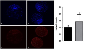
In this study, blastocyst quality was evaluated by the expression of candidate genes involved in embryo apoptosis and pluripotency (Figure 3). Also, the presence and quantity of Oct-4 protein was analyzed by immunofluorescence (Figure 4).
mRNA expression of parthenogenetic embryos developed from oocytes in vitro matured with EGF and melatonin. (A) BAX/BCL2: BCL2 associated X/B-cell lymphoma 2 and (B) OCT4: octamer-binding transcription factor 4. a,b,cDifferent superscripts indicate differences within a gene and between treatments (P < 0.05). Data are expressed as mean ± SEM. Non-parametric Mann–Whitney test was used. Treatments: EGF, epidermal growth factor; Mel, melatonin. Data are normalized to genes GAPDH, PPIA and YWHAZ.

Immunofluorescence analysis of OCT4 protein in parthenogenetic blastocysts. OCT4 signal intensity in group EGF + Mel (B′) was stronger than in Control group (A′). (A, B) Hoechst 33342 nuclear staining. Densitometric analysis of OCT4 fluorescence confirmed a difference [P < 0.05; analysis of variance (ANOVA)] in the EGF+Mel group compared with the Control. Magnification ×200. a,bDifferent superscripts indicate difference (P < 0.05).

BAX/BCL2 ratios were determined, and no differences were found among treatments (P > 0.05). Our study also found that the expression of BAX alone was significantly higher in the EGF+Mel group compared with other groups (P < 0.05, data not shown). OCT4 expression was significantly elevated in the EGF+Mel treatment compared with all other groups (P < 0.05).
This higher mRNA expression was confirmed by densitometric analysis of immunofluorescence to detect OCT4 protein (P < 0.05; Figure 4) with day 7 blastocysts from the EGF+Mel group (B and B′) showing a brighter fluorescence signature than Control (A and A′).
Discussion
Despite all the efforts, IVM conditions continue to need improvement (Rizos et al., Reference Rizos, Ward, Duffy, Boland and Lonergan2002). On the one hand, COCs used for IVM are usually derived from smaller growing antral follicles in which the cumulus EGF signalling network is still developing (Richani and Gilchrist, Reference Richani and Gilchrist2018). On the other hand, in vitro conditions are likely to disrupt this network (Richani and Gilchrist, Reference Richani and Gilchrist2018). Conversely, it is known that higher oxygen tension for most IVM conditions compared with the in vivo environment increases ROS production and CCs may spontaneously undergo apoptosis with negative consequences for oocyte competency (Ikeda et al., Reference Ikeda, Imai and Yamada2003) and therefore the quality of the resulting blastocyst. Adverse IVM conditions will then affect embryo development by disturbing CC gene expression (Tesfaye et al., Reference Tesfaye, Ghanem, Carter, Fair, Sirard, Hoelker, Schellander and Lonergan2009) and oocyte mRNA abundance within the developing COC (Watson et al., Reference Watson, De Sousa, Caveney, Barcroft, Natale, Urquhart and Westhusin2000). The addition of synthetic EGF is being recognized as a way for improving cytoplasmic maturation of oocytes cultured in vitro (De La Fuente et al., Reference De La Fuente, O’Brien and Eppig1999), and promoting CC expansion as efficiently as FSH alone (Tirone et al., Reference Tirone, D’Alessandris, Hascall, Siracusa and Salustri1997). Furthermore, the inclusion of EGF in a medium containing FSH gave better results than using FSH alone (Lonergan et al., Reference Lonergan, Carolan, Van Langendonckt, Donnay, Khatir and Mermillod1996; De La Fuente et al., Reference De La Fuente, O’Brien and Eppig1999), working in concert to regulate oocyte maturation and optimize its competency (Farin et al., Reference Farin, Rodriguez, Alexander, Hockney, Herrick and Kennedy-Stoskopf2007). Studies have also shown that CCs have low or undetectable levels of LH receptor (Peng et al., Reference Peng, Hsueh, LaPolt, Bjersing and Ny1991), and other stimulatory factors, such as EGF or EGF-like peptides, have the potential to induce the expression of CC-associated genes. Furthermore, the addition of antioxidants in vitro has been demonstrated to decrease ROS, preventing CC DNA fragmentation (Tao et al., Reference Tao, Zhou, Xia, Wang, Wu and Fu2004), improving the developmental competence of oocytes (Kwak et al., Reference Kwak, Cheong, Jeon, Lee, Choi, Jeung and Hyun2012; Wang et al., Reference Wang, Tian, Zhang, He, Ji, Li, Tan and Liu2014). Among different factors, Mel has been proven to be an efficient ROS scavenger, acting during nuclear maturation via CCs and directly in the oocyte by binding to its receptors MTNR1A and MTNR1B (El-Raey et al., Reference El-Raey, Geshi, Somfai, Kaneda, Hirako, Abdel-Ghaffar, Sosa, El-Roos and Nagai2011) or accelerating the formation of maturation-promoting factor (Shi et al., Reference Shi, Tian, Zhou, Wang, Gao, Zhu, Zeng, Tian and Liu2009).
The expression of certain CC expansion genes is associated with oocyte competency and is considered as a predictor of oocyte/embryo quality (Assidi et al., Reference Assidi, Dieleman and Sirard2010; Lager et al., Reference Lager, Kocabas, Otu, Ruppel, Langerveld, Schnarr, Suarez, Jarrett, Conaghan, Rosa, Fernandez, Rawlins, Cibelli and Crosby2013; Uyar et al., Reference Uyar, Torrealday and Seli2013). In our study, out of four oocyte/embryo candidates to marker genes, only GREM1 expression seemed to be affected by the proposed treatments in IVM. Gremlin 1 is a highly conserved glycoprotein belonging to the Cerberus and Dan subfamily of secreted bone morphogenetic protein (BMP) antagonists and it is involved in embryogenesis, organ development, tissue differentiation and cancer (Park et al., Reference Park, Sung, Choi, Kim, Kim and Surh2020). In reproduction, GREM1 expression in granulosa cells has been related to follicular progression (Sudo et al., Reference Sudo, Avsian-Kretchmer, Wang and Hsueh2004; Myers et al., Reference Myers, Tripurani, Middlebrook, Economides, Canalis and Pangas2011), also increased expression of GREM1 was observed in CCs from matured buffalo oocytes compared with immatures ones (Bhardwaj et al., Reference Bhardwaj, Ansari, Pandey, Parmar, Chandra, Kumar and Sharma2016). A direct relationship between EFG/melatonin and GREM1 has not yet been described, but it could be considered as a possibility once it has been demonstrated that GREM1 physically interacts with EGF receptors in breast cancer cells and thereby activates EGFR signalling (Park et al., Reference Park, Sung, Choi, Kim, Kim and Surh2020). Further experiments must be done to address this interaction.
Oxidative stress affects the oocyte and CC’s potential to produce a viable embryo by negatively affecting cellular functions, damaging proteins, lipids, and nucleic acids (Guérin et al., Reference Guérin, El Mouatassim and Ménézo2001). In our study, in contrast with the study performed by Rodrigues-Cunha et al. (Reference Rodrigues-Cunha, Mesquita, Bressan, Collado, Balieiro, Schwarz, de Castro, Watanabe, Watanabe, de Alencar Coelho and Leal2016) we demonstrated a decrease in CuZnSOD transcript levels when adding Mel to the IVM medium compared with the Control. Also, contrary to other studies indicating Mel’s ability to reduce the expression of glutathione peroxidases (Fischer et al., Reference Fischer, Slominski, Zmijewski, Reiter and Paus2008; Galano et al., Reference Galano, Tan and Reiter2011), our study showed that the expression of GPX1 in CCs was not altered after the IVM medium was supplemented with Mel, compared with the other treatment groups. We postulate that the presence of Mel in IVM either combined with EGF or not, could be acting to decrease pro-oxidant by-products when interacting with ROS and acting efficiently as a scavenger of ROS, not requiring the CCs to increase their antioxidant machinery through CuZnSOD expression. We believe that more studies need to be done to determine the mechanism by which Mel could be affecting CCs and possibly impacting blastocyst production and ROS levels in the medium.
Mammalian parthenogenetic embryos undergo normal in vitro preimplantation development (Surani and Barton, Reference Surani and Barton1983), avoiding the largely known effect of bull sperm in cleavage rates, developmental kinetics, blastocysts yields and blastocysts quality (please refer to review by Vandaele et al., Reference Vandaele, Mateusen, Maes, de Kruif and Van Soom2006). Although some studies have found differences in terms of trophectoderm cell number, percentage of embryos on day 3, apoptosis, and necrosis index, as well as the expression of some genes related to metabolism, de novo methylation, apoptosis, and cell proliferation between blastocysts produced in vitro and those produced by parthenogenesis (Gómez et al., Reference Gómez, Caamaño, Bermejo-Alvarez, Díez, Muñoz, Martín, Carrocera and Gutiérrez-Adán2009a; Gómez et al., Reference Gómez, Gutiérrez-Adán, Díez, Bermejo-Alvarez, Muñoz, Rodriguez, Otero, Alvarez-Viejo, Martín, Carrocera and Caamaño2009b), others have shown biological similarities between bovine parthenotes and IVF embryos (Labrecque and Sirard, Reference Labrecque and Sirard2011; Naturil-Alfonso et al., Reference Naturil-Alfonso, Saenz-de-Juano, Peñaranda, Vicente and Marco-Jiménez2011) which make them an good option in many biotechnological investigations (Muñoz et al., Reference Muñoz, Penarossa, Caamaño, Díez, Brevini and Gómez2015; Choi et al., Reference Choi, Lee, Lee, Yum, Lee, Lim and Jang2016; Kaneda et al., Reference Kaneda, Takahashi, Yamanaka, Saito, Taniguchi, Akagi, Watanabe and Takashi2017; Sakatani et al., Reference Sakatani, Yamanaka, Balboula and Takahashi2017). Although the positive effect of the use of EGF in IVM on IVF produced blastocyst rate has been largely studied (Abeydeera et al., Reference Abeydeera, Wang, Cantley, Rieke, Prather and Day1998; Sirisathien et al., Reference Sirisathien, Hernandez-Fonseca and Brackett2003), some contradictory data exist regarding embryo production after treatment with Mel during IVM (Takada et al., Reference Takada, Junior, Mingoti, Balieiro, Cipolla-Neto and Coelho2012; Remião et al., Reference Remião, Lucas, Domingues, Silveira, Barther, Komninou, Basso, Jornada, Beck, Pohlmann, Junior, Seixas, Campos, Guterres and Collares2016; Rodrigues-Cunha et al., Reference Rodrigues-Cunha, Mesquita, Bressan, Collado, Balieiro, Schwarz, de Castro, Watanabe, Watanabe, de Alencar Coelho and Leal2016). Our results on parthenogenetic embryos found improved cleavage rates when IVM was supplemented with EGF, Mel or both, compared with the Control group (Figure 2). Moreover, blastocysts rates were higher when supplemented with any of the treatments; this differed from the studies performed by Kang et al. (Reference Kang, Koo, Kwon, Park, Jang, Kang and Lee2009) and also Do et al. (Reference Do, Shibata, Taniguchi, Nii, Nguyen, Tanihara, Takagi and Otoi2015), who did not find any difference in cleavage and blastocyst rate after supplementation with the same concentration of Mel. Furthermore, in contrast with Do et al. (Reference Do, Shibata, Taniguchi, Nii, Nguyen, Tanihara, Takagi and Otoi2015) who proposed that to increase blastocyst rate in porcine, Mel had to be supplemented during IVC (Do et al., Reference Do, Shibata, Taniguchi, Nii, Nguyen, Tanihara, Takagi and Otoi2015), our results demonstrate that supplementation during IVM could also be effective.
The evaluation of the quality of the resulting blastocyst is key to determining if the embryo has the capacity to implant and develop successfully to term. In accordance with Banwell and colleagues (Reference Banwell, Lane, Russell, Kind and Thompson2007) our study demonstrated that IVM conditions were able to influence the transcription signature pattern of the resulting blastocyst. With regards to apoptosis, for example, it is widely accepted that the intracellular balance between pro-apoptotic (i.e. BAX) and anti-apoptotic (i.e. BCL2) proteins indicates a cell’s tendency to undergo apoptosis (Duan et al., Reference Duan, Ou, Li, Su, Ou, Yang, Yue and Ban2005). Our results (Figure 3) showed that the BAX/BCL2 ratio (mRNA expression) was not different among the groups studied but, if analyzed separately, we observed an increased BAX expression in EGF+Mel but no differences in BCL2 expression among treatments (data not shown). Although the study by Watson et al. (Reference Watson, De Sousa, Caveney, Barcroft, Natale, Urquhart and Westhusin2000) did not find any correlation between the oocyte environment and further apoptotic index in blastocysts, studies have shown that the addition of supplements to the IVM medium could affect the apoptotic index as revealed by terminal deoxynucleotidyl transferase (TdT) dUTP nick-end labelling (TUNEL) assay (Warzych et al., Reference Warzych, Peippo, Szydlowski and Lechniak2007). Ultimately the assessment of apoptosis by protein expression markers would be necessary to accurately determine the blastocyst’s apoptotic status after Mel treatments during IVM.
Conversely, our study demonstrated that relative mRNA expression of OCT4 was greatly increased (P < 0.05) following EGF+Mel treatment compared with the other treated groups (Figure 3). These results were confirmed by immunofluorescence (Figure 4) in which the higher expression of OCT4 in parthenotes from EGF+Mel was clearly observed. OCT4 expression determines the fate of ESCs in vitro (Niwa et al., Reference Niwa, Miyazaki and Smith2000), then our study demonstrates that parthenotes produced under IVM supplemented with EGF+Mel could be a potential good candidate for embryonic stem cells (ESC) isolation and use, without the ethical problems of using in vitro or in vivo produced blastocysts. There is no consensus about the existence of a pivotal cell marker indicating blastocyst quality but OCT4 mRNA has been shown to be highly expressed in competent oocytes and blastocysts (Schultz, Reference Schultz2002; Amarnath et al., Reference Amarnath, Li, Kato and Tsunoda2007). Therefore, we believe that IVM treatment supplemented with EGF+Mel in this study was able to produce presumptive good quality blastocysts, although other cell markers related to blastocyst quality should be investigated in the future.
Our study concludes that supplementation of bovine IVM medium with EGF and Mel somehow affects CC gene expression and further improves in vitro embryo development. With regards to Mel, in particular, we believe that its addition to IVM medium could scavenger ROS, giving the oocyte a better environment in which to develop up to the blastocyst stage. However, the combinatory use of EGF and Mel did not add the expected extra positive effect in these parameters, but interestingly rendered better quality embryos based on the expression of genes known to be related to pluripotency. Further studies are necessary to elucidate which molecular mechanisms are underlying this combinatory positive effect on embryo pluripotency.
Financial support
This work was supported by the Toronto Zoo Foundation and MITACS-Accelerate (www.mitacs.ca) programme.
Declaration of interest
The authors declare no conflicts of interest.
Ethical standards
Not applicable.