Introduction
Cationic antimicrobial peptides (CAP) are active peptides capable of killing pathogenic microorganisms in body fluid, an important component of the innate immune system. Studies on CAP application are becoming hot spots in veterinary medicine, pharmacology, physiology, etc. Wang et al. (Reference Wang, Lin and Xu2004b) reported the differences of expression of CAP pr-39 in various developmental stages of pigs. Zhang et al. (Reference Zhang, Bi, Cao, Ma and Chen2005) cloned the chicken beta-defensin gene and analysed its sequence. Li et al. (Reference Li, Zhang and Li2001) analysed the cow lectoferritin gene. Wang et al. (Reference Wang, Han and Huang2004a) analysed the mouse lectoferritin gene. Huang and Wang (Reference Huang and Wang2005) expressed mouse lectoferritin heterologously in Escherichia coli. Shi et al. (Reference Shi, Yang, Zhang and Cheng2006) expressed heterologously the fusion protein composed of melittin and green fluorescence protein in insect cells. Zhao et al. (Reference Zhao, Liu, Zheng, Lu and Zheng2006) expressed heterologously human lectoferritin in tomoto. As yet there are few reports about using CAP to create a target-sensitive toxin to kill target cells.
Immunotoxin (IT) kills specifically carcinoma cells. IT is composed of a target part – antibody and cytokine – which can specially bind to antigen or receptors overexpressed on carcinoma cells, and a cytotoxic part, which can kill carcinoma cells when the target part co-exists covalently (Pennell and Erickson, Reference Pennell and Erickson2002). In normal conditions, epidermal growth factor receptor (EGFR) is widespread, but at low density (5000–10 000/molecules/cell) on mammal cell surfaces; it is blocked and unable to bind to its ligand. When the body is hurt, EGFRs become exposed and bind to their ligands to start healing, before being blocked again. Due to out of control gene expression, EGFRs can become abnormally exposed or overexpressed, resulting in abnormal cell proliferation and carcinomas (Gullick, Reference Gullick1991). On the surface of many kinds of carcinomas, the EGFRs are overexpressed with normal form and steady phenotype, tightly binding to ligands such as epidermal growth factor (EGF) and transforming growth factor-α (TGFα). Davies and Chamberlin (Reference Davies and Chamberlin1996) reported that EGFRs of over 40% of mastocarcinomas, 75% of ovary carcinomas, 81% of cervix carcinomas, 71% of oesophageal carcinomas and 50% of liver carcinomas overexpress 100 times more than normal and are exposed on the cell surface. Many kinds of carcinoma EGFR overexpression imply high metastasis rates and bad pronosis (Harris et al., Reference Harris, Eichholtz, Hiles, Page and O'Hare1999; Kersemaekers et al., Reference Kersemaekers, Fleuren and Kenter1999). EGFR is expected to be a target site for anticarcinoma ITs (Modi and Seidman, Reference Modi and Seidman2002).
Up to now, the cytotoxic part of ITs has been mainly from plant and microorganisms, such as ricin, diphtheria toxin (DT) and pseudomonas exotoxin (PE), the cytotoxic effect of which is to inhibit protein synthesis in target cells (Lambert et al., Reference Lambert, Goldmacher and Blattler1991; Choe et al., Reference Choe, Bennett and Fujii1992; Kreitman, Reference Kreitman2001). An internalization step is necessary for establishing the toxin's active efficiency. Melittin (MEL) is from bee venom, a typical membrane-lytic CAP which destroys target cell membranes without an internalization step. Using MEL as the cytotoxic part of IT, without an internalization step, will significantly raise cytotoxic activity.
In this study we used MEL as the cytotoxic part and mouse (Mus musculus) epidermal growth factor (MEGF) as the target part, incorporating the two domains by a linker sequence into a single-molecule IT heterologously expressed in E. coli. The linker is flexible enough to make the two domains, MEGF and MEL, independently active, because the sequence of the linker is from the highly flexible immunoglobin hinge region. The positive charges of the cationic region of the MEL domain were neutralized by MEGF (the pI of MEGF is 4.6), which makes the MEGFMEL unable to bind to negatively charged normal cells. The MEGF domain could lead MEGFMEL binding to the carcinoma cells overexpressing EGFR, so that the amphipathic alpha helix of the MEL domain could then specifically destroy the carcinoma cell membrane.
Materials and methods
Plasmid and strains
Recombinant plasmids pGEMTmegf and pUCTmel were developed in our laboratory (He et al., Reference He, Ruan, Niu, Chen, Yu and Fu2002; Ruan et al., Reference Ruan, Niu, Zhao, He and Fu2005). Plasmid pET30a and E. coli BL21 were from our laboratory collection. T4 DNA ligase and restriction endonuclease enzymes (EcoRI, BamHI, SacI and HindIII) were purchased from Shanghai Sangon Co. Ltd (Shanghai, China). E. coli was grown in Luria-–Bertani (LB) medium (1% tryptone, 0.5% yeast extract, 0.5% sodium chloride) containing 50 μg/ml ampicillin. The linker sequence was synthesized by Shanghai Sangon Co. Ltd as follows:
linker1: 5′-C CGT (GGT)4 TCT (GGT)4 TCT (GGT)3 G-3′
linker2: 3′-TC GAG GCA (CCA)4 AGA (CCA)4 AGA (CCA)3 CCT AG-5′
Linker1 and linker2 became a double-strand linker with the cosmid sites SacI at the 5′ end and BamHI at 3′ end after renaturation.
Primers MEGF-P1 (matching with the MEGF 5′ region) and MEL-P2 (matching with the melittin 3′ region) were synthesized by Shanghai Sangon Co. Ltd, with the following sequences:
MEGF-P1: 5′-CCCGAATTCAATAGTTACCCAGGATGCCCAT-3′ (EcoRI site underlined)
MEL-P2: 5′-CCCAAGCTTTTACTGTTGCCTCTTACGTTT-3′ (HindIII site underlined).
Construction of megfmel chimeric DNA
The megf sequence, with the sites EcoRI at the 5′ end and SacI at the 3′ end, was generated by digesting the PCR product with EcoRI and SacI restriction enzymes (megf/EcoRI+SacI).
The mel sequence with the sites BamHI at the 5′ end and HindIII at the 3′ end was generated by digesting the PCR product with BamHI and HindIII restriction enzymes (mel/BamHI+HindIII).
The megf/EcoRI+SacI and mel/BamHI+HindIII were ligated by linker (SacI cosmid site at 5′ end and BamHI cosmid site at 3′ end) with T4 DNA ligase. Then, the megfmel fusion gene was generated by PCR with primers MEGF-P1 and MEL-P2.
Cloning of chimeric construct pETmegfmel
The megfmel PCR product was ligated to T-vector before digestion by EcoRI and SacI restriction enzymes. The digested megfmel fragment was subcloned into the plasmid pET30a, which was previously digested with the same restriction endonuclease enzymes, to develop the chimeric construct pETmegfmel. This construct was sequenced to confirm correct DNA sequence after PCR and digestion verification. The primer for sequencing was the universal primer of the pET vector: pET30-Pc (5′-TAATACGACTCACTATAGGG-3′).
Expression and purification of the chimeric protein
The plasmid pETmegfmel was transformed into E. coli BL21 cells to generate the recombinant strain BL21-pETmegfmel, while pET30a was transformed into E. coli BL21 cells to generate the strain BL21-pET30a as the negative control. The transformed bacteria were plated on LB plates. The bacterial cultures were induced with iso-propyl β-d-thiogalactopyranoside (IPTG; final concentration was 0.50 mmol/l) when the optical density of the BL21-pETmegfmel and BL21-pET30a reached 0.5 OD600. The expression product was the chimeric protein MEGFMEL with 50 amino acid residues of pET30a at its N-terminal end and at its C-terminal end. The chimeric protein MEGFMEL was purified using the thawing and freezing method (Ruan et al., Reference Ruan, Niu, Zhao, He and Fu2005) and detected on a 15% SDS–PAGE gel (Wang and Fan, Reference Wang and Fan2000).
Quantification of the purified protein
The concentration of the target protein was measured by the Folin-phenol method according to Zhou (Reference Zhou1991).
Bioactivity assay of the recombinant expressed product
We evaluated the cytotoxic activity of the product through a [3H]thymine deoxyribonucleoside (Tdr) incorporation assay, with A431 as the target cells. The whole procedure was as follows:
The concentration of A431 cells was adjusted to 5×10⁴/ml with RPMI-1640 medium, then 10 μCi [3H]Tdr were added to 1 ml target cell solution and the culture was shaken for 6 h at 37°C to thoroughly incorporate [3H]Tdr into the daughter cells' DNA. A431 cells were then washed three times with RPMI-1640 medium and their concentration adjusted to 5×10⁴/ml. Fifty microlitres of the cell suspension were added to the 96-well culture plate, followed by the gradient dilution of recombinant chimeric protein MEGFMEL in each well. The BL21-pET30a expressed product was used as the negative control, while 1.0 mol/l NaOH served as positive control.
Cells in the 96-well plate were incubated for 12 h at 37°C. A volume of 50 μl culture medium containing 1.0 mg/ml trypsinase and 10 μg/ml DNase was transferred to each well. The positive control received, in addition, 1.0 mol/l HCl to neutralize NaOH. The plate was finally incubated for 1 h at 37°C to ensure the disintegration of dead cells and their DNA to release the [3H]Tdr.
The counts per minute (cpm) in the supernatant and deposit in each well were determined and the cytotoxic activity was calculated. The cytotoxic activity was calculated using the formula: [cpm in supernatant/(cpm in supernatant+cpm in deposit)]×100%. The concentration of the protein of interest when cytotoxic activity was 50% gave the LD50 (median lethal dose).
Statistical analysis
The data analyses were performed using SAS/Statistic Version 6.12 software program (SAS Institute Inc., Cary, North Carolina, USA). The F-test was used to determine the statistically significant differences between the two groups in each parameter.
Results
Vector pETmegfmel verification
The vector pETmegfmel was sequenced to verify the DNA sequence after PCR and digestion processes. The sequencing result confirmed the existence of the DNA insert with the sequence shown in Fig. 1.
Sequence of the DNA insert in pETmegfmel.

Electrophoresis assay and concentration of the chimeric protein MEGFMEL
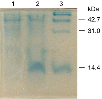
After the preliminary purification used in this assay, the recombinant expressed product of BL21-pETmegfmel migrated with an apparent molecular size of 14.0 kDa, as expected (Fig. 2, lane 2), while nothing was generated in the negative control strain BL21-pET30a (Fig. 2, lane 1). This result confirmed the success of the recombinant expression of chimeric protein MEGFMEL. The purity quotient of the product was 68%, according to grey scale scanning (Fig. 2). The calculated concentration of the chimeric protein MEGFMEL was 63.45 μg/ml.
Detection of the chimeric protein MEGFMEL by SDS-PAGE electrophoresis. Lanes: 1, BL21-pET30a; 2, BL21-pETmegfmel; 3, marker. (See online for a colour version of this figure.)

Chimeric protein MEGFMEL bioactivity
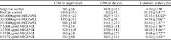
The recombinant chimeric protein MEGFMEL was diluted with distilled water to give the following concentrations: 60, 30, 15, 7.5, 3.75, 1.875 and 0.9375 μg/ml. Cytotoxic activity assays of the chimeric protein MEGFMEL were performed with A431 as target cells using [3H]Tdr incorporation assay (Table 1).
Cytotoxic activity of the recombinant chimeric protein MEGFMEL to A431 cells

CPM, counts per minute.
** , Significant difference compared to negative control at P<0.01.
The results in Table 1 show that the cytotoxic activity of the recombinant chimeric protein MEGFMEL to the A431 cells was significant. In addition, the cytotoxic activity was evaluated by further dilutions between 60 and 30 μg/ml. The LD50 of the protein was estimated as 52.6 μg/ml (cytotoxic activity 50.39±5.45%). Because of the presence of some impure protein in the recombinant product, the LD50 of the recombinant MEGFMEL should actually be much lower. A higher protein purification could possibly increase the cytotoxic activity.
Discussion
The carcinoma cells A431 overexpress EGFR on the surface. They are perfect target cells for the assay of ITs targetting EGFR. However, up to now, the cytotoxic part of ITs has been from plants and microorganisms (PE, DT, ricin, etc.). The strong immunogenicity and essential internalization step affect the efficiency of these ITs (Lambert et al., Reference Lambert, Goldmacher and Blattler1991; Choe et al., Reference Choe, Bennett and Fujii1992; Kreitman, Reference Kreitman2001). CAP is becoming a potential toxin for IT construction due to its strong lethal effect and membrane-lytic cytotoxicity mechanism. In this study, a membrane-lytic IT, the chimeric protein MEGFMEL (composed of MEGF as the target part and MEL as the cytotoxic part), was constructed to neutralize carcinoma cells through EGFR overexpression. In vitro activity measurement showed that MEGFMEL had a strong lethal effect on A431 carcinoma cells.
The N-terminal end of the recombinant chimeric protein MEGFMEL is built up of 50 amino acid residues of pET30a. Previous studies have demonstrated that changes of the amino acid residues of the epidermal growth factor, except 4–48, have no influence on its activity (Burgess et al., Reference Burgess, Lloyd and Smith1988). Our previous study showed that the activity of MEGF (57% purity) with an additional pET30a sequence at the N-terminal end was 30% of that of the standard MEGF (Ruan et al., Reference Ruan, Niu, Zhao, He and Fu2005). The present study has proved that MEGF with an additional pET30a sequence at the N-terminal end can ensure the affinity of the recombinant chimeric protein MEGFMEL to EGFR.
Some problems, however, should be considered to ensure clinical application of the IT. These problems include maintaining the half-life in vivo, keeping the immunogenicity as low as possible, improving safety testing, etc. The results in this study showed that using CAP as the cytotoxic part to construct an EGFR-targetting IT is possible. It is expected that a human CAP source (human beta defensins, hBDs) will be developed as the cytotoxic part and human epidermal growth factor (hEGF) as the target part to build more efficient ITs.
Acknowledgements
This work was supported by the Zhejiang Natural Science Foundation and Hangzhou Science and Technology Plan.