Methylation of a subset of DNA cytosine residues is tightly associated with gene regulation. Generally, DNA methylation occurs in the cytosine-guanine sequence (CpG) in adult cells; however, recent genome-wide methylation analysis revealed that a subset of methylation identified in embryonic stem cells was in a non-CpG context (Refs Reference Ramsahoye1, Reference Lister2). Approximately 70% of CpGs in the genome are methylated. However, short stretches (up to 2 kb) of CpG-rich regions, which are found in the promoters of around 60% of genes, are generally unmethylated and called ‘CpG islands’ (CGIs) (Refs Reference Bestor3, Reference Cross and Bird4). CGIs are also found in non-promoter regions: around 50% of CGIs are not coincident with annotated promoters, and their biological function is not well understood (Ref. Reference Illingworth5). In contrast to methylation in non-promoter CGIs, methylation in promoter CGIs is closely linked to alterations of chromatin structure and is associated with gene silencing. In mammals, DNA methylation is the most stable epigenetic modification known and is involved in many biological phenomena such as X-chromosome inactivation in females, imprinting, inactivation of repetitive elements and epigenetic reprogramming during mammalian development (Ref. Reference Bird6).
Epigenetic silencing is described as a ‘third pathway’ in Knudson's model of tumour-suppressor gene inactivation and can affect gene expression without accompanying genetic changes (Ref. Reference Jones and Laird7); therefore, studies over the past two decades have focused on aberrant DNA methylation in neoplasia. Cumulative analyses revealed that cancer cells generally demonstrate global hypomethylation and regional hypermethylation at selected gene-associated CGIs simultaneously. Aberrant hypermethylation in CGI promoters of critical tumour-suppressor genes has been described, and association between this abnormality and tumour initiation and progression is now widely recognised (Ref. Reference Jones and Baylin8).
Recently, two important advances in this field have contributed to its popularity as a research area. First, two nucleoside inhibitors of DNA methylation, 5-azacytidine (AZA) and 5-aza-2'-deoxycytidine (DAC) were established as new treatments for the myelodysplastic syndromes (MDSs), which are heterogeneous haematopoietic disorders characterised by dysplasia of one or more of the myeloid blood cell lineages and an increased risk of acute myeloid leukemia (Refs Reference Issa9, Reference Shen10). In addition to these methylation inhibitors, another type of epigenetic drug, histone deacetylase inhibitors, which lead to the accumulation of acetylation in histones, have been found to be effective for treatment of lymphomas (Refs Reference Yoo and Jones11, Reference Marks and Xu12). Since numerous studies indicate that all malignancies have both genetic and epigenetic abnormalities, targeting epigenetics is a reasonable and potentially effective approach for cancer treatment. The second advance is in the area of genome-scale DNA methylation analysis technology (Refs Reference Beck and Rakyan13, Reference Laird14). Previous DNA methylation profiling, such as two-dimensional gel electrophoresis (restriction landmark genome scanning or RLGS), has been used widely, but is technically challenging (Refs Reference Hayashizaki15, Reference Kawai16). Recent microarray-based technology and more powerful and flexible next-generation sequencing technology have improved the process. These recent developments in DNA methylation analysis techniques can be performed on a genome scale and entire methylomes can be characterised at single-base-pair resolution (Ref. Reference Lister2).
A link between the above two advances has important implications for cancer diagnostics and treatment. Characterisation of DNA methylation profiling of cancer patients might be of great benefit to determine strategies for optimising epigenetic treatment. In this review, DNA methylation profiling and its clinical implications are discussed.
Significance of DNA methylation in cancer
Cancer cells show loss of global methylation and gain of regional methylation simultaneously. Aberrant distribution of DNA methylation is found in almost all cancer cells. Global hypomethylation may cause genomic instability through impairment of appropriate chromatin structure (Refs Reference Okano17, Reference Eden18, Reference Cadieux19). In addition, hypomethylation in CGI promoter regions is associated with the derepression of certain cancer-associated genes, such as melanoma antigen genes (MAGEs) (Ref. Reference De Smet20). Loss of imprinting (LOI), leading to pathological biallelic expression of IGF2 (which typically activates the maternally silenced allele), is another well known cause of tumorigenesis (Ref. Reference Sakatani21). Although incompletely studied, abnormal gene activation by hypomethylation might mediate the early aberrant clonal expansion of cells and the development of tumours.
Aberrant hypermethylation in promoter CGIs has been comprehensively studied in human cancers, although mechanisms regulating the establishment of de novo DNA methylation remain poorly understood. This DNA methylation in promoter CGIs links to gene silencing in a subset of cancer-related genes (Ref. Reference Baylin22). Many cancers appear to have genes that are silenced by DNA methylation leading to loss of function. Since several molecular pathways are involved in tumour formation, simultaneous silencing of many genes by epigenetic mechanisms could be a strong driving force for transformation (Refs Reference Jones and Baylin23, Reference Ting, McGarvey and Baylin24). The frequency of DNA methylation in cancerous tissues varies between different genes. In addition, targets of DNA methylation differ depending on the cell of origin of cancers (Ref. Reference Kondo and Issa25). One plausible hypothesis to explain selectivity is that DNA methylation occurs in CGIs randomly, with specificity subsequently happening through the process of selection. However, it seems unlikely that all DNA methylation arises in a random fashion, and some methylation might reflect abnormalities in programmes of epigenetic control, which results in methylation patterns that are generated through selective advantage (Ref. Reference Jones and Baylin8). Indeed, in genome-wide studies, DNA methylation does not always target tumour-suppressor genes, and many of the hypermethylated genes are already repressed in normal tissues before transformation (Ref. Reference Keshet26). Thus, it also appears likely that CGIs differ in their intrinsic susceptibility to de novo methylation in cancer cells (Ref. Reference Feltus27).
Recently, links between polycomb group protein (PcG)-mediated histone H3K27 trimethylation (H3K27me3) and DNA methylation in cancer were described. The conventional view is that the mark imposed by polycomb repressive complex 2 (PRC2) during development predisposes some genes for later de novo methylation (Refs Reference Ohm28, Reference Widschwendter29, Reference Schlesinger30). This model appears to be applicable to a set of genes prone to gain DNA methylation during tumorigenesis. However, the presence of H3K27me3 does not always lead to de novo methylation, and H3K27me3 target genes can be methylated in one cancer and unmethylated in another cancer from the same tissue type (Refs Reference Kondo31, Reference Gal-Yam32). Another credible model has been reported recently, in which the existence of stalled RNA polymerase on promoter regions might be a cue to determine the epigenetic fate of promoter DNA methylation (Ref. Reference Takeshima33). Although this model is also applicable to a set of genes, several mechanisms that establish de novo DNA methylation appear to be involved in each gene. Further analyses are still required to elucidate how DNA methylation arises in selected CGI promoters.
Significance of the CpG island methylator phenotype
Recent studies in colorectal cancers have indicated the existence of an accumulation of high rates of aberrant promoter hypermethylation in a subset of cases known as CpG island methylator phenotype (CIMP) positive (Refs Reference Toyota34, Reference Shen35). The mechanisms responsible for causing CIMP tumours is not known; however, CIMP-positive colorectal cancers exhibit distinct genetic and clinical features, including high rates of mutations of BRAF and KRAS, low rates of TP53 mutations, a specific histology (mucinous, poorly differentiated), a proximal location and characteristic clinical outcomes (Ref. Reference Issa9). Furthermore, the existence of different classes of CIMP tumours (e.g. CIMP1 and CIMP2) has been shown recently, raising the possibility that sporadic colorectal cancers arise from distinct parallel pathways, in which the type of genetic and epigenetic instability ultimately explain the characteristic carcinogenesis pathways altered (see below) (Refs Reference Shen35, Reference Ogino36, Reference Yagi37). Given these distinct clinicopathological features, CIMP-related carcinogenesis might proceed through a specific pathway in which the epigenetic changes that occur in premalignant cells determine subsequent genetic changes, thereby fostering the progression of these clones (Ref. Reference Baylin and Ohm38).
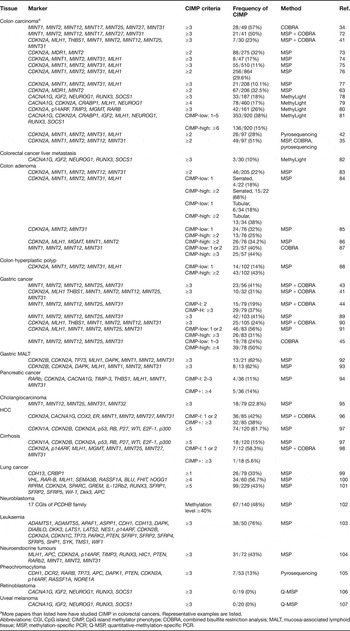
The criteria for defining CIMP tumours, especially in tumour types other than colorectal cancers, are in development, which has contributed to some of the controversies in this field of study. CIMP was originally proposed when DNA methylation in colorectal cancers was found to have a bimodal distribution, with a group of cancers showing an accumulation of high rates of aberrant promoter hypermethylation and unique biological or clinical features. Published studies on CIMP in human neoplasia (focusing on tumours other than colorectal cancers) are listed in Table 1. Since it is not clear which panel of markers will be the most accurate for identifying CIMP in each tissue type, the frequency and significance of CIMP tumours is still controversial (Refs Reference Issa39, Reference Issa40). In addition, the methodology for detection of aberrant DNA methylation in cancer cells is another pragmatic issue. The use of methylation-specific PCR (MSP), a highly sensitive and nonquantitative technique to measure methylation, might sometimes fail to find CIMP in certain tumours (Ref. Reference Issa39). Indeed, almost every research group that quantitatively studied the methylation status of the genes originally used to define CIMP has confirmed that there is concordant methylation in colon and gastric cancers (Refs Reference Shen35, Reference Kanai41, Reference An42, Reference Toyota43, Reference Kim44, Reference Kusano45). Although DNA methylation in gastric cancers does not show as clear a bimodal distribution as found in colorectal cancers, the aggregate data does suggest that a group of CIMP-positive gastric tumours probably arises via a process that is distinct from that of non-CIMP (or less DNA-methylation-dependent) tumours. Better characterisation of this unique molecular subgroup of tumours defined by excessive rates of aberrant promoter methylation will require uniform methodology and marker panels for each type of cancer analysed.
CpG island methylator phenotypes in human malignancies

aMore papers than listed here have studied CIMP in colorectal cancers. Representative examples are listed.
Abbreviations: CGI, CpG island; CIMP, CpG island methylator phenotype; COBRA, combined bisulfite restriction analysis; MALT, mucosa-associated lymphoid tissue; MSP, methylation-specific PCR; Q-MSP, quantitative-methylation-specific PCR.
Methodologies for DNA methylation profiling
It is now widely accepted that DNA methylation marks of individual genes or panels defining CIMP are associated with the clinical outcome of cancers. Thus, there is an interest in studying cancers with appropriate panels of markers to develop molecular classification systems relevant to diagnosis and therapy, as has been shown in the examples of colorectal cancer and myelodysplastic syndrome (Refs Reference Shen10, Reference Shen35). Recent technological progress now enables us to study DNA methylation profiles comprehensively. The new techniques provide a large amount of information that might be of help to better understand the role of epigenetic changes in the pathogenesis of human cancers. Using these techniques, one can translate the data on aberrant DNA methylation in cancer into clinical practice, which could result in a better outcome for patients.
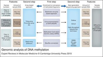
Some of the available techniques and their features are described in Figure 1. Methylated cytosine is indistinguishable from unmethylated cytosine by standard sequencing approaches. Therefore, before analysis by high-throughput applications, enrichment for methylated DNA or sodium bisulfate conversion are generally used to overcome this problem. Two methods are widely used to enrich for methylated DNA: affinity purification and restriction-enzyme digestion. There are two commonly used affinity purification methods – methylated DNA immunoprecipitation (MeDIP) and the methylated-CpG island recovery assay (MIRA) (Refs Reference Weber46, Reference Zhang47, Reference Rauch48). In MeDIP, an antibody specific for methylated cytosine is used to immunocapture methylated genomic fragments (Refs Reference Weber46, Reference Zhang47). The resulting enriched fraction can be subjected to standard DNA detection; thus, MeDIP can be combined with microarrays or large-scale sequencing analysis. However, to apply this technique to a large number of samples, the large amount of starting DNA and the high cost of the antibody can be a problem. MIRA is based on the high affinity of the complex of methyl-binding domain protein 2b (MBD2b) and MBD3L1 to methylated DNA (Ref. Reference Rauch48). Methylated DNA fragments can be captured and purified on an MBD column, and then subjected to analysis by microarrays or sequencing. Differential restriction enzyme digestion is the classical method for detecting methylated DNA fragments. The most widely used methylation-sensitive restriction enzymes are HpaII and SmaI, which have methylation-insensitive isoschizomers (MspI for HpaII) or neoschizomers (XmaI for SmaI) (Refs Reference Khulan49, Reference Estecio50, Reference Shen51, Reference Gao52, Reference Omura53). A combination of these enzymes followed by ligation-mediated PCR enables the enrichment of methylated DNA fragments.
Genomic analysis of DNA methylation. The enrichment of methylated DNA or bisulfite-converted DNA is followed by array-based analysis or high-throughput sequencing analysis. The former is easy to study in a large number of samples owing to the multiplex array platform, whereas the latter has difficulties dealing with large numbers of samples. Since bisulfite-converted DNA has a reduced sequence complexity, several approaches are needed to apply this technique to high-throughput sequencing analysis, such as RRBS (reduced representation bisulfite sequencing), BC-seq (bisulfite conversion followed by capture and sequencing), BSPP (bisulfite padlock probe) and WGSBS (whole-genome shotgun bisulfite sequencing).

Sodium bisulfate treatment of DNA is now a fundamental method for DNA methylation analysis (Ref. Reference Clark54). This treatment causes deamination of unmethylated cytosine to uracil, while leaving methylated cytosine intact. After PCR amplification, which converts uracil to thymine, the cytosine-to-thymine signal ratio is directly proportional to the level of DNA methylation at any given position. This method has been combined with pyrosequencing analysis (Ref. Reference Colella55), restriction-enzyme digestion (combined bisulfite restriction analysis or COBRA) (Ref. Reference Xiong and Laird56) or amplified with specific primers (MSP) (Ref. Reference Herman57) and is a very useful option to analyse the DNA methylation status in selected genes. However, this method is difficult to combine with genome-wide approaches. After bisulfate conversion, cytosines at non-CpG sites are all converted to thymine, which results in a reduction of sequence complexity. However, recently, an array approach (Illumina BeadsArray technology) was described that appears to overcome this problem (Ref. Reference Bibikova58). To adapt bisulfite-converted samples to next-generation sequencing platforms, more efficient approaches have been developed that use microarray capture (Ref. Reference Albert59), size-fractionation after restriction enzyme digestion (reduced representation bisulfite sequencing or RRBS) (Ref. Reference Meissner60) or other types of sequence capture (Refs Reference Hodges61, Reference Li62). These techniques effectively enable the adaptation of the bisulfite-converted DNA to the next-generation sequencing platforms. Bioinformatics and biostatistics are also important challenges when bisulfite-converted DNA is sequenced by next-generation sequences, because sequence-based analyses produce more comprehensive massive data sets, which require a practical alignment to a reference genome. Once sequencing costs come down and convenient bioinformatic analyses are easily accessible, it will also be possible to routinely analyse the whole genome by shotgun bisulfite sequencing.
Analysis of DNA enriched for methylated regions can be analysed by microarrays or by next-generation sequencing platforms. Microarray analysis has been successfully used to study large genomic regions. However, this technique suffers from limitations such as cross-hybridisation and lower resolution (analysed regions are limited by the probes) compared with next-generation sequencing. However, microarray analysis is now relatively economical and applicable to a large number of samples, especially because multiplexed array platforms are available. In addition, any probe can be easily selected and mounted on the multiplex platform, which allows the study of different regions [focusing on 5'-untranslated regions (UTRs), 3' UTR, coding exons etc.]. The next-generation sequencing platform is a more powerful technique to analyse DNA methylation genome-wide and can overcome some of the weaknesses of a microarray-based approach, with current limitations being cost and throughput. Because of its accuracy and abundant information recovered, genome-wide DNA methylation analysis will shift from microarrays to next-generation sequencing in the near future.
Clinical implications of DNA methylation profiling for human malignancies
DNA methylation in particular genes is associated with clinical outcome. For example, methylation of MLH1, which encodes a mismatch repair protein whose loss leads to microsatellite instability (MSI), is associated with a better prognosis in colorectal cancer (Refs Reference Shen35, Reference Van Rijnsoever63). DNA methylation in the O6-methylguanine DNA methyltransferase gene (MGMT), which encodes a DNA-repair enzyme, is associated with sensitivity to alkylating agents in brain tumours (Ref. Reference Esteller64). Aberrant DNA methylation of the checkpoint with forkhead-associated and ring finger (CHFR), a mitotic checkpoint gene, is associated with sensitivity to microtubule inhibitors in colorectal and gastric cancers (Refs Reference Toyota65, Reference Satoh66). Methylation of death-associated protein kinase (DAPK) is associated with early recurrence of bladder cancer (Ref. Reference Tada67). In addition to the relationship between DNA methylation in single genes and clinical outcome, recent integrated analysis between genetic and epigenetic changes in colorectal cancer demonstrated that these cancers can be divided into three molecularly distinct subclasses: (1) CIMP1: MLH1 and BRAF mutations and rare KRAS and TP53 mutations; (2) CIMP2: KRAS mutations and rare MLH1, BRAF or TP53 mutations; (3) CIMP-negative: high rate of TP53 mutations and lower rates of MSI or mutations of BRAF or KRAS (Ref. Reference Shen35). CIMP1 colorectal cancer tends to have a good prognosis, whereas CIMP2 colorectal cancer is resistant to chemotherapy (5-FU or cetuximab) and generally has poor prognosis (Ref. Reference Issa40). These findings imply that understanding the genetic and epigenetic alterations that promote human cancers and the identification of appropriate biomarkers are prerequisite for successful cancer therapy.
Very recently, genome-wide DNA methylation profiling using a restriction-enzyme-based approach (HpaII tiny fragment enrichment by ligation-mediated PCR or HELP) reveals the existence of distinct DNA methylation patterns in acute myeloid leukaemia (AML) and identifies new, biologically and clinically relevant defined AML subgroups (Ref. Reference Figueroa68). The study identified a 15-gene DNA methylation classification that is capable of predicting overall survival in AML patients, even in patients for whom clinical biomarkers are not currently available. Similar studies using genome-wide DNA methylation analysis have been done in hepatocellular carcinoma, colorectal cancer and mesothelioma (Refs Reference An42, Reference Gao52, Reference Goto69, Reference Arai70).
Since genetic alterations are generally irreversible, but epigenetic alterations underlying tumorigenesis can be modulated by drug treatment, typing or profiling of DNA methylation in each cancer might be useful for therapeutic benefit. The DNA methylation inhibitors AZA and DAC, are now used for treatment of myelodysplastic syndrome. It is important to recognise that although the inhibition of DNA methylation is the molecular effect of these drugs, their downstream effects on neoplastic behaviour are nonspecific (Ref. Reference Issa and Kantarjian71). This nonspecificity makes the prediction of clinical outcome difficult. Thus, the responses might be predominantly apoptosis mediated and rapid in some patients, whereas in others, a slow differentiation effect is prominent. To understand the effects of DNA methylation inhibitors more precisely, genome-wide DNA methylation profiling before and after treatment may be useful. This approach could identify novel markers for successful epigenetic therapy.
Conclusions
The contribution of aberrant DNA methylation to carcinogenesis was first reported almost two decades ago. Early cancer research focused on the fact that neoplastic cells have aberrant gene expression via DNA methylation, which results in deregulation of functional pathways. The focus is now shifting to understand aberrant genome-wide DNA methylation in human malignancies, because current knowledge indicates that all malignancies have a mixture of genetic and epigenetic defects. Epigenetic dysregulation of gene expression is theoretically reversible by small molecules. The epigenetic potential of such DNA methylation inhibitors might be limited by other epigenetic mechanisms, such as histone modifications, which are also worth exploring as therapeutic targets. Deciphering epigenetic information, including DNA methylation, in each cancer, and applying this information to the selection of appropriate therapy, including epigenetic therapy, is likely to transform cancer treatment in the future.
Acknowledgements
We appreciate the peer-reviewers' thoughtful comments on this review. Research in Y.K.'s laboratory is supported by grants from a Grant-in-Aid for Cancer Research from the Ministry of Health, Labour and Welfare of Japan, and a Grant-in-Aid for Scientific Research from the Japan Society for the Promotion of Science.