Introduction
The zona pellucida (ZP) is a multilaminar coat that is synthesized, secreted and assembled by an oocyte during oogenesis (Gook et al., Reference Gook, Martic, Borg and Edgar2004). In humans, it is composed of four glycoproteins (Lefièvre et al., Reference Lefièvre, Conner, Salpekar, Olufowobi, Ashton, Pavlovic, Lenton, Afnan, Brewis, Monk, Hughes and Barrat2004). The ZP plays physiologically important roles during fertilization and subsequent embryonic development, including binding of spermatozoa (Tsubamoto et al., 1999), prevention of polyspermic penetration (Moos et al., Reference Moos, Faundes, Kopf and Schultz1995), protection of embryonic cells in the fallopian tube (Herrler & Beier, Reference Herrler and Beier2000) and the hatching of embryos from the ZP (Gonzales & Bavister, Reference Gonzales and Bavister1995).
The hardness of the ZP is often defined as its resistance to enzymatic solubility or to mechanical force. The time required for a protease solution to fully digest the ZP has been reported to be greater in fertilized zygotes than that in unfertilized oocytes (Schiewe et al., Reference Schiewe, Araujo, Asch and Balmaceda1995; Manna et al., Reference Manna, Rienzi, Greco, Sbracia, Rahman, Poverini, Siracusa and De Felici2001). Therefore, change in the chemical hardness of the ZP associated with exocytosis of cortical granules (CG) during fertilization can be detected by a protease-based assay. Measurements for the physical hardness of the ZP have been performed using capillary suction apparatus (Drobnis et al., Reference Drobnis, Andrew and Katz1988), micropipette aspiration (Khalilian et al., Reference Khalilian, Navidbakhsh, Valojerdi, Chizari and Yazdi2010) and micro tactile sensor (Sun Reference Sun, Wan, Roberts, Bischof and Nelson2003; Murayama et al., Reference Murayama, Mizuno, Kamakura, Fueta, Nakamura, Akaishi, Anzai, Watanabe, Inui and Omata2006; Wacogne et al., Reference Wacogne, Pieralli, Roux and Gharbi2008) and have made it possible to indicate that significant changes (including increase in ZP hardness) occur between the oocyte and the embryo. Regardless of the assay type, it remains controversial whether cryopreservation of oocytes and embryos induces ZP hardening (Matson et al., Reference Matson, Graefling, Junk, Yovich and Edirisinghe1997; Vanderzwalmen et al., Reference Vanderzwalmen, Bertin, Debauche, Standaert, Bollen, Roosendaal, Vandervorst, Schoysman and Zech2003). There are some practical limitations for utilization of either chemical or physical assays in estimating the hardness of human ZP. Chemical assays induce irreversible changes that affect the developmental ability of oocytes or embryos, physical assays are technically difficult to perform or require special equipment, which is not commercially available.
Birefringence observed under a polarized light microscope is a method for non-invasive visualization and quantitative analysis of meiotic spindles and the ZP (Oldenbourg, Reference Oldenbourg1996). While many attempts have been made to investigate whether birefringence parameters of the spindles and/or the ZP are predictive of the developmental ability of human oocytes (Shen et al., 2006; Rama Raju et al., Reference Rama Raju, Prakash, Krishna and Madan2007; Montag et al., Reference Montag, Schimming, Köster, Zhou, Dorn, Rösing, Van Der Ven and Ven der Ven2008; Madaschi et al., Reference Madaschi, Aoki, de Almeida Ferreira Braga, de Cássia Sávio Figueira, Semião Francisco, Iaconelli and Borges2009) and structural density of organelles (Pelletier et al., Reference Pelletier, Keefe and Trimarchi2004; Shen et al., Reference Shen, Stalf, Mehnert, Eichenlaub-Ritter and Tinneberg2005; Kilani et al., Reference Kilani, Cooke, Kan and Chapman2006), only a limited number of reports have addressed the potential correlation between birefringence parameters and the hardness of the ZP. Recently, Gu et al. (Reference Gu, Lu, Lin and Lu2010) compared the birefringence of the ZP in human embryos before and after cryopreservation and found no significant difference between them. Kilani et al. (Reference Kilani, Cooke, Kan and Chapman2006) reported that the age of woman and a prolonged culture period may influence the birefringence parameters.
The objective of the present study was to investigate whether a non-invasive method for measurement of the birefringence parameter was useful for estimating the hardness of the human ZP. Correlations of a few birefringence parameters with ZP hardness, estimated either by enzymatic digestion time (Experiment 1) or maximum expansion rate (Experiment 2), and the fertilization- or hatching-dependent change in a birefringence parameter (Experiment 3), were investigated.
Materials and methods
Oocytes/embryos used for ZP analysis
Informed consent was obtained from all patients participating in this study. Cumulus–oocyte complexes (COCs) were retrieved by follicle aspiration from ovaries stimulated according to a gonadotropin-releasing hormone antagonist protocol. Four to 5 h after retrieval, COCs were inseminated according to the conventional in vitro fertilization (IVF) protocol or were denuded in a human recombinant hyaluronidase solution (ICSI Cumulase®; MediCult) and subjected to a piezo-intracytoplasmic sperm injection (ICSI) protocol (Yanagida et al., Reference Yanagida, Katayose, Yazawa, Kimura, Konnai and Sato1999). After confirming the presence of two pronuclei 18 to 20 h post-insemination (defined as fertilized normally), oocytes were cultured for up to 7 days in a potassium simplex optimized medium with amino acids (KSOMAA) (Global®; IVF Online; Biggers et al., Reference Biggers and Racowsky2002) containing 0.5% (w/v) human serum albumin (HSA; Irvine Scientific) at 37 °C in a humidified air of 5% O2, 5% CO2 and 90% N2.
Quantitative analysis of the ZP under polarized light microscope
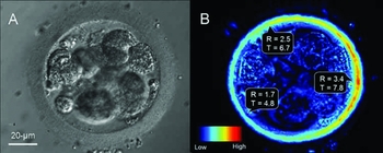
To image the ZP for subsequent quantitative analysis, each sample (oocyte or embryo) was placed in a 5-μl microdroplet of HEPES-buffered human tubal fluid (Modified HTF-HEPES; Irvine Scientific) containing 0.5% HSA and overlaid with mineral oil in a glass-bottomed Petri dish (Willco; World Precision Instruments) at 37 °C. The samples were observed at a magnification of × 400 using an inverted microscope (IX-71; Olympus) equipped with the Oosight™ imaging system (Cambridge Research & Instrumentation), consisting of a circular polarizer and interference filter and a personal computer running software for intuitive imaging and analysis. The values for retardance (R) and thickness (T) were automatically recorded and measured by the Zona Finder™ tool or the line-scanning tool in the analysis software (Fig. 1). The mean values for R and T were calculated for a single section of the entire ZP, unless specific site(s) of the ZP were selected for measurement.
Bright field image (A) and birefringence image (B) of human zona pellucida under a polarized light microscope. R: retardance (nm), T: thickness (μm). The inner half area of the zona appears to have the higher retardance value, compared with the outer half area. Within a zona pellucida, R and T values are variable.

Experimental design
Experiment 1: Correlations of birefringence parameters (R, T and R × T) with chemical hardness of ZP were investigated using a traditional chemical approach. Briefly, developmentally arrested embryos (n = 12), which had not been transferred to patients in their ICSI cycles, were exposed to 20-μl microdrops of phosphate-buffered saline containing 0.1% (w/v) protease (Sigma) and covered with mineral oil at 37 °C. The samples were sequentially observed under a magnification of × 400 until the ZP was no longer visible. The hardness of the ZP was defined as the time required for the protease to complete the digestion process.
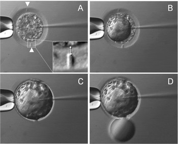
Experiment 2: Correlations of birefringence parameters (R, T and R × T) with physical hardness of the ZP were investigated in a model experiment for sham hatching (Fig. 2). First, ZP from IVF-derived and developmentally arrested embryos (n = 11) were drilled at two different sites using a laser system (ZILOS-tk™), which caused a decrease in birefringence parameter values (Fig. 2(A)). The mechanical puncture of the ZP was expected to occur at one of the two drilled sites after full expansion by increasing the internal pressure (Fig. 2(B)). Without these drilled sites, accurate estimation of birefringence parameters at the punctured site of the ZP would be impossible because the birefringence profile of each ZP is heterogeneous (Fig. 1(B)), and puncture of the ZP occurs at random sites that are not always within the focused field. The internal pressure was increased by microinjection with Fluorinert™ electronic fluid (Sumitomo 3M) using a pipette, with an outer diameter of <1 μm, connected to a piezo-driven micromanipulator (PMM-150FU; PrimeTech). Following penetration of the ZP by several piezo-pulses (speed 3, intensity 3) and gentle injection of the Fluorinert™ fluid, the ZP was extended maximally (Fig. 2(C)) and finally punctured (Fig. 2(D)). The hardness of the ZP was defined as the maximum expansion rate: the percentage of increase of the inner diameter of the ZP compared with the original diameter before injection.
A model experiment to investigate correlation between birefringence parameter and tolerance of zona pellucida to physical expansion. Each zona carries two partially laser-drilled sites (arrowheads) where values for retardance and thickness can be measured ( ) (A). As Fluorinert™ electronic fluid is microinjected (B), the zona expands (C) and finally punctures at either one of the two drilled sites (D).

Experiment 3: Changes in a birefringence parameter (R × T) due to fertilization or hatching were investigated using viable oocyte/embryo samples; no samples were observed more than once. To compare the fertilization-dependent change in the R × T values, denuded oocytes at metaphase II stage that were prepared for ICSI (n = 23) and ICSI- or IVF-derived embryos that were confirmed to be fertilized normally by the presence of two pronuclei (n = 29 or n = 20, respectively) were used. The birefringence of the embryos was observed before thinning of ZP was initiated. To compare the hatching-dependent changes in the R × T values, the following stages were used: morulae to early blastocysts (n = 9), expanding blastocysts (n = 4), and hatched blastocysts (n = 4). These embryos had been removed from clinical use because of their poor quality. Artificial shrinkage of the blastocoel cavity in expanding blastocysts was induced to collect the accurate data for birefringence parameters because the presence of trophectoderm cells in contact with the ZP affects quantitative analysis.
Statistical analysis
All measurements were performed by the same technician. Pearson's correlation coefficient (r) was calculated for relationships between birefringence parameters of ZP texture (R, T and R × T) and its hardness parameters for the ZP (enzymatic digestion time and maximum expansion rate). Changes in R × T values of its ZP among unfertilized oocytes and fertilized embryos by IVF or ICSI, and among unexpanded, expanding and hatched embryos were analysed by two-tailed Student's t-test. A p-value < 0.05 was considered statistically significant.
Results
Experiment 1
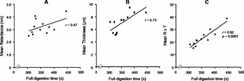
Correlations between birefringence parameters (R, T and R × T) of ZP texture and full digestion time by protease are shown in Fig. 3(A), (B) and (C), respectively. The time required for protease to solubilize the ZP ranged from 305–447 s, and it significantly correlated with the mean R × T values. However, no significant correlation between digestion times and mean R or T values was observed.
Correlations of retardance (A), thickness (B) and retardance × thickness (C) of zona pellucida with the time required for full enzymatic digestion in 0.1% protease solution.

Experiment 2
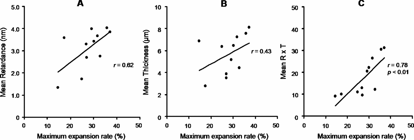
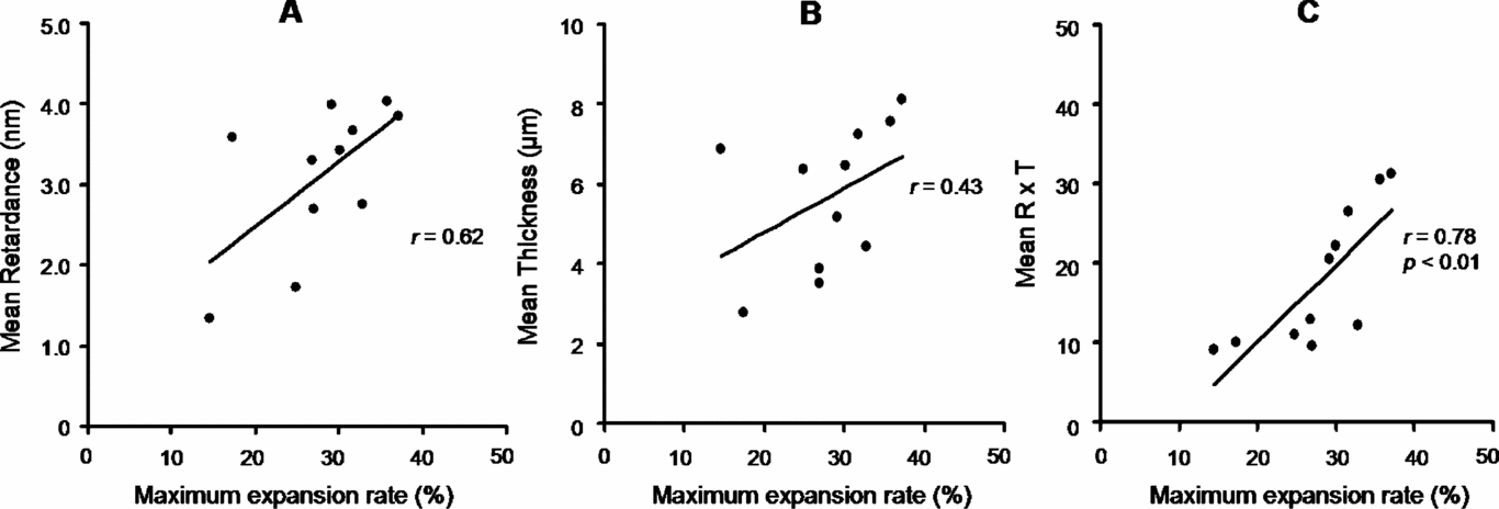
Correlations between birefringence parameters (R, T and R × T) in the punctured site of ZP by sham-hatching treatment and its maximum expansion rate are shown in Fig. 4(A), (B) and (C) respectively. The maximum expansion rate of the ZP ranged from 15% to 38%, which significantly correlated with the mean R × T values. No significant correlation was observed between the maximum expansion rate and mean R or T values, as in Experiment 1.
Correlations of retardance (A), thickness (B) and retardance × thickness (C) at the punctured drilled-sites with the maximum expansion rate of the zona pellucida induced by Fluorinert™ fluid microinjection.
Experiment 3
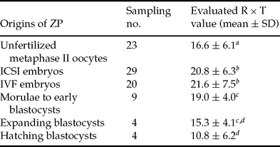
The fertilization- or hatching-dependent change in the birefringence parameter (R × T) is shown in Table 1. The R × T values of the ZP in IVF- and ICSI-derived embryos were significantly higher than those values found in unfertilized metaphase II oocytes. In addition, the R × T values after in vitro hatching of viable blastocysts significantly decreased compared with those of unexpanded morula and early blastocysts, while those of the expanding blastocysts had intermediate values.
Effect of fertilization by ICSI and IVF or in vitro hatching on the birefringence profile of human zona pellucida (ZP).
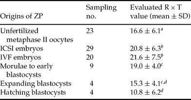
a vs. b; c vs. dDifferent superscripts indicate significant difference at p < 0.05.
Discussion
A non-invasive birefringence parameter, R × T, of human ZP correlated significantly with its ZP hardness, which was estimated chemically by the times required for protease to solubilize ZP (Fig. 3) and physically by the maximum expansion rate of the ZP after microinjection of inactive fluid (Fig. 4). The Oosight™ imaging system employed here presents an alternative method for assaying ZP hardness. Furthermore, it is more preferable than the protease-based chemical ZP hardness assay because it overcomes the disadvantage of the chemical assay – the irreversibility of the dissolved ZP. The Oosight™ imaging system can also be used as an alternative to some other physical methods that have been proposed for the measurement of ZP hardness (Drobnis et al., Reference Drobnis, Andrew and Katz1988; Sun, Reference Sun, Wan, Roberts, Bischof and Nelson2003; Murayama et al., Reference Murayama, Mizuno, Kamakura, Fueta, Nakamura, Akaishi, Anzai, Watanabe, Inui and Omata2006; Wacogne et al., Reference Wacogne, Pieralli, Roux and Gharbi2008; Khalilian et al., Reference Khalilian, Navidbakhsh, Valojerdi, Chizari and Yazdi2010), all of which require special equipment or instruments or are technically difficult to perform. To date, without examining correlations with results in previously used assays for ZP hardness, the birefringence parameters have been used to indicate that ZP birefringence increases with a woman's age and prolonged period of in vitro culture (Kilani et al., Reference Kilani, Cooke, Kan and Chapman2006). It has also been reported that the ZP birefringence failed to detect any changes due to cryopreservation of human embryos (Gu et al., Reference Gu, Lu, Lin and Lu2010) as a part of a controversial discussion on the incidence of the change (Matson et al., Reference Matson, Graefling, Junk, Yovich and Edirisinghe1997; Vanderzwalmen et al., Reference Vanderzwalmen, Bertin, Debauche, Standaert, Bollen, Roosendaal, Vandervorst, Schoysman and Zech2003).
The mean R × T values in IVF- and ICSI-derived embryos were significantly higher than those in unfertilized metaphase II oocytes (Table 1), suggesting that hardening of human ZP can be detected by the Oosight™ imaging system. Furthermore, it is suggested that the mechanical stimulus of sperm injection during an ICSI regimen as well as spontaneous sperm penetration during an IVF regimen can induce CG reaction-dependent changes in ZP hardness. We found no difference in the mean R × T values between IVF- and ICSI-derived embryos, which is in accordance with the results from a protease-based chemical ZP hardness assay (digestion time; 48.9 ± 2.7 and 45.3 ± 3.4 min, respectively) reported by Manna et al. (Reference Manna, Rienzi, Greco, Sbracia, Rahman, Poverini, Siracusa and De Felici2001). As the ZP birefringence of fertilized zygotes was not always observed at the time of the two pronuclei confirmation (the latest observation at day 6), the possible effect of a prolonged culture period on ZP hardening, if any, cannot be ruled out and needs to be further investigated. The protease-based chemical ZP hardness, the most commonly used index for ZP hardness, has been used to indicate the CG reaction-dependent ZP hardening induced by spontaneous fertilization (Schiewe et al., Reference Schiewe, Araujo, Asch and Balmaceda1995; Manna et al., Reference Manna, Rienzi, Greco, Sbracia, Rahman, Poverini, Siracusa and De Felici2001) and oocyte cryopreservation (Matson et al., Reference Matson, Graefling, Junk, Yovich and Edirisinghe1997; Larman et al., Reference Larman, Sheehan and Gardner2006). Some investigators measuring physical ZP hardness have also reported the ZP hardening due to fertilization (Drobnis et al., Reference Drobnis, Andrew and Katz1988; Sun, Reference Sun, Wan, Roberts, Bischof and Nelson2003; Murayama et al., Reference Murayama, Mizuno, Kamakura, Fueta, Nakamura, Akaishi, Anzai, Watanabe, Inui and Omata2006; Wacogne et al., Reference Wacogne, Pieralli, Roux and Gharbi2008; Khalilian et al., Reference Khalilian, Navidbakhsh, Valojerdi, Chizari and Yazdi2010).
In the present study (Experiment 2), the maximum expansion rate was used as a parameter for physical ZP hardness under a sham-hatching model by microinjection of Fluorinert™ electronic fluid. Two sites were preliminarily drilled in each ZP (located at the 0 and 6 o'clock positions; Fig. 2(A)) to observe the punctured site on a focused field. As these drills are likely to be a sort of assisted hatching (AH) treatment (Mantoudis et al., Reference Mantoudis, Podsiadly, Gorgy, Venkat and Craft2001), puncturing of ZP in sham-hatching treatment occurred earlier than that in microinjected intact ZP without laser drill sites or the ZP of viable blastocysts (data not shown), but the one-to-one correlation between ZP birefringence of the punctured site and the maximum expansion rate of ZP can be identified. Thus far, Yang's modules, either by mechanical aspiration treatment (Drobnis et al., Reference Drobnis, Andrew and Katz1988; Khalilian et al., Reference Khalilian, Navidbakhsh, Valojerdi, Chizari and Yazdi2010) or by micro-electromechanical system-based cellular force sensors (Murayama et al., Reference Murayama, Mizuno, Kamakura, Fueta, Nakamura, Akaishi, Anzai, Watanabe, Inui and Omata2006; Wacogne et al., Reference Wacogne, Pieralli, Roux and Gharbi2008), have been estimated as measures for the mechanical behaviour of ZP. A significant decrease in R × T value between unexpanded and hatched embryos (Table 1) suggested that hardness of human ZP may be irreversibly affected by embryo-derived proteolytic activity and/or mechanical force with expansion of the blastocoel cavity. The birefringence parameter that was supported by high correlations with chemical and physical ZP hardness would be predictive for occurrence of in vitro hatching, and therefore provides a suggestion for application of AH treatment to the ZP. AH treatment can be applied to the location with a low R × T value within a single ZP, in addition to applying the following selection criteria: patients who are ≥38 years of age or those with an elevated FSH level or embryos with ≥15 μm ZP thickness or those with a high degree of fragmented cells (Cohen et al., Reference Cohen, Elsner, Kort, Malter, Massey, Mayer and Wiemer1990, Reference Cohen, Alikani, Trowbridge and Rosenwaks1992).
In conclusion, hardness of human ZP can be estimated non-invasively by birefringence-based microscopic observation with a high correlation to estimates determined by enzymatic digestion time and maximum expansion rate. Increase in ZP hardness by IVF and ICSI was detected and kinetics of the birefringence parameter around the hatching can provide useful information of ZP for AH treatment.
Acknowledgements
The authors wish to thank Ms Mako Korekane and Mr Takao Hara (Yamashita Ladies’ Clinic, Hyogo, Japan) for their technical assistance.