
Introduction
Avian haemosporidians (Apicomplexa: Haemosporida) belonging to the genera Plasmodium, Haemoproteus and Leucocytozoon are diverse blood parasites affecting birds worldwide (Valkiūnas, Reference Valkiūnas2005). Bird hosts infected with avian haemosporidians can suffer from anaemia, weight loss and sickness (Merino et al., Reference Merino, Moreno, José Sanz and Arriero2000; Palinauskas et al., Reference Palinauskas, Valkiūnas, Bolshakov and Bensch2008, Reference Palinauskas, Žiegytė, Šengaut and Bernotienė2018) and be subject to high mortality rates in susceptible populations. Avian infections can be common, with many bird individuals tolerating chronic infections for life after surviving the acute malaria phase (Asghar et al., Reference Asghar, Hasselquist and Bensch2011). Such infections can have significant effects on life history traits and may act as selective agents in wild bird populations (Asghar et al., Reference Asghar, Hasselquist and Bensch2011).
Avian haemosporidians spend part of their life cycles in bird hosts and dipteran vectors, with highly interdependent ecological relationships. Therefore, accurate detection of avian haemosporidians is important in addressing questions regarding host–vector–parasite interactions, ecology and evolution (Santiago-Alarcon et al., Reference Santiago-Alarcon, Palinauskas and Schaefer2012; Sehgal, Reference Sehgal2015; Pacheco et al., Reference Pacheco, Cepeda, Bernotienė, Lotta, Matta, Valkiūnas and Escalante2018a). Moreover, accurate detection is also needed for understanding the epidemiology of infections and developing disease and vector control strategies, especially in places where avian conservation is of concern.
The detection of blood parasites can be challenging. Accurate identification of avian haemosporidians from blood smears is difficult as it requires a high level of taxonomic expertise to differentiate minute characters across taxa while accounting for the range of morphological features resulting from various developmental stages (Valkiūnas et al., Reference Valkiūnas, Kazlauskienė, Bernotienė, Bukauskaitė, Palinauskas and Iezhova2014).
Since the 2000s, molecular techniques have been widely used in the detection and characterization of avian blood parasites, including a widely used nested polymerase chain reaction (PCR) protocol to amplify a fragment of the mitochondrial cytochrome b (cytb) gene (Bensch et al., Reference Bensch, Stjernman, Hasselquist, Örjan, Hannson, Westerdahl and Pinheiro2000; Hellgren et al., Reference Hellgren, Waldenström and Bensch2004). The amplified products are subsequently sequenced via the traditional Sanger approach and are used as the barcode for lineage identification by matching identified sequences available in the public MalAvi database (Bensch et al., Reference Bensch, Hellgren and Pérez-Tris2009). A recently published protocol incorporates the use of three different primer sets targeting Haemoproteus, Plasmodium and Leucocytozoon, respectively, in a multiplex PCR (Ciloglu et al., Reference Ciloglu, Ellis, Bernotienė, Valkiūnas and Bensch2019). The resulting amplicons yield products of different size for each genus, allowing for rapid genus-level identification. However, the current repertoire of molecular methods does not allow for haemosporidian lineage identification in the amplified products from bird individuals co-infected by multiple parasite lineages belonging to the same genus. In capillary sequencing outputs, amplified genes from co-infected individuals often show up as jumbled chromatograms, sometimes with double peaks (‘double base calling’) (Marzal et al., Reference Marzal, Bensch, Reviriego, Balbontin and De Lope2008), making it virtually impossible to distinguish blood parasite strains apart.
As it is difficult to recover fully from parasitic infections, and considering the relatively long lifespan of birds, co-infections with multiple blood parasite strains are thought to be common in wild birds, although rarely detected (Valkiūnas, Reference Valkiūnas2005; Bensch et al., Reference Bensch, Waldenström, Jonzén, Westerdahl, Hansson, Sejberg and Hasselquist2007). Crucially, co-infections by multiple parasites of the same haemosporidian genus may be more common than multi-generic mixed infections (Silva-Iturriza et al., Reference Silva-Iturriza, Ketmaier and Tiedemann2012). In cases where multiple blood parasite infections are suspected due to the presence of double peaks, the fragments are usually subjected to an additional tedious cloning step and separately sequenced (Pérez-Tris and Bensch, Reference Pérez-Tris and Bensch2005; Silva-Iturriza et al., Reference Silva-Iturriza, Ketmaier and Tiedemann2012). The presence of co-infections can also be ascertained by examining blood smear slides of the affected bird hosts (Valkiūnas et al., Reference Valkiūnas, Kazlauskienė, Bernotienė, Bukauskaitė, Palinauskas and Iezhova2014), although many co-infections are overlooked this way (Xuan et al., Reference Xuan, Kaewlamun, Saiwichai, Thanee, Poofery, Tiawsirisup, Channumsin and Kaewthamasorn2021). Additionally, if blood samples are stored exclusively in buffers for molecular study, verification of co-infected samples via microscopy is no longer possible as blood smear slides cannot be prepared.
With the advance of next-generation sequencing (NGS), high-throughput sequencing of heterogeneous barcodes can be carried out simultaneously at a fraction of the cost of Sanger sequencing (Meier et al., Reference Meier, Wong, Srivathsan and Foo2016; Yeo et al., Reference Yeo, Srivathsan and Meier2020). Employing NGS methods in the avian haemosporidian screening process provide an added advantage of retrieving multiple parasite lineages in co-infected birds. Even so, to the best of our knowledge, NGS has not been applied to avian haemosporidian detection and identification.
In the present study, a PCR assay and workflow suitable for avian haemosporidian screening on an NGS Illumina platform was developed. The detection rates of this assay were compared against detections using the first primer pair (HaemNF1 and HaemNR3) of the widely used nested PCR protocol (Hellgren et al., Reference Hellgren, Waldenström and Bensch2004), and sequence similarities of the cytb barcodes recovered from both methods were evaluated, allowing for an assessment of the reliability of the NGS method in detecting co-infections. The potential implications of previous practices of exclusively relying on Sanger sequencing were discussed, followed by a critical evaluation of the accuracy of using short NGS barcodes as compared to longer Sanger reads in avian haemosporidian identification.
Materials and methods
Sample selection
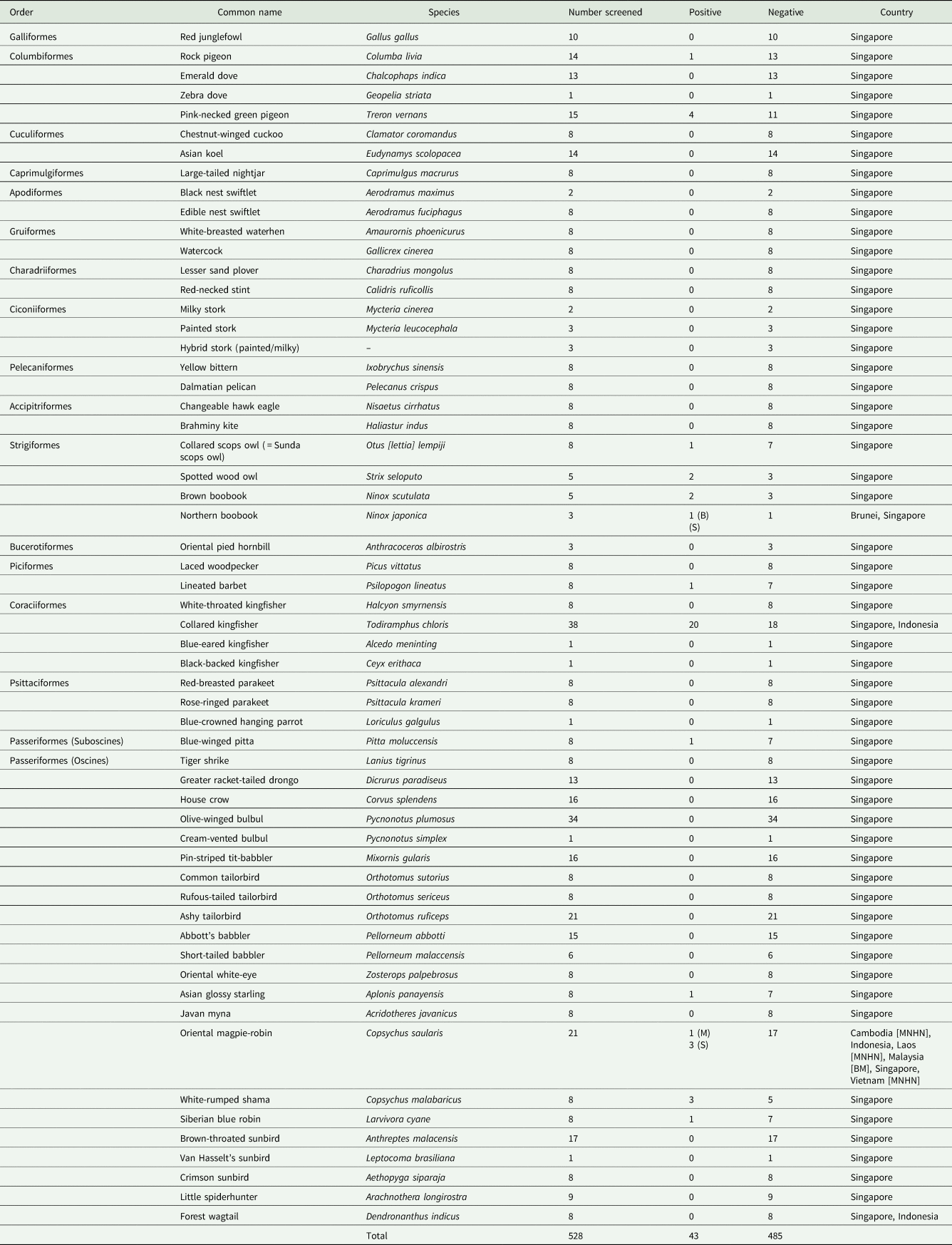
Since 2013, collections of avian blood and tissue (pectoral muscle) samples have been ongoing in Singapore, and a substantial collection has been amassed. Blood samples have mostly been obtained from mist netting at various locations under requisite permit, while tissue samples have been obtained through subsampling bird carcasses submitted to our laboratory by the public or by various agencies and organizations (see Acknowledgements for details). These bird carcasses are exclusively the result of window strikes or death by natural causes. Tissue samples were included in the screening as they have been found to be as reliable as blood samples in the detection of avian haemosporidians (Fecchio et al., Reference Fecchio, Collins, Bell, García-Trejo, Sánchez-González, Dispoto, Rice and Weckstein2019). In addition to the Singaporean samples, a smaller number of blood samples from Brunei (Sadanandan et al., Reference Sadanandan, Tan, Schjølberg, Round and Rheindt2015), Cambodia, Laos, Indonesia (Rheindt et al., Reference Rheindt, Gwee, Baveja, Ferasyi, Nurza, Rosa and Haminuddin2020), Malaysia and Vietnam – all collected for previous projects – were also used (Appendix 1; see Acknowledgements for lending institutions). All samples used in this study were preserved directly in molecular-grade absolute ethanol and stored at −20°C. To ensure screening of a wide range of species for avian haemosporidians, we selected birds from across various orders for which there was a minimum of eight unique individual samples available, making exceptions for rare species or species of particular interest. A total of 528 individuals representing 58 bird species across 17 orders were screened (Appendix 1).
Primer design
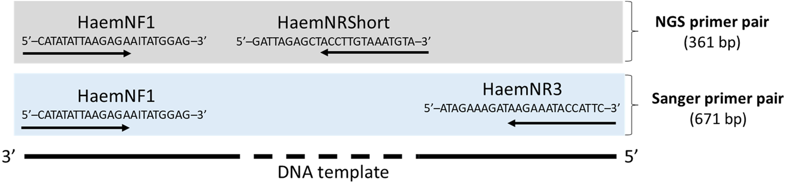
To enable the detection and sequencing of multiple avian haemosporidian strains in a co-infected blood sample via NGS, a reverse primer was designed (HaemNRShort, 5′-GATTAGAGCTACCTTGTAAATGTA-3′). To do so, 114 cytb sequences across a wide range of Haemoproteus, Plasmodium, Leucocytozoon and Hepatocystis strains (Pacheco et al., Reference Pacheco, Matta, Valkiūnas, Parker, Mello, Stanley, Lentino, Garcia-Amado, Cranfield, Pond and Escalante2018b) were downloaded and a sufficiently conserved region was targeted. The Oligonucleotide Properties Calculator (http://biotools.nubic.northwestern.edu/OligoCalc.html) was used to ensure that the designed primer satisfies standard requirements such as optimal primer length, GC content and lack of potential hairpin formation. The primer sequence was then checked against cytb sequences of avian species to preclude cross-amplification. This new primer, in combination with a widely used published forward primer (HaemNF1; Hellgren et al., Reference Hellgren, Waldenström and Bensch2004), produces a cytb barcode fragment of 367 base pairs, which is suitable for sequencing on an Illumina NGS platform (Fig. 1). In order to sequence the barcodes on an NGS platform and demultiplex them successfully in the bioinformatic stage, a one-step PCR reaction with unique primer-tag combinations following Meier et al. (Reference Meier, Wong, Srivathsan and Foo2016) was implemented.
Schematic illustration of the directions and combinations of the two different primer pairs used for mitochondrial cytochrome b gene amplification in this study. The next-generation sequencing (NGS) primer pair consists of the forward primer HaemNF1 and the newly designed reverse primer HaemNRShort. The Sanger primer pair (HaemNF1 and HaemNR3) was designed by Hellgren et al. (Reference Hellgren, Waldenström and Bensch2004).

Molecular work
DNA extractions were carried out with the DNeasy® Blood & Tissue Kit (Qiagen, GmbH, Hilden, Germany) to obtain genomic DNA from the avian samples. To screen and detect avian haemosporidians using the traditional approach, PCR was carried out. Positive and negative controls were included in every batch of PCR. The reaction volumes of 25.0 μL were set up as follows: 2.5 μL 10× Taq Buffer with (NH4)2SO4, 2 mm MgCl2, 0.2 mm dNTP Mix, 0.0002 mm of each primer (Sanger primer pair), 0.625 units of DreamTaq DNA polymerase, at least 5 ng of DNA template and sterilized millipore water to make up the volume. An initial denaturation step (94°C, 3 min), 30 cycles of denaturation (94°C, 30 s), annealing (50°C, 40 s) and extension (72°C, 1 min) and a final extension step (72°C, 10 min) were carried out using the Mastercycler nexus gradient (Eppendorf, Hamburg, Germany). To ensure repeatability and to ascertain if there were any false-negative or false-positive results, triplicate screening with the Sanger primer pair was conducted for 40 randomly selected samples (including samples without avian haemosporidian infections). Subsequently, amplification with the NGS primer pair was carried out for the samples which had tested positive for avian haemosporidians on the basis of the Sanger primer pair. Tests for haemosporidian infection were considered positive when a band was seen in a 2% agarose gel after visualization with a UV transilluminator (Syngene, Synoptics Limited, Cambridge, UK).
Samples amplified with the Sanger primer pair were cleaned up using ExoSAP-IT® and cycle-sequenced with the BigDye® Terminator v3.1 Cycle Sequencing Kit (Applied Biosystems Inc., Foster City, CA, USA). Sequencing was carried out on an Applied Biosystems 3130XL Genetic Analyzer. Samples amplified with the NGS primer pair were pooled in equimolar proportions and sequenced on an Illumina MiSeq 300 bp paired-end platform (Novogene, Beijing, China).
Bioinformatics
Sanger sequences were examined and edited using CodonCode™ Aligner version 8.0.2 (LI-COR, Inc., Lincoln, NE, USA) for ends trimming, resolution of ambiguous bases and to assemble contigs from forward and reverse strands. Double peaks (double base calling) in the sequences were scored as such when they occurred in both the forward and reverse strand chromatograms at the same base pair positions, and were replaced with corresponding ambiguity codes.
Quality checks for the Illumina raw read files were carried out using FastQC (Andrews, Reference Andrews2010), and adaptors were removed using Cutadapt (Martin, Reference Martin2011). Demultiplexing and subsequent steps such as dereplication, denoising and taxonomic assignment were carried out using the OBITools suite which has been optimized for metabarcoding (Boyer et al., Reference Boyer, Mercier, Bonin, Le Bras, Taberlet and Coissac2016). The final output files contained unique sequences obtained from each sample with their respective counts.
To avoid incorporating possible contamination in downstream analysis, only haemosporidian sequences of the top two highest counts (referred to as dominant and subdominant sequences subsequently) were analysed for each bird individual. A ratio of subdominant sequence count over dominant sequence count was calculated across all the positively infected bird samples. Samples with low coverage (dominant count lower than 1000) were excluded. The resulting cytb sequences were aligned using MAFFT version 7 (Katoh and Standley, Reference Katoh and Standley2013). To determine if sequences obtained from both Sanger and NGS platforms match, they were visualized in AliView (Larsson, Reference Larsson2014) and bases with ambiguity codes within the Sanger sequences were cross-examined against NGS sequences at the same base positions.
To check for the presence of pseudogenes, all cytb sequences were translated into amino acid sequences and the reading frames were permutated to check for the presence of stop codons in AliView. To assess sequence divergence, pairwise p-distances between the Sanger and the dominant and subdominant NGS sequences, respectively, were calculated with MEGA11 (Tamura et al., Reference Tamura, Stecher and Kumar2021).
Comparing the performance of short and long cytb sequences in lineage identification
To compare the accuracy of lineage identification with short NGS sequences (~330 bp), all Sanger and NGS cytb sequences generated were matched against curated sequences from the global avian haemosporidian MalAvi database (Bensch et al., Reference Bensch, Hellgren and Pérez-Tris2009). Among the NGS sequences, only dominant barcodes were included in the analysis, except in cases where the subdominant NGS sequence matched the Sanger sequence (D2179, D2003 and F1102). From here onwards, we referred to them as ‘homologous NGS sequences’. The Basic Local Alignment Search Tool (BLAST) v2.12 (Altschul et al., Reference Altschul, Gish, Miller, Myers and Lipman1990) was used at an expect value (e-value) cut-off of 1 × 10⁻⁵. A comparison of the top identity match(es) (%), sequence length and number of mismatches between the NGS and Sanger sequences was carried out. If possible, species names were assigned to perfect matches when such taxonomic information was available in MalAvi. In most other cases, the cytb barcodes were assigned the so-called lineage names. Lineages on the MalAvi database are defined based on the currently implemented ‘one base pair rule’ method, whereby a single base pair difference in the cytb barcode is interpreted as species-level differentiation (Galen et al., Reference Galen, Nunes, Sweet and Perkins2018). Such practice may lead to an overestimation of avian haemosporidian diversity (Hellgren et al., Reference Hellgren, Atkinson, Bensch, Albayrak, Dimitrov, Ewen, Kim, Lima, Martin, Palinauskas, Ricklefs, Sehgal, Valkiūnas, Tsuda and Marzal2014; Palinauskas et al., Reference Palinauskas, Bernotienė, Žiegytė, Bensch and Valkiūnas2017; Galen et al., Reference Galen, Nunes, Sweet and Perkins2018). Therefore, a more conservative approach was taken in this case, considering variations of up to two base pair differences as the same lineage (Martinsen et al., Reference Martinsen, Paperna and Schall2006; Ricklefs et al., Reference Ricklefs, Outlaw, Svensson-Coelho, Medeiros, Ellis and Latta2014).
Results
Avian haemosporidian detection with Sanger primer pair
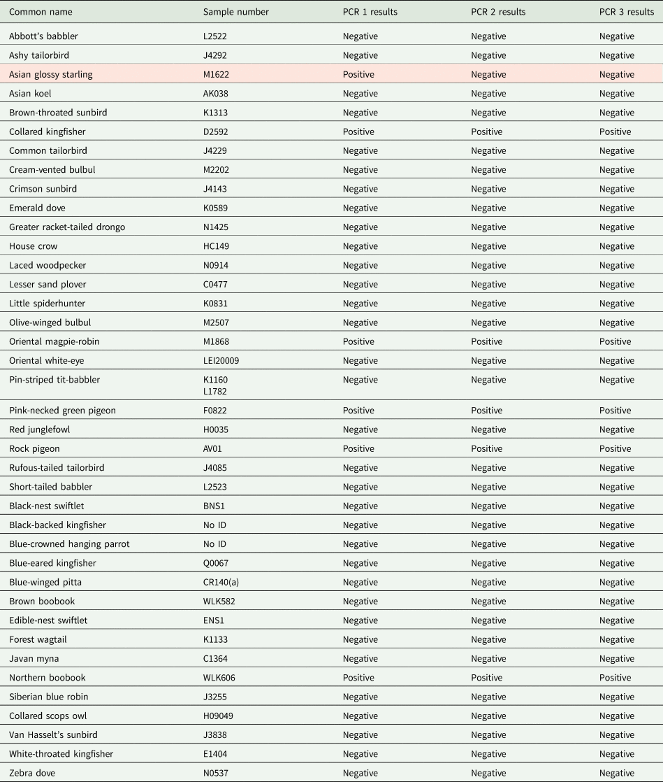
Out of 528 bird samples screened, a total of 43 were positive for avian haemosporidians (8.14%) on the basis of gel electrophoresis. All PCR triplicates of 40 samples that had been randomly selected from a total of 528 samples produced consistent results, with the exception of one sample (M1622) (97.5%) (Appendix 2). This outcome indicated that screening is highly repeatable, with a low incidence of false positives or negatives. Out of the 43 positive samples, two were not sequenced successfully (L2344 and L2339) on the Sanger sequencing platform as the chromatograms were jumbled.
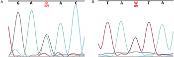
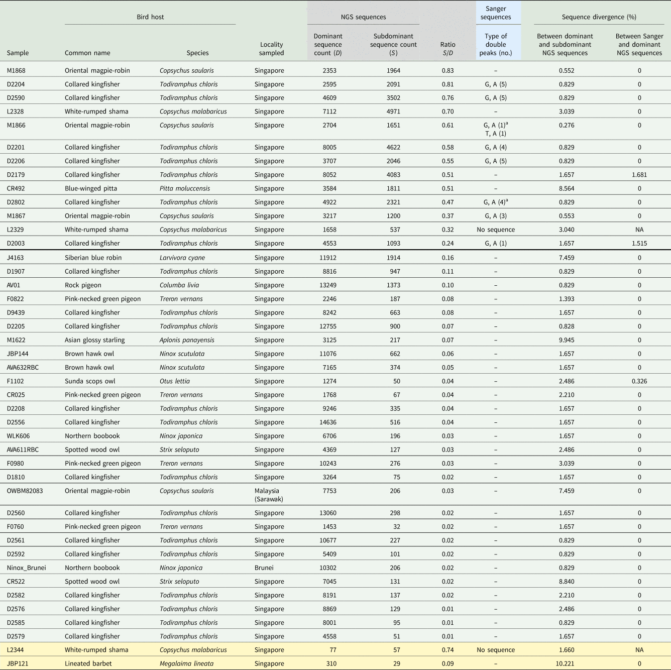
Examination of the Sanger chromatograms of successfully sequenced individuals revealed that eight samples (19.0%) contained double peaks in their cytb chromatograms (Fig. 2, Table 1), indicating the likely presence of co-infecting avian haemosporidian strains. Among them, A/G type double peaks were the most common (Table 1).
Two examples of double peaks detected in the chromatograms of the Sanger sequences of a single individual. (A) An A/G (green and black) double peak at position 309 and (B) an A/T (green and red) double peak at position 89.

Summary of 43 blood samples testing positive for avian haemosporidian infection on the basis of gel electrophoresis, including details on cytochrome b sequences obtained from next-generation sequencing (NGS) (grey) and from Sanger sequencing (blue)

For NGS data, counts are shown for dominant (D) and subdominant (S) sequence counts only. Samples are ordered according to their count ratio, as calculated by dividing subdominant sequence counts over dominant sequence counts (S/D). The thick black line indicates the 0.2 ratio threshold used, with samples above the line considered to be co-infected. Sequence divergences were calculated using the p-distance method. Samples highlighted in yellow were excluded from downstream analyses owing to low sequence coverage (<1000× in dominant NGS sequence count).
a Sanger double peaks in which one out of the two called bases does not match the dominant or subdominant NGS sequences.
Avian haemosporidian detection with NGS primer pair
The newly developed NGS primer was successful in the detection of avian blood parasites: all samples testing positive with the Sanger primer set were successfully amplified with the NGS primer set as well. Two samples (L2344 and JBP121; Table 1) were excluded from downstream analysis due to low coverage (<1000 times coverage for the dominant sequence).
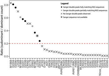
The cytb sequences obtained from the Sanger method always matched with either the dominant or subdominant NGS sequences, even in cases where the Sanger double peak is not reflected fully in the NGS sequences. In most cases (92.7%), the Sanger sequences matched with the dominant NGS sequence (Fig. 3; Table 1, last column). No stop codons were found in all the sequences.
Descending ratio of subdominant next-generation sequence (NGS) counts over dominant NGS counts, with information on the presence or absence of congruence between NGS and Sanger sequences. A total of 41 samples which were successfully amplified (showing up as positive in gel electrophoresis) and which exhibited >1000× in the dominant NGS sequence count are shown here. The red dotted horizontal line indicates the 0.2 ratio threshold used, and samples above the line are considered co-infected.

Detection of co-infections using both methods
Based on the comparison of Sanger and NGS-based detection patterns, we considered a subdominant to dominant NGS sequence ratio of 0.2 to be the lower limit at which haemosporidian co-infections can be conclusively diagnosed (Fig. 3). For samples with ratios below 0.2, we only considered their dominant sequences as representative of the infecting haemosporidian in order to avoid incorporating contaminant sequences in downstream analysis. A total of 13 samples were found to be co-infected using this NGS-based threshold (Fig. 3, Table 1). Among these samples, eight exhibited double peaks in the Sanger chromatograms. Four samples did not show signs of double peaks and the one remaining sample exhibited a jumbled, illegible trace chromatogram (L2339). The ambiguity codes of Sanger double peaks of two samples did not fully match the dominant and subdominant NGS sequences at the same base positions (Fig. 3, Table 1). For example, in sample M1866, a Sanger double peak of A/G was detected, but both dominant and subdominant NGS sequences only reflected G at that base position.
Among the eight co-infected samples containing double peaks in the chromatograms, a large majority (7) exhibited a signature of mixed infections involving highly similar haemosporidian lineages based on the detection of p-distance sequence divergences of <1% between the dominant and subdominant NGS sequences. In contrast, the haemosporidian lineages involved in the remaining five mixed infections (detected through the NGS method alone) were less similar.
Comparing lineage identification performance between short NGS and longer Sanger reads
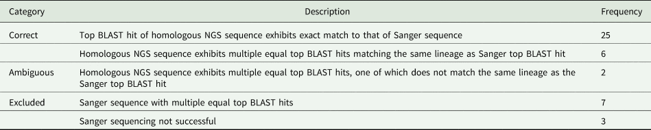
Samples which were not successfully Sanger or NGS sequenced (3) were excluded from this analysis. Similarly, cases in which a Sanger sequence produced multiple equal top BLAST hits to the MalAvi database (7) were also excluded. An overwhelming proportion of homologous NGS cytb barcodes exhibited an identical match to those of the Sanger barcodes (93.93%) (Table 2). Only in two instances, having a longer Sanger sequence improved the accuracy of lineage identification whereas the shorter homologous NGS sequences produced ambiguous BLAST matches.
A comparison of the Basic Local Alignment Search Tool (BLAST) results of short NGS and longer Sanger barcodes when matched against the global avian haemosporidian (MalAvi) database

Frequency refers to the number of samples matching the description.
Discussion
The detection and lineage identification of avian haemosporidians has long been challenging, even more so in cases of mixed infections. Mixed infections may not always manifest themselves as ‘double peaks’ in Sanger chromatograms and are reported to be highly underestimated (Valkiūnas et al., Reference Valkiūnas, Kazlauskienė, Bernotienė, Bukauskaitė, Palinauskas and Iezhova2014). In this study, a PCR assay suitable for use with an NGS platform (Illumina) was developed and a metabarcoding workflow was employed to allow for the detection of multiple avian haemosporidian lineages within individual birds. The NGS workflow reflected the same number of infections as the Sanger workflow, but detected 62.5% more cases of co-infections based on the 0.2 threshold applied to subdominant over dominant sequence count ratios.
The 0.2 threshold applied here is based on a conservative evaluation of subdominant to dominant NGS sequence count ratios (Fig. 3). Among the samples which exhibited double peaks in the Sanger chromatograms, the lowest subdominant to dominant NGS sequence count ratio was 0.24. Naturally, applying a cautious threshold of 0.2 might mean missing out on mixed infections where the parasitaemia of the secondary (or even tertiary) co-infection is substantially lower than the primary infection. In fact, the detection of multiple infections with a ratio of less than 0.2 which had a match between Sanger sequence and subdominant sequence points to such instances of co-infection (Table 1). The detection of such mixed infections is challenging because it is difficult to differentiate low secondary parasitaemia from background sequence contamination. Unfortunately, as the samples used here were stored directly in molecular-grade ethanol, we were unable to determine the level of parasitaemia in the infected individuals on the basis of blood smear slides. Hence, our identification of 13 co-infections among 43 total infections must be considered a conservative estimate.
With the exception of one case (D2003) in which double peaks were observed in the Sanger chromatograms, the sequence divergences of the dominant and subdominant sequences in co-infected cases were less than 1% (Table 1). In contrast, the majority of co-infections that were identified through NGS sequencing alone involved species with less similar sequence profiles (>1.5% p-distance). This pattern suggests that mixed infections involving dissimilar haemosporidian lineages are more likely to be overlooked when Sanger PCR assays are used, or are discarded as uninterpretable mixed traces (Tenney et al., Reference Tenney, Wu, Langton, Klueh, Quatrano and Brent2007; Dmitriev and Rakitov, Reference Dmitriev and Rakitov2008).
The newly developed NGS assay based on Illumina short reads yields shorter fragments (330+ bp) compared to the widely used Sanger primer pair which produces cytb fragments of around 470 bp, with unknown effects on lineage identification accuracy. In the present study, the NGS barcodes largely converged on the same haemosporidian species or lineages when compared to Sanger barcodes. Shorter barcodes may sometimes produce ambiguous identifications (multiple equal matches) in instances where longer Sanger barcodes are able to yield a more precise lineage identification, although this occurred rarely (6.06%) in the present study. Shorter barcodes have been shown to exhibit a higher amplification success rate across a wide range of animal and plant taxa (Meusnier et al., Reference Meusnier, Singer, Landry, Hickey, Hebert and Mehrdad2008; Françoso and Arias, Reference Françoso and Arias2013; Little, Reference Little2013), and this may provide an added advantage in detecting avian haemosporidians at low parasitaemia. Future studies should rigorously test the performance of NGS barcodes by comparing them with full-length cytb barcodes in silico and determine if the shorter barcodes are informative across positions and lengths.
Based on the screening results, only congeneric infections were detected. The widely used nested PCR protocol often preferentially amplifies Haemoproteus DNA in mixed infections of Plasmodium and Haemoproteus (Bernotienė et al., Reference Bernotienė, Palinauskas, Iezhova, Murauskaitė and Valkiūnas2016; Ciloglu et al., Reference Ciloglu, Ellis, Bernotienė, Valkiūnas and Bensch2019). Future studies can be carried out to test the NGS assay against samples which have been screened morphologically for blood parasites, or against samples with known parasitaemia loads.
This NGS assay has several advantages over currently available detection methods and exhibits improvements in detecting mixed infections when compared with Sanger sequencing. The use of the NGS assay allows co-infecting haemosporidians within an avian host to be identified effectively. Barcoding on NGS platforms also lowers per-sample sequencing costs when compared to Sanger sequencing (Yeo et al., Reference Yeo, Srivathsan and Meier2020), especially when screening a large number of samples in the order of hundreds to thousands. Additionally, shorter barcodes have been shown to amplify significantly better than longer barcodes (Yeo et al., Reference Yeo, Srivathsan and Meier2020). There are multiple published metabarcoding pipelines that can be employed in analysing such sequences (Edgar, Reference Edgar2010; Boyer et al., Reference Boyer, Mercier, Bonin, Le Bras, Taberlet and Coissac2016; Callahan et al., Reference Callahan, McMurdie, Rosen, Han, Johnson and Holmes2016), further increasing the ease of detecting avian haemosporidians and their mixed infections. This method is also particularly suited for samples stored in appropriate buffers for molecular work, but without available blood smear slides to assess mixed infection status and/or parasitaemia loads. The application of NGS-based barcoding methods can hence enhance parasite identification and reduce erroneous inferences of co-infections based on artefacts in Sanger sequencing, especially when co-infections are a common occurrence.
Data
The cytb barcodes obtained from both Sanger sequencing and the NGS-based method have been uploaded to Genbank (OM649638–OM649760). The full length cytb barcodes have also been deposited in the avian haemosporidian barcode database MalAvi.
Acknowledgements
We are grateful to the past and present members of the Avian Evolution Lab (National University of Singapore) for acquiring and providing the avian blood and tissue samples screened in this study. We thank L. Neves and his team from Jurong Bird Park and the Animal Concerns Research and Education Society (ACRES) for kindly helping in acquisition and granting us access to bird carcasses from Singapore. We are grateful to Kelvin Lim at the Lee Kong Chian Natural History Museum for providing samples. We would also like to acknowledge the National Parks Board Singapore (NPRP13-019-7) for approval to access research sites within Singapore and for logistical support. Samples from Brunei were obtained from a previous collaboration with K. Schjølberg from Brunei Darussalam (Sadanandan et al., Reference Sadanandan, Tan, Schjølberg, Round and Rheindt2015). We are thankful to the Indonesian government's research and science agencies and ministries (LIPI and RISTEK), in particular Dr Dewi Prawiradilaga, for granting a permit to collect the Indonesian samples for this work (313/SIP/FRP/E5/Dit.KI/X/2018). We would also like to thank J. Fuchs from the Muséum National d'Histoire Naturelle and S. Birks from the Burke Museum for providing samples.
Author contributions
D.N.H. and H.Y. conceived and designed the study. D.N.H. conducted the lab work and analysis. H.Y. carried out the bioinformatic steps and analysis. D.N.H. and H. Y. wrote the article. F.E.R. was involved in editing the manuscript and supervision of the project.
Financial support
This work was supported by a Wildlife Reserves Singapore Conservation Fund (R-154-000-C28-592) from Mandai Nature awarded to F.E.R. from the National University of Singapore, Department of Biological Sciences.
Conflict of interest
None.
Ethical standards
This study received approval from the National University of Singapore (NUS) Institutional Animal Care and Use Committee (B16-00572) and laboratory work was conducted in accordance with NUS's Office of Safety, Health and Environment regulations.
Appendix 1
List of birds screened and the associated avian haemosporidian infection prevalence

Appendix 2
Triplicate screening results of 40 randomly selected samples