Implications
Ruminants, which sustain the livelihood of millions of people in the world, are capable of producing human food using fibrous feedstuffs that cannot be utilised directly by monogastric animals including man. A negative aspect is, however, the production in the rumen of the greenhouse gas methane. The methanogens, producers of methane, utilise the end products of fermentation from other rumen microbes, particularly H2, as substrate. Available information indicates that decreasing the number of H2 producers such as protozoa and some fibrolytic microbes and/or increasing the number and activity of non-methanogenic, H2 utilisers are promising ways to reduce methane emissions.
Introduction
Livestock sustain the livelihood of millions of people in the world in both developing and developed countries. Up to 12% of the world’s population, in particular the rural poor, are highly dependent on domestic animals for their sustenance (Thornton et al., Reference Thornton, Herrero, Freeman, Mwai, Rege, Jones and McDermott2007; FAO Newsroom, 2009). The rapid economic growth observed in some regions of the world also brings increases in income for a large part of the population, and this translates into higher consumption of animal products per capita. This increase in consumption is in addition to the normal estimated rate of increase in the world population. Indeed, global consumption of meat is expected to rise from 229 to 465 million tonnes and milk from 580 to 1043 million tonnes by 2050 (FAO, 2006). Much of this increase is projected to occur in China and India (Grigg, Reference Grigg1999; Delgado et al., Reference Delgado, Rosegrant, Steinfeld, Ehui and Courbois2001). This increased demand brings with it challenges in terms of global resource usage and food security (World Bank, 2008). Clearly, there is a need to develop sustainable systems of animal production that do not compete directly with mankind for foodstuffs. The efficiency of production of meat, milk and other products from ruminants depends on the basis on which efficiency is considered. Gill et al. (Reference Gill, Smith and Wilkinson2010) estimated that only between 6% and 26% of dietary energy was recovered in ruminant products. However, when calculated as human-edible efficiency (human-edible energy contained in the product divided by human-edible inputs), these values increased to between 65% and 374% recovery dependent on the production system and reflect the ability of ruminants to utilise fibrous feedstuffs not readily utilised by monogastrics, including man. Gill et al. (Reference Gill, Smith and Wilkinson2010) conclude, and we would concur, that when used to transform fibrous feedstuffs produced on land not suitable for primary cropping or by-products of the food industry, ruminants can be net contributors to the global supply of human-edible food. Unfortunately, however, ruminant production carries with it a significant environmental cost (Steinfeld et al., Reference Steinfeld, Gerber, Wassenaar, Castel, Rosales and de Haan2006) both at the local and global levels. Although locally this is mainly associated with intensive operations that contaminate the air, land or water with nitrogenous compounds and phosphorous, the global effect is predominantly due to the contribution to the emissions of greenhouse gases (GHGs), which occurs in both intensive and extensive systems. Global estimates of the contribution of livestock agriculture to GHG emissions vary between 9% and 18% of anthropogenic emissions, dependent on whether only direct emissions are accounted for or whether emissions associated with the production of feeds, fuel usage, land use change, transport of products, etc. are also included in the calculation (Steinfeld et al., Reference Steinfeld, Gerber, Wassenaar, Castel, Rosales and de Haan2006; IPPC, 2007). Methane emissions represent between 30% and 50% of the total GHG emitted from the livestock sector with enteric methane from ruminant production systems representing by far the most numerically important source being responsible for approximately 80% of the methane emissions from the sector (Gill et al., Reference Gill, Smith and Wilkinson2010).
Methane is produced in the gastrointestinal tract of ruminants and in particular within the rumen by a specialised group of microbes, the methanogenic archaea (Janssen and Kirs, Reference Janssen and Kirs2008; Liu and Whitman, Reference Liu and Whitman2008). Because methanogens are ultimately responsible for methane production in ruminants, a considerable research effort is being directed to characterise these microbes (Attwood et al., Reference Attwood, Kelly, Altermann and Leahy2008). However, in the complex microbial web of the gastrointestinal tract many other microorganisms also have an important influence on methane production, either by promoting an environment suitable for methanogen survival or by producing the substrates utilised by the methanogens. The aim of this paper is to highlight the relationship between methanogenesis and some of these latter microbial groups and bring attention to some particular functional groups that could play a role in developing strategies to decrease methane emissions from ruminant livestock.
Rumen microbiota
The microbial community inhabiting the rumen is extremely diverse. The majority of these microbes have not yet been cultivated; however, using molecular biology methods it has been estimated that, for the bacteria alone there are at least between 300 and 400 phylotypes (Edwards et al., Reference Edwards, McEwan, Travis and Wallace2004; Larue et al., Reference Larue, Yu, Parisi, Egan and Morrison2005; Yu et al., Reference Yu, Yu and Morrison2006). In addition, protozoa, fungi, methanogenic archaea and bacteriophages also contribute to the diversity and functioning of the rumen microbial ecosystem. These microbial rumen symbionts, which have co-evolved with their animal hosts for millions of years, are highly specific and perform metabolic functions that are essential for the development, health and nutrition of the animal.
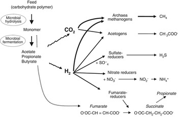
The rumen is an anaerobic, methanogenic environment where CO2 and H2 produced from the fermentation of feeds, which are the main electron acceptor and donor, respectively, within the system (Figure 1). In this type of habitat, the degradation of organic matter is accomplished by the concerted cooperation of different groups of anaerobes. Plant structural carbohydrates, proteins and other organic polymers contained in feeds ingested by the animal are degraded to their monomer components by primary anaerobic fermenters. These monomers are then further converted into volatile fatty acids, CO2 and H2 by both the primary fermenters and other microbes that do not have the capacity to hydrolyse complex polymers by themselves (secondary fermenters). Methanogens are at the bottom of this trophic chain and use the end products of fermentation as substrates (Figure 1). The synthesis of methane contributes to the efficiency of the system in that it avoids increases in the partial pressure of H2 to levels that might inhibit the normal functioning of microbial enzymes involved in electron transfer reactions, particularly NADH dehydrogenase, resulting in NADH accumulation, and ultimately reduce rumen fermentation. The capturing of the H2 produced by one microbial species by another is normally referred as interspecies H2 transfer (Wolin et al., Reference Wolin, Miller and Stewart1997) and is a process that in many cases involves a syntrophic relationship between two microbes.
Schematic microbial fermentation of feed polysaccharides and H2 reduction pathways in the rumen.

As discussed below the type of diet and the presence of electron acceptors other than CO2 in the rumen will have a major effect on the presence and activity of H2 producers and utilisers. This is because pathways other than methanogenesis can also consume H2 and thus potentially compete with and decrease methanogenesis in the rumen.
Rumen methanogens
Methanogens belong to the domain archaea. The diversity of archaea found in the rumen have been recently reviewed by Janssen and Kirs (Reference Janssen and Kirs2008). Most archaea identified in the rumen belong to known methanogen clades with a predominance of Methanobrevibacter spp. The pooled data from several surveys show that the Methanobrevibacter clade accounts for nearly two-thirds of rumen archaea. The remaining one-third was composed, of roughly equal parts by phylotypes belonging to Methanomicrobium and the rumen cluster C (Janssen and Kirs, Reference Janssen and Kirs2008). However, in some studies the proportion of these different groups was inversed or differed greatly (Janssen and Kirs, Reference Janssen and Kirs2008 and references therein), and it is not clear whether the major differences in archaeal distribution between studies are due to methodological differences or truly reflect differences due to animals and/or diets. Most rumen methanogens do not contain cytochromes and although they are less efficient at obtaining energy through the production of methane than their cytochrome-containing relatives of the order Methanosarcinales (Thauer et al., Reference Thauer, Kaster, Seedorf, Buckel and Hedderich2008), they are better adapted to the environmental conditions prevailing in the rumen. They have a lower threshold for H2 partial pressure, a fast doubling time, that can be as short as 1 h, and they develop better at the mesophilic temperature and near neutral pH of the rumen (Thauer et al., Reference Thauer, Kaster, Seedorf, Buckel and Hedderich2008).
There are three major substrates used by methanogens to produce methane: CO2, compounds containing a methyl group or acetate (Liu and Whitman, Reference Liu and Whitman2008). In the rumen, the predominant pathway is the hydrogenotrophic using CO2 as the carbon source and H2 as the main electron donor (Hungate, Reference Hungate1967). Formate is also an important electron donor used by many rumen hydrogenotrophic methanogens and may account for up to 18% of the methane produced in the rumen (Hungate et al., Reference Hungate, Smith, Bauchop, Yu and Rabinowitz1970). Methylamines and methanol produced in the rumen can also be used by methylotrophic methanogens of the order Methanosarcinales and Methanosphaera spp. from the order Methanobacteriales (Liu and Whitman, Reference Liu and Whitman2008). The contribution of these substrates to methanogenesis has not been measured, but it is likely to be small as the methanogens able to perform this conversion are not predominant members of the rumen methanogenic population (Janssen and Kirs, Reference Janssen and Kirs2008). Methane is also produced from acetate via the aceticlastic pathway and this pathway appears to be limited to members of the order Methanosarcinales (Liu and Whitman, Reference Liu and Whitman2008). Oppermann et al. (Reference Oppermann, Nelson and Brown1961) measured the increase in radio-labelled methane following infusion of 14C-acetate in vivo and concluded that this pathway was not important in the rumen. Methanosarcina grown on acetate had a very slow growth rate in vitro suggesting they would not thrive under normal rumen conditions and retention times (Stewart et al., Reference Stewart, Flint and Bryant1997). This was confirmed by molecular biology based culture-independent methods that retrieved only low numbers of Methanosarcina from the rumen (Janssen and Kirs, Reference Janssen and Kirs2008).
Methanogens are found associated with the rumen liquid and solid phases and also with the rumen epithelium. Information concerning the latter group is still scarce (Shin et al., Reference Shin, Choi, Lim, Hong, An, Cho, Kim, An, Kang and Lee2004; Pei et al., Reference Pei, Mao, Cheng and Zhu2010), but the rumen epithelium seems to sustain high concentration of methanogens and some novel phylotypes (Pei et al., Reference Pei, Mao, Cheng and Zhu2010). It is not known whether this methanogenic community has a significant role in rumen methanogenesis but methanogens attached to the gut epithelium have been described in termites (Leadbetter and Breznak, Reference Leadbetter and Breznak1996), and in such a microaerobic environment they are capable of producing methane and reducing oxygen at the same time (Tholen et al., Reference Tholen, Pester and Brune2007). Notwithstanding the possible contribution of epithelial methanogens, it is generally assumed that methanogens within the rumen milieu, be it in the free liquid, is associated with the solid digest or indeed attached to the rumen protozoa that make the major contribution to rumen methanogenesis.
Protozoa and methanogenesis
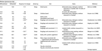
The rumen protozoa were first described in Gruby and Delafond (Reference Gruby and Delafond1843) and with their striking appearance it was assumed that they must be important for the welfare of their host. However, despite the fact that protozoa make up a large portion of the rumen biomass, their role in ruminal fermentation and their contribution to the metabolism and nutrition of the animal remains an area of considerable controversy (Williams and Coleman, Reference Williams and Coleman1992). Both flagellated and ciliated protozoa have been described in the rumen with ciliate protozoa by far the more prominent belonging to two orders: the Entodiniomorphida and the Vestibuliferida (Holotrich; Williams and Coleman, Reference Williams and Coleman1992). By classical morphological criteria, more than 250 species of ciliates have been described which live in the rumen of various feral and domesticated ruminants (Williams and Coleman, Reference Williams and Coleman1992). Ciliated protozoa can account for a substantial fraction of both the biodiversity and the biomass in the rumen and up to 100 billion ciliates belonging to more than 20 species may populate the rumen of a single cow (Williams and Coleman, Reference Williams and Coleman1992). Rumen ciliated protozoa are metabolically very active, able to influence fermentation of feeds and other rumen microbial populations and, consequently, to affect the amount and proportion of the end products from rumen fermentation including methane (Williams and Coleman, Reference Williams and Coleman1992; Eugène et al., Reference Eugène, Archimède and Sauvant2004). Protozoa engulf organic matter, particularly bacteria, into digestive vacuoles where hydrolysis and fermentation take place. The main volatile fatty acids produced are acetate and butyrate (Williams and Coleman, Reference Williams and Coleman1992; Hillman et al., Reference Hillman, Williams and Lloyd1995). Rumen protozoa are not essential to the animal to survive and defaunation (the removal of protozoa from the rumen using a wide variety of chemical and physical techniques) has been widely used to study the role of ciliate protozoa in rumen function (Williams and Coleman, Reference Williams and Coleman1992). However, the results obtained do need to be interpreted with some caution as defaunation also leads to major changes in the rumen microbial population (Williams and Coleman, Reference Williams and Coleman1992). In a meta-analysis of published data, Eugène et al. (Reference Eugène, Archimède and Sauvant2004) concluded that defaunation resulted in an increase in the molar proportion of propionate in the rumen (P < 0.05) and a decrease in concentrations of butyrate (P < 0.05) and acetate (P = 0.08). On the basis of stoichiometry, such a shift in rumen volatile fatty acid production should result in a decrease in methane production as less metabolic H2 will be available as a substrate for methanogenesis (Demeyer, Reference Demeyer1991). The effect of defaunation on methane production are less clear; Hegarty (Reference Hegarty1999) summarised published in vivo and in vitro trails and showed that the removal of protozoa from the rumen would result in a 13% decrease in methane production. We have repeated and updated this analysis using only in vivo studies (Table 1), again taken as an average over all studies defaunation resulted in a 10.5% decrease in methane emissions. However, a great deal of variability can be seen within the available data, with some studies recording increased emissions of methane following defaunation (Table 1). Although undoubtedly much of the variation can be explained by methodological differences including the method used to obtain protozoa-free animals, there is also evidence of a dietary effect and more study is required to help fully explain the relationship between defaunation and methanogenesis.
The effect of defaunation on methane production as measured in in vivo experiments

*, ** Indicate significant differences (P < 0.05 and P < 0.01, respectively) as reported in the original reference.
To further explore the relationship between methane production and rumen protozoa we analysed all available in vivo data in which measurements of methane emissions, intake and rumen protozoa numbers were recorded. A total of 17 publications and unpublished trials from our laboratory corresponding to 21 experiments fitted this criterion. The data set contained a range of dietary treatments that were tested for their modulating effect on methanogenesis. A total of 65 treatments were kept for analysis of which 21 were controls and 44 were experimental diets. The experimental factors tested in these diets were lipid supplementation (n = 23) of up to 7% of offered feed dry matter, plants or plant extracts rich in tannins or saponins (n = 16), and diets rich in starchy concentrates (30% to 50% of offered feed dry matter, n = 5). Cattle and small ruminants (sheep and goat) were equally represented (48% and 52%, respectively). The majority of the data (85%) were from production trials and the remaining were from trials in which animals were fed at maintenance. Methane production was measured in chambers (63% in open chambers or respiratory calorimeters) or with the SF6 (sulphur hexafluoride) tracer method (37%) of Johnson et al. (Reference Johnson, Huyler, Westberg, Lamb and Zimmerman1994).
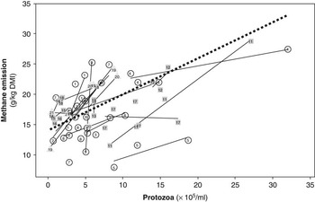
Data obtained over this broad range of conditions were analysed using the general linear procedure included in Minitab Version 14 (Minitab Inc., State college, PA, USA) to evaluate the relationship between methane yield (g/kg dry matter intake (DMI)) and concentration of protozoa (105/ml of rumen liquid; Figure 2). The linear model included the effect of the study and of the dietary treatment as fixed effect (Sauvant et al., Reference Sauvant, Schmidely, Daudin and St-Pierre2008). We observed that the number of protozoa explained 47% of the variability in methane emissions (P < 0.0001) suggesting that protozoa played a catalytic role in rumen methanogenesis under the experimental conditions studied. The extent of the decrease in methane yield averaged 0.6 g methane/kg DMI per reduction of 105 protozoal cell/ml and does not appear to be affected by the type of dietary treatment studied (P = 0.14), but methane yield indicated by the intercept of the regressions (not shown) differed slightly between the treatments (P = 0.07). This difference is probably due to the nature of the dietary treatments and corresponding controls and to the different experimental conditions associated with each one of them, for example, mainly small ruminants for plant extracts and only productive cattle for starch-rich concentrates. Within the data set there are a few contrasting results that are not large enough to affect the tendency, but that should be noted. Guan et al. (Reference Guan, Wittenberg, Ominski and Krause2006) observed a significant decrease in protozoal numbers for a high-concentrate diet compared with a low-concentrate diet without any modification in the amount of methane emitted. A large variation in methane emission without any effect on protozoal numbers was observed in some trials using condensed tannins (Animut et al., Reference Animut, Puchala, Goetsch, Patra, Sahlu, Varel and Wells2008), starch-rich concentrates (Martin et al., Reference Martin, Ferlay, Chilliard and Doreau2007a and unpublished data) and lipids (Machmüller and Kreuzer, Reference Machmüller and Kreuzer1999, Machmüller et al., Reference Machmüller, Soliva and Kreuzer2003a). This analysis confirms the important influence that rumen protozoa have on methanogenesis under certain conditions. Where possible we suggest that protozoal measurements should be done in all animal trials in which methane mitigation options are tested to corroborate this finding. Protozoa may also be important for the success of other antimethanogenic strategies. For instance, the temporary mitigation effect of ionophores in a cattle trial has been directly associated with the recovery of protozoal numbers in animals following 4 to 6 weeks of continuous treatment (Guan et al., Reference Guan, Wittenberg, Ominski and Krause2006).
Methane emissions and rumen protozoa concentration in ruminants receiving diets rich in starchy concentrates (△), lipids (○) and plant extracts (□): review of available literature. Data from Machmüller and Kreuzer, Reference Machmüller and Kreuzer1999 (7); Machmüller et al., Reference Machmüller, Ossowski and Kreuzer2000 (8); Sliwinski et al., Reference Sliwinski, Kreuzer, Wettstein and Machmüller2002 (17); Lovett et al., Reference Lovett, Lovell, Stack, Callan, Finlay, Conolly and O’Mara2003 (6, 20); Machmüller et al., Reference Machmüller, Soliva and Kreuzer2003a and Reference Machmüller, Soliva and Kreuzer2003b (9, 10); Hess et al., Reference Hess, Beuret, Lotscher, Hindrichsen, Machmüller, Carulla, Lascano and Kreuzer2004 (14, 15, 16); Lila et al., Reference Lila, Mohammed, Kanda, Kurihara and Itabashi2005 (18); Guan et al., Reference Guan, Wittenberg, Ominski and Krause2006 (21); Jordan et al., Reference Jordan, Lovett, Hawkins, Callan and O’Mara2006a (5); Jordan et al., Reference Jordan, Kenny, Hawkins, Malone, Lovett and O’Mara2006b (4); Martin et al., Reference Martin, Ferlay, Chilliard and Doreau2007a and unpublished data (19); Martin et al., Reference Martin, Dubroeucq, Micol, Agabriel and Doreau2007b and unpublished data (1); Pen et al., Reference Pen, Takaura, Yamaguchi, Asa and Takahashi2007 (12); Animut et al., Reference Animut, Puchala, Goetsch, Patra, Sahlu, Varel and Wells2008b (11), Beauchemin et al., Reference Beauchemin, McGinn, Benchaar and Holtshausen2009 (3); Holtshausen et al., Reference Holtshausen, Chaves, Beauchemin, McGinn, McAllister, Odongo, Cheeke and Benchaar2009 (13); Martin et al. personal communication (2). The black dashed line represents the regression after taking into account the effect of study: methane (g/kg dry matter intake) = 13.95 (s.e. = 0.87) + 0.6×10⁻⁵ (s.e. = 0.11) × protozoa (105/ml); (R 2 = 0.65; R 2 adjusted = 0.47; P < 0.001).

A number of mechanisms by which protozoa could enhance methanogenesis are possible in which protozoa produce H2, serve as hosts for methanogens and also protect them from oxygen toxicity. H2 is a fermentation by-product that is produced in large quantities by the protozoa in a specialised organelle equivalent to the mitochondria of aerobic eukaryotes: the hydrogenosome. This H2 is utilised by methanogens that are found inside (Finlay et al., Reference Finlay, Esteban, Clarke, Williams, Embley and Hirt1994) or in close association with protozoal cells (Stumm et al., Reference Stumm, Gijzen and Vogels1982). This interaction is a typical example of interspecies H2 transfer that favours both the methanogens and the protozoa (Hillman et al., Reference Hillman, Lloyd and Williams1988; Ushida et al., Reference Ushida, Newbold and Jouany1997). Removal of H2 allows the fermentation of organic matter to proceed mainly to acetate and CO2 at the expense of butyrate and lactate production resulting in more efficient ATP production by the protozoan host.
Intracellular methanogens are found inside the most common protozoal species accounting for 1% to 2% of the host cell volume (Finlay et al., Reference Finlay, Esteban, Clarke, Williams, Embley and Hirt1994). It has been shown that in other environments including freshwater ecosystems and also in the gastrointestinal tract of invertebrates and amphibians, the endosymbiont methanogens within protozoa are protozoan genera-specific (van Hoek et al., Reference van Hoek, van Alen, Sprakel, Leunissen, Brigge, Vogels and Hackstein2000). However, there is still no solid evidence that the same phenomenon occurs in rumen ciliates (Janssen and Kirs, Reference Janssen and Kirs2008). Tóthová et al. (Reference Tóthová, Piknová, Kišidayová, Javorský and Pristaš2008) have found that a specific methanogen, which differed from other rumen methanogens, could be isolated from in vitro cultures of the rumen ciliate Entodininium caudatum, but it is not clear whether this is an artefact of the culture technique. Others (Sharp et al., Reference Sharp, Ziemer, Stern and Stahl1998; Ohene-Adjei et al., Reference Ohene-Adjei, Teather, Ivan and Forster2007) have detected changes in the methanogenic phylotypes retrieved in the presence or absence of protozoa, but again it is not clear to what extent this could be due to a specific association of methanogens with individual protozoal genera and to what extent it reflects the wider changes in rumen metabolism associated with defaunation and selective refaunation (Williams and Coleman, Reference Williams and Coleman1992).
In contrast to intracellular methanogens, the methanogens attached to the cell surface appear to be less numerous and less ubiquitous, as at a given time only 30% to 50% of protozoal cells being observed do have extracellular-associated methanogens (Vogels et al., Reference Vogels, Hoppe and Stumm1980; Finlay et al., Reference Finlay, Esteban, Clarke, Williams, Embley and Hirt1994). The extent of the association is influenced by diet and time of feeding (Stumm et al., Reference Stumm, Gijzen and Vogels1982; Tokura et al., Reference Tokura, Ushida, Miyazaki and Kojima1997). Adherence of methanogens to protozoa is low after feeding and appears to be related to the relative abundance of H2 in the medium (Stumm et al., Reference Stumm, Gijzen and Vogels1982). Most Entodiniomorphs seem able to carry methanogens on their surface, but no episymbiotic methanogens were observed associated with Holotrichs protozoa (Vogels et al., Reference Vogels, Hoppe and Stumm1980). Nevertheless, the Holotrich Isotricha prostoma isolated from the rumen produced large amount of methane indicating that extracellular, as opposed to intracellular, methanogens are not crucial (Ushida and Jouany, Reference Ushida and Jouany1996). Tokura et al. (Reference Tokura, Ushida, Miyazaki and Kojima1997) used mixed protozoa isolated from the rumen at different times of the day and reported that the number of both intra- and epi-associated methanogens per protozoal cell was less than three per cell before feeding and increasing to 100 to 1000 per cell after 1 h of feeding. Taken together, these data suggest that intracellular methanogens are more important both in number and activity than the externally associated methanogens.
A single protozoan may produce between 0.5 and 4.5 nmol H2/day (Williams and Coleman, Reference Williams and Coleman1992; Finlay et al., Reference Finlay, Esteban, Clarke, Williams, Embley and Hirt1994; Ushida and Jouany, Reference Ushida and Jouany1996) with a production of methane that ranges from trace amounts up to 3 nmol/day (Ushida and Jouany, Reference Ushida and Jouany1996; Tokura et al., Reference Tokura, Ushida, Miyazaki and Kojima1997). There is some discrepancy in these figures as using stoichiometric calculations, the H2 produced per protozoan is not enough to synthesise all the methane emitted. Protozoa also produce considerable amounts of formate (Tokura et al., Reference Tokura, Ushida, Miyazaki and Kojima1997), which can be partly used as a substrate for methane production. In addition, the methodology used in these different works might have contributed to this variation. Not all protozoal genera will have the same role in methanogenesis and despite their importance there is still scant information on the individual contribution to methane emissions. The cellulolytic protozoan Polyplastron was a weak producer, Epidinium caudatum was intermediate and I. prostoma and Entodinium caudatum were high producers (Newbold et al., Reference Newbold, Lassalas and Jouany1995; Ushida and Jouany, Reference Ushida and Jouany1996; Ranilla et al., Reference Ranilla, Jouany and Morgavi2007). When single protozoa were incubated at densities normally found in the rumen Entodinium sp. was identified as the most important protozoal genera contributing to methane emissions (Ranilla et al., Reference Ranilla, Jouany and Morgavi2007). However, these results were obtained in vitro and need to be confirmed in animal trials.
As mentioned at the beginning of this section, rumen protozoa are metabolically very active and influence rumen function in several ways. In addition to their role in methane emissions, the presence of protozoa is positively related to fibre digestion and is negatively associated with the outflow of microbial protein from the rumen (Williams and Coleman, Reference Williams and Coleman1992; Eugène et al., Reference Eugène, Archimède and Sauvant2004). All these aspects should be considered in any strategy aiming to control the protozoal population in the rumen. Williams and Withers (Reference Williams and Withers1993) indicated that when the numbers of protozoa were low their effect on fibre hydrolysis was still positive but microbial protein synthesis seemed not to be affected. Incidentally, the results presented in Figure 2 also indicate the benefits of low protozoal populations on methane emissions. Methods, other than dietary, are needed to control the protozoal load and evaluate the effect of reduced protozoal populations on emissions and animal performances.
Plant fibre degradation and H2 production
Fibrolytic microorganisms play a pivotal role in the rumen ecosystem. Indeed, they are at the first level of the microbial trophic chain transforming plant cell wall polysaccharides from feeds into volatile fatty acids, CO2 and H2. They are thus considered as a major microbial functional group in the rumen with a population estimated at ≈109 cells/ml digesta either by culture or by molecular methods (Mosoni et al., Reference Mosoni, Chaucheyras-Durand, Bera-Maillet and Forano2007). Most members of this group produce H2 as a main end product of fermentation, which under normal physiological conditions, is in turn rapidly used by methanogens. This interspecies H2 transfer between H2 producers and utilisers is fundamental in the functioning of anaerobic ecosystems including the rumen (Wolin et al., Reference Wolin, Miller and Stewart1997). If this transfer is affected, the build-up of H2 in the milieu inhibits the reoxidation of co-enzymes involved in redox reactions within bacterial cells, ultimately depressing the fermentation processes. This is the reason why methanogenesis is intimately linked to degradation of plant fibre in the rumen.
Interspecies H2 transfer has been well described in vitro, especially between cellulolytics and methanogens (Wolin et al., Reference Wolin, Miller and Stewart1997). One of the consequences of this interaction is a shift in fermentative metabolic pathways of the H2 producers. For example, the fibrolytic bacterium Ruminococcus albus produces ethanol, acetate, H2 and CO2 in monoculture in vitro, whereas it does not produce ethanol when co-cultured with a methanogen (Wolin et al., Reference Wolin, Miller and Stewart1997). When R. albus is co-cultivated with a methanogen, H2 never accumulates in the culture as it is transformed into methane, and acetate is the main metabolite produced. In the presence of methanogens, as in the rumen, R. albus will thus mainly produce acetate from cellulose, and cellulose degradation is increased in vitro (Pavlostathis et al., Reference Pavlostathis, Miller and Wolin1990). Other bacterial species such as Ruminococcus flavefaciens and all the rumen fungi and protozoa produce H2 and have been shown to interact positively with methanogens (Latham and Wolin, Reference Latham and Wolin1977; Joblin et al., Reference Joblin, Naylor and Williams1990; Williams et al., Reference Williams, Withers and Joblin1994). As described above for R. albus, in addition to changing the end products of fermentation, an increase in cellulolysis was also observed when anaerobic fungi are cultured in association with methanogens (Latham and Wolin, Reference Latham and Wolin1977; Joblin et al., Reference Joblin, Naylor and Williams1990). This synergic effect was also measured in vivo, in the rumen of gnotobiotic lambs with controlled microbiota (Fonty et al., Reference Fonty, Williams, Bonnemoy, Morvan, Withers and Gouet1997); the establishment of a methanogenic strain in the rumen of lambs (previously lacking methanogens) induced an increase in the population of R. flavefaciens, as well as in straw degradation and glycoside hydrolase activity.
Recent in vitro work investigated the effects of methanogen inhibition on fibrolytic populations in complex rumen microbiota (Table 2). Inhibitors of the methane pathway, bromochloromethane (BCM) and bromoethanesulfonate (BES) led to a disappearance of the methanogen populations and to a strong decrease in methane production. BCM led to a decrease in R. flavefaciens populations with a concomitant proliferation of Fibrobacter and fungi with no effect on total bacteria and protozoa (Goel et al., Reference Goel, Makkar and Becker2009). In contrast, BES had no effect on R. flavefaciens and decreased significantly the fungal population while increasing Fibrobacter (Guo et al., Reference Guo, Liu, Zhu and McSweeney2007). These different effects of BCM and BES on the ruminococcal and fungal populations are not easily explained, and could be due to different experimental conditions. The addition of different saponins, used as defaunating agents, led to a marked decrease in protozoa, as expected, but sometimes also in fungi and methanogens (Table 2). In Guo et al. (Reference Guo, Liu, Lu, Zhu, Denman and McSweeney2008), the methanogen populations were not affected by the addition of tea saponins, but the methanogenic activity, estimated by quantification of the expression of the mcrA gene (coding for the enzyme of the terminal step of the methanogenesis pathway), clearly decreased. Methane production was slightly affected by saponins (<10% decrease), which correlates with in vivo data (see the previous section: Protozoa and methanogenesis). The Fibrobacter population tend to increase (Table 2; Wina et al., Reference Wina, Muetzel, Hoffmann, Makkar and Becker2005; Goel et al., Reference Goel, Makkar and Becker2008; Guo et al., Reference Guo, Liu, Lu, Zhu, Denman and McSweeney2008). To summarise the results of these in vitro experiments, the inhibition of methanogens was correlated to a decrease in methane synthesis, but it also led to an inhibition of the Ruminococci and the fungi in some of them. The inhibition of methanogenesis is expected to increase the partial pressure of H2 leading to an inhibition of H2-producing microorganisms such as the Ruminococci and the fungi. The data collected in Table 2 suggest that in the complex rumen microbiota the mechanisms involved in H2 balance are not straightforward. Nevertheless, fibre digestibility was not affected in these experiments (Table 2), except in Wina et al. (Reference Wina, Muetzel, Hoffmann, Makkar and Becker2005). The increase in Fibrobacter populations observed in these studies may have allowed an efficient fibre degradation to be maintained. Thus, promoting non-H2-producing fibrolytic organisms such as Fibrobacter succinogenes might be an alternative way to decreasing methane emissions in the rumen without impairing fibre digestibility.
Effect of several additives on methane production, microbial populations, fibre digestibility and acetate/propionate ratio in rumen incubations in vitro Footnote a

Rfl = Ruminococcus flavefaciens; Ral = Ruminococcus albus; Fsu = Fibrobacter succinogenes; BCM = bromochloromethane; BES = bromoethanesulfonate.
a All the additives were added to batch mixed cultures in vitro except BCM 2 that was added to continuous cultures.
Cellulolytic bacterial species, methanogens and fungi were estimated by quantification of their rrs gene by relative quantitative PCR, except in the work of Wina et al. (Reference Wina, Muetzel, Hoffmann, Makkar and Becker2005) in which they were estimated by quantification of 16S RNA using dot blots, and in Guo et al. (Reference Guo, Liu, Lu, Zhu, Denman and McSweeney2008), in which methanogens were estimated by PCR quantification of the mcrA gene.
Protozoa were quantified by cell counting or by rrs gene PCR quantification (only in Guo et al., Reference Guo, Liu, Lu, Zhu, Denman and McSweeney2008).
*, ** Indicate significant differences (P < 0.05 and P < 0.01, respectively) as reported in the original reference.
The effect of the composition of the fibrolytic community on methane production has also been investigated. In vitro incubation of rumen content from gnotobiotic lambs harbouring H2-producing (Ruminococci and fungi) or non-H2 producing (Fibrobacter) fibrolytic microorganisms and methanogens indicated that methane was produced in higher quantities by the inoculum containing the H2-producing cellulolytic microbiota (Chaucheyras-Durand et al., Reference Chaucheyras-Durand, Masseglia, Fonty and Forano2008). These results suggest that the composition of the fibrolytic community (producers or non-producers of H2) may have an impact on H2 accumulation and subsequent methane production. However, in an in vivo study with gnotobiotic lambs, inoculation of a methanogenic strain increased both the Ruminococci and the Fibrobacter populations (Mosoni et al., Reference Mosoni, Gagen, Denman, McSweeney and Forano2008).
Earlier study has shown that numbers of fibrolytic bacteria were positively correlated to that of methanogens in the rumen of various animals, including cattle, sheep, deer and llama, and in the horse caecum (Morvan et al., Reference Morvan, Bonnemoy, Fonty and Gouet1996). This correlation was explained because the major fibrolytic species such as R. albus and R. flavefaciens produce H2 as a main fermentation end product. In contrast, the same authors reported that in the rumen of buffalo the numbers of methanogens were comparatively lower than that of fibrolytic bacteria. This may be because Fibrobacter, a bacterium that does not produce H2, was the dominant fibrolytic bacterium in the rumen of buffalos (Morvan et al., Reference Morvan, Dore, Rieu-Lesme, Foucat, Fonty and Gouet1994). Unfortunately, methane emissions were not measured in the study. Ionophores such as monensin have been shown to cause a moderate inhibition of methane production, and a similar mechanism was proposed because monensin is known to inhibit the Ruminococci without affecting F. succinogenes (Chen and Wolin, Reference Chen and Wolin1979). The relationship between cellulolytic microorganisms and methane production thus merits further investigation as promoting non-H2-producing fibrolytic microbes could be a way to decrease H2 – and consequently methane- production while keeping active rumen fibre degradation.
In conclusion, although some in vitro experiments tend to show a relationship between methanogens or methane production and the ratio of H2-producing/non-H2-producing fibrolytic bacteria, which could be explained on the basis of interspecies H2 transfer, in vivo experiments are needed to explore more deeply these relationships in the rumen.
Reductive acetogenesis
The conversion of H2 and CO2 into acetate through reductive acetogenesis occurs in many microbial anaerobic ecosystems including the gastrointestinal tract of non-ruminant animals, where it can co-exist with methanogenesis (Klieve and Ouwerkerk, Reference Klieve and Ouwerkerk2007). A decreased emission of methane associated with an increase in acetate available to the host animal is a desirable condition that, combined with the observation that reductive acetogenesis is the main electron sink observed in many non-ruminant herbivores (Klieve and Ouwerkerk, Reference Klieve and Ouwerkerk2007), has stimulated the study to promote this pathway in the rumen (Lopez et al., Reference Lopez, McIntosh, Wallace and Newbold1999). Active homoacetogens become established in the rumen of newborn animals as early as 20 h after birth (Morvan et al., Reference Morvan, Dore, Rieu-Lesme, Foucat, Fonty and Gouet1994). Methanogens became established soon after, as they are detected within 1 to 3 days after birth, but when methanogens’ numbers increase, before reaching adult concentrations at 3 weeks of age in lambs, the concentration of homoacetogens decrease (Morvan et al., Reference Morvan, Dore, Rieu-Lesme, Foucat, Fonty and Gouet1994; Skillman et al., Reference Skillman, Evans, Naylor, Morvan, Jarvis and Joblin2004). In adult animals, densities of homoacetogens range from 103 to 107/g (Le Van et al., Reference Le Van, Robinson, Ralph, Greening, Smolenski, Leedle and Schaefer1998; Fonty et al., Reference Fonty, Joblin, Chavarot, Roux, Naylor and Michallon2007). Numbers are affected by diet and also by the technique used to cultivate and enumerate these bacteria, but they are at least 10-fold less numerous than the methanogens (Le Van et al., Reference Le Van, Robinson, Ralph, Greening, Smolenski, Leedle and Schaefer1998; Fonty et al., Reference Fonty, Joblin, Chavarot, Roux, Naylor and Michallon2007). In lambs isolated at birth with a controlled rumen microbiota not harbouring methanogens, homoacetogen numbers were 107 to 108/g and reductive acetogenesis represented up to 25% of the total fermentation output (Fonty et al., Reference Fonty, Joblin, Chavarot, Roux, Naylor and Michallon2007). However, the H2 recovery calculated from the stoichiometry of VFA production was less than 50%, indicating a probable loss of H2 as gas. Introduction of methanogens into the rumen of these animals eliminated reductive acetogenesis altogether and restored H2 recovery to normal values, near 100% (Fonty et al., Reference Fonty, Joblin, Chavarot, Roux, Naylor and Michallon2007). Homoacetogens are outcompeted by methanogens because they are less efficient at obtaining energy from the oxidation of H2 and have both a lower affinity and a higher threshold for the substrate (Hoehler et al., Reference Hoehler, Alperin, Albert and Martens1998; Le Van et al., Reference Le Van, Robinson, Ralph, Greening, Smolenski, Leedle and Schaefer1998; Weimer, Reference Weimer1998). The partial pressure of H2 in the rumen is normally lower than the threshold of reductive acetogenesis. Isolates from the rumen, in addition to being autotrophic are also heterotrophic and it has been proposed that they rely mainly on organic substrates for their survival in the rumen ecosystem (Le Van et al., Reference Le Van, Robinson, Ralph, Greening, Smolenski, Leedle and Schaefer1998). Attempts to boost reductive acetogenesis by increasing the numbers of natural rumen homoacetogens or by addition of non-rumen isolates have not been successful in several in vitro and in vivo trials (Demeyer et al., Reference Demeyer, Fiedler and DeGraeve1996; Immig et al., Reference Immig, Demeyer, Fiedler, Van Nevel and Mbanzamihigo1996; Nollet et al., Reference Nollet, Demeyer and Verstraete1997 and Reference Nollet, Mbanzamihigo, Demeyer and Verstraete1998; Le Van et al., Reference Le Van, Robinson, Ralph, Greening, Smolenski, Leedle and Schaefer1998; Lopez et al., Reference Lopez, McIntosh, Wallace and Newbold1999).
Bacteria using alternative H2 sinks
Some bacteria present in the rumen can respire anaerobically. These bacteria are capable of using electron acceptors other than CO2 to oxidise H2, hence decreasing its availability to form methane (Figure 1). Sulphate-reducing bacteria, Denitribacterium detoxificans and Wolinella succinogenes utilise sulphate, nitrocompounds and nitrate, respectively, as electron acceptors (Weimer, Reference Weimer1998; Anderson et al., Reference Anderson, Rasmussen, Jensen and Allison2000; Simon, Reference Simon2002). Normally these bacteria are not dominant in the ecosystem, but their numbers can increase if the right potential electron acceptor is provided in the diet (Figure 1). The reduction of sulphate is not desirable as the end product, H2 sulphide, is toxic to the host animal (Gould et al., Reference Gould, Cummings and Hamar1997). Nitrocompounds have been shown to reduce methanogenesis (Anderson et al., Reference Anderson, Carstens, Miller, Callaway, Schultz, Edrington, Harvey and Nisbet2006). However, the main mechanism of action of nitrocompounds seems to be mediated by the inhibition of formate dehydrogenases and/or H2 dehydrogenases (Anderson et al., Reference Anderson, Krueger, Stanton, Callaway, Edrington, Harvey, Jung and Nisbet2008) and, paradoxically, increases in the numbers of D. detoxificans, which uses H2 to reduce these compounds, may reduce the abating potential of nitrocompounds (Anderson et al., Reference Anderson, Carstens, Miller, Callaway, Schultz, Edrington, Harvey and Nisbet2006, Gutierrez-Banuelos et al., Reference Gutierrez-Banuelos, Anderson, Carstens, Slay, Ramlachan, Horrocks, Callaway, Edrington and Nisbet2007). In contrast, one strain of D. detoxificans was able to utilise nitrate and reduced methane production in vitro when this substrate was added (Anderson and Rasmussen, Reference Anderson and Rasmussen1998).
Nitrate, like sulphur, can be considered as an alternative electron sink. The thermodynamics of the conversion of nitrate into NH3 is more favourable than the formation of methane and can effectively replace methanogenesis in the rumen if nitrate is available. However, in the rumen nitrate is rapidly reduced into nitrite and the rate of reduction of nitrite into NH3 is slower (Iwamoto et al., Reference Iwamoto, Asanuma and Hino1999). Nitrite accumulation is undesirable as it is extremely toxic to the host animal. In spite of this, increases in the number of bacteria capable of reducing nitrite have been shown to reduce both methane production and nitrite toxicity. Wolinella succinogenes, a rumen bacterium that grows by respiratory nitrate ammonification (Simon, Reference Simon2002), has the ability to reduce nitrate into NH3 with little accumulation of nitrites. Wolinella was shown to reduce methane production in vitro but only if nitrate was added to the incubation media (Iwamoto et al., Reference Iwamoto, Asanuma and Hino2002). These effects were also demonstrated in vivo using strains of Escherichia coli with high nitrate/nitrite-reducing activity (Sar et al., Reference Sar, Mwenya, Santoso, Takaura, Morikawa, Isogai, Asakura, Toride and Takahashi2005a and Reference Sar, Mwenya, Santoso, Takaura, Morikawa, Isogai, Asakura, Toride and Takahashi2005b).
Methanotrophy, that is, the oxidation of methane, was reported to account for <0.5% of rumen methane production in vitro (Kajikawa et al., Reference Kajikawa, Valdes, Hillman, Wallace and Newbold2003). This oxidation was found to be anaerobic and associated with sulphate reduction. In contrast, putative methane-oxidising bacteria, evidenced by PCR amplification with specific primers, were found attached to the rumen epithelium (Mitsumori et al., Reference Mitsumori, Ajisaka, Tajima, Kajikawa and Kurihara2002) where conditions may favour aerobic oxidation of methane. This metabolic pathway has yet to be quantified in vivo. In terms of benefit to the host, the final product of this pathway, CO2, does not supply any energy, and the nitrogen and energy contained in the biomass of methanotrophs would be negligible. Capnophily, that is, the ability to use CO2, is also present in the rumen with many rumen isolates from common bacterial species such as Prevotella, Fibrobacter, Ruminococcus, Lachnospira and Succinomonas requiring CO2 to grow (Dehority, Reference Dehority1971). Capnophilic bacteria also use H2 to produce organic acids, particularly succinic acid as final products, but the influence of this metabolic conversion on H2 balance is not known. Mannheimia succiniciproducens, a rumen bacterium whose genome has been sequenced, produces succinic acid as the main metabolic product and consumes CO2 and H2 in the process (Hong et al., Reference Hong, Kim, Lee, In, Choi, Rih, Kim, Jeong, Hur and Kim2004). This and other similar bacteria may have an effect on net H2 and methane balance if their numbers and activity are sufficient in the rumen.
Concluding remarks
The microbiota and the host animal have co-evolved for millions of years and the production of methane is a mechanism that improves the fermentation process. There is not a clear relationship between the number of methanogens and methanogenesis in the rumen (Nollet et al., Reference Nollet, Mbanzamihigo, Demeyer and Verstraete1998; Machmüller et al., Reference Machmüller, Soliva and Kreuzer2003a and Reference Machmüller, Soliva and Kreuzer2003b; Yanez-Ruiz et al., Reference Yanez-Ruiz, Hart, Martin-Garcia, Ramos and Newbold2008; Mosoni et al. personal communication), except for trials when specific inhibitors of methanogenesis were used (i.e. BSM; Denman et al., Reference Denman, Tomkins and McSweeney2007). It is reasonable to think that the reduction in methane emissions without an associated change in the number of methanogens must be attributed to changes in the rumen microbiota and the availability of H2. In this study, we show that there is direct correlation between the number of protozoa and methane production. Indeed, some of the most successful strategies to reduce emissions have to some extent a negative effect upon protozoa. The balance between H2 producers and non-H2 producers in the fibrolytic community can be shifted under experimental conditions to reduce methanogenesis without altering their fibre-degrading capability. Communities predominantly composed of non-H2 producers have been observed in some ruminant species under natural conditions indicating the viability of the approach. Finally, the use of an alternative electron sink such as nitrate needs to be revisited and tested under long periods of supplementation to allow increases in the number of nitrate utilisers in the rumen and avoid toxicity. Nitrate supplementation could be a feasible alternative in N-poor diets.
Decreasing rumen methanogenesis is possible, with some strategies already available and many others that are being tested. It is possible to modify the microbial ecosystem to decrease its production of methane, but the manipulation of microbial components of the system has to be considered in an integrated way. In the next few years, advances brought about through rumen metagenomic projects and the utilisation of new ‘omics’ technologies will broaden our understanding of the mechanisms involved in methanogenesis and other metabolic H2-consuming and releasing processes, and will help the scientific community to find new targets for mitigation.
Acknowledgement
We are grateful to M. Eugène for her input and help in the analysis of the protozoa/methane data.